
Pattern analysis of retinal maps for the diagnosis of optic nerve diseases by optical coherence tomography

a technology of optical coherence tomography and retinal maps, applied in the field of ophthalmology, can solve the problems of difficult clinical examination detection of nfl bundle defects, difficult to easily utilize knowledge in diagnostic methods, and difficult to achieve high-quality construction

Active Publication Date: 2008-12-18
USC STEVENS UNIV OF SOUTHERN CALIFORNIA
View PDF10 Cites 91 Cited by
  • Summary
  • Abstract
  • Description
  • Claims
  • Application Information

AI Technical Summary

Benefits of technology

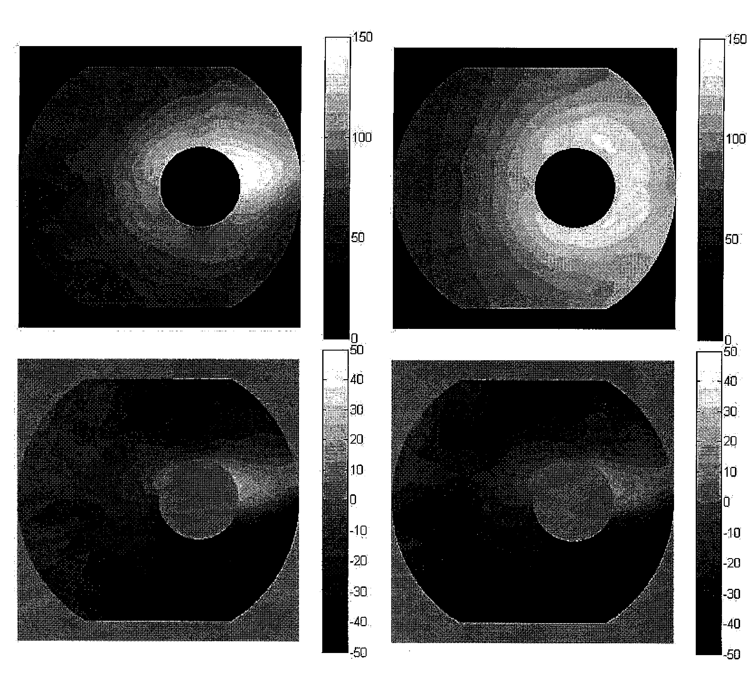
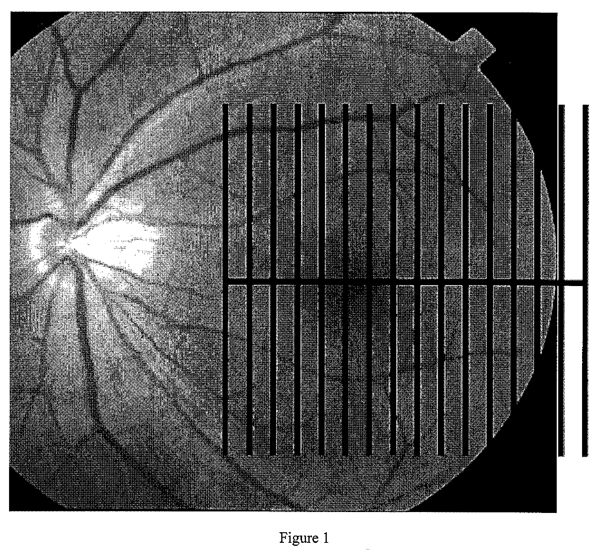
[0022]In the methods of the present invention, the inventors have discovered that the quality of the macular maps and the subsequent construction of the thickness maps are highly dependent on the scanning patterns employed. Accordingly, inventors of the present invention have devised novel scanning patterns for achieving rapid macular scans that can facilitate the construction of high quality three-dimensional thickness maps.

Problems solved by technology

While this principle is simple in theory, it is not easy in practice.
Unfortunately, in practice, this knowledge cannot be easily utilized in diagnostic methods.
Likewise, NFL bundle defects are difficult to detect on clinical examination.
Although red-free fundus photography is capable of detecting changes in the vascular system and nerve fibers of the retina, the technique is rarely used in clinical practice.
However, since a significant loss to RGC population can occur prior to detectable visual field deficits, and this structural loss can precede detectable function loss by up to 5 years, current methods for clinical diagnosis of glaucoma are not adequate for early detection of the disease.

Method used


Image

  • Pattern analysis of retinal maps for the diagnosis of optic nerve diseases by optical coherence tomography
  • Pattern analysis of retinal maps for the diagnosis of optic nerve diseases by optical coherence tomography
  • Pattern analysis of retinal maps for the diagnosis of optic nerve diseases by optical coherence tomography

Examples


example

Detection of Macular Ganglion Cell Loss in Glaucoma

Methods

1. Clinical Study

[0098]Participants in the prospective Advanced Imaging for Glaucoma Study (AIGS) between the periods of 2003 and 2007 were included. These participants were classified into four groups: normal (N), perimetric glaucoma (PG), glaucoma suspect (GS) and pre-perimetric glaucoma (PPG). Only the data from the baseline visit was used. The GS group was not used in this study because the members' glaucoma status was indeterminate. We used only data from AIGS centers that employed FD-OCT during the study period. The eligibility criteria for the three groups analyzed are briefly described below.

[0099]The N group participants had intraocular pressure (IOP) of less than 21 mm Hg for both eyes, a normal Humphrey SITA 24-2 visual field (VF) [mean deviation (M) and pattern standard deviation (PSD) within 95% limits of the normal reference and a glaucoma hemifield test (GHT) within 97% limits], a central corneal thickness ≧500...


PUM


Abstract

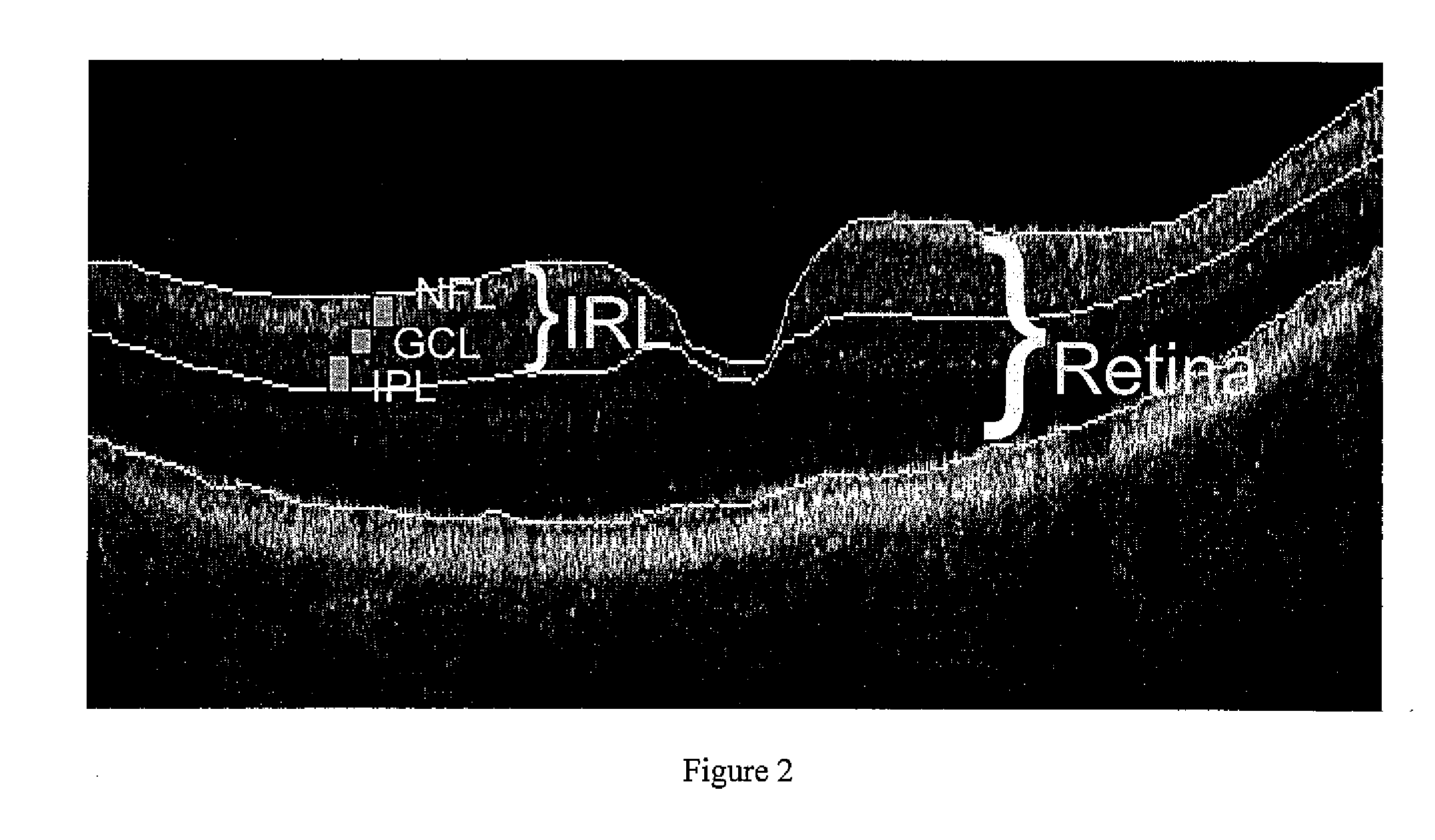
Methods for analyzing retinal tomography maps to detect patterns of optic nerve diseases such as glaucoma, optic neuritis, anterior ischemic optic neuropathy are disclosed in this invention. The areas of mapping include the macula centered on the fovea, and the region centered on the optic nerve head. The retinal layers that are analyzed include the nerve fiber, ganglion cell, inner plexiform and inner nuclear layers and their combinations. The overall retinal thickness can also be analyzed. Pattern analysis are applied to the maps to create single parameter for diagnosis and progression analysis of glaucoma and optic neuropathy.

Description

CROSS-REFERENCE TO RELATED APPLICATIONS[0001]This application claims the benefit of U.S. Provisional Application No. 60 / 944,449 filed Jun. 15, 2007, entitled “PATTERN ANALYSIS OF RETINAL MAPS FOR THE DIAGNOSIS OF OPTIC NERVE DISEASES BY OPTICAL COHERENCE TOMOGRAPHY”. The benefit under 35 USC §119(e) of the United States provisional application is hereby claimed. The above priority application is hereby incorporated herein by reference.STATEMENT REGARDING FEDERALLY SPONSORED RESEARCH AND DEVELOPMENT[0002]The present invention is made, at least in part, with the support of NIH grant R01 EY013516. The government has certain rights in the invention.FIELD OF THE INVENTION[0003]The invention pertains to the field of ophthalmology. More particularly, the invention pertains to methods for acquiring and analyzing optical coherence tomography images to detect optic nerve diseases.BACKGROUND OF THE INVENTION[0004]All optic neuropathies primarily affect the inner layers of the retina. In partic...

Claims


Application Information

Patent Timeline
IPC IPC(8): A61B3/10G06K9/00
CPCA61B5/0066G06K2209/05G06K2209/051G06T7/0012G06T2207/30041A61B3/102A61B3/12A61B3/024G06V2201/03G06V2201/031
Inventor HUANG, DAVIDTAN, OU
Owner USC STEVENS UNIV OF SOUTHERN CALIFORNIA
Learn More