sCD Fingerprints

a fingerprint and histopathological technology, applied in the field of scd fingerprints, can solve the problems of insufficient accuracy of histopathological approach, high cost, impracticality, etc., and achieve the effect of complicating the cd fingerprint obtained

Inactive Publication Date: 2008-12-25
CAMBRIDGE ENTERPRISE LTD +1
View PDF11 Cites 9 Cited by
  • Summary
  • Abstract
  • Description
  • Claims
  • Application Information

AI Technical Summary

Benefits of technology

[0025]One skilled will appreciate that often more than one disease state may be present in an individual at a given time. This may complicate the CD fingerprint obtained, such that the fingerprint is an aggregate fingerprint of several disease states. The effect of multiple disease states (composites) in an individual may be minimised if the reference fingerprint for any given disease state is generated from several or many individuals. Importantly, composites may generate their own patterns and be used as reference in their own right.
[0051]One skilled in the art will appreciate that the methods of the present aspect of the invention can be used to test potential therapeutic agents suitable for the prophylaxis and / or treatment of diseases. An agent of therapeutic potential will affect the sCD profile or ‘fingerprint’ of the disease: If several fingerprints are taken at various stages of a disease and compared with those obtained from samples in which an individual has been treated with a potential therapeutic agent, then the effect on one or more sCD fingerprints can readily be assessed.
[0057]Generally, individual sCD levels will be elevated in a disease state as compared with a ‘normal’ non-diseased individual. Occasionally however, the level of an individual sCD will decrease in a disease state as compared with a normal non-diseased individual. However, according to the present invention, it is the changes of the profile of a number of sCDs (that is a fingerprint) during a disease which provides an accurate measure of the effect of one or more agents on a disease state in an individual.

Problems solved by technology

In many cases, a classic histopathological approach may not be sufficient to produce accurate diagnosis and any delay in confirming the diagnosis would have financial and morbidity repercussions for the healthcare institution and most importantly for the individual.
Disease states and disease staging are also determined by different imaging techniques such as X-rays, nuclear magnetic resonance (NMR), CT analysis and others, however, these are expensive and impractical when dealing with large numbers of individuals, or when it is necessary to monitor disease progression closely, or in health institutes or clinical situations where such equipment is unavailable.
Furthermore such investigations are impractical for individuals because it would result in such individuals obtaining high radiation doses.
For this reason such tests cannot be carried out serially and are thus of little use in monitoring drug responses and monitoring disease progression.
Although there are several genetic assays available to assess gene mutations, the identification of specific genetic changes may not always be a direct indicator of a disease or disorder and thus cannot be relied upon as an accurate prognostic indicator.
Again, however, prior attempts to develop a diagnostic assay for complex disease conditions or disorders based on the identification of single antigen or very small numbers of antigens have not been uniformly successful.
However, classical biochemical methods are limited, for example an elevated cholesterol in serum indicates hypercholesterolaemia but does not definitively indicate atherosclerosis.
A further disadvantage of biochemical methods of diagnosis is that they generally permit the measurement of only one or two indicator / s of disease in any one test.
Moreover, if several tests are performed in an attempt to provide a more complete picture, this inevitably increases the number of variables which complicates interpretation.
Furthermore, for many diseases there are no reliable biochemical markers, especially for diseases of global importance such as breast cancer, colorectal cancer and lung cancer.
In the case of solid tumours such as colorectal cancer, a number of carcinoembryonic antigen (CEA) markers have been identified, however they have poor sensitivity and very low specificity.
There is still, for example, no marker for acute appendicitis and consequently, a great many patients undergo unnecessary invasive surgery.
It has been estimated that more than 40,000 unnecessary appendicitis operations occur each year due to misdiagnosis with associated costs of $700 million.
It is generally accepted however that the change in levels of any one sCD is not specific to a given disease state and cannot therefore usefully be used in the diagnosis of disease states.
However, there are several disadvantages with this technique.
Such techniques have many disadvantages associated with them, for example that of background noise and the difficulty of measuring antigen levels accurately.
Such methods only allow semiquantitative determination of the relative densities of sub-populations of cells of distinct immunophemotypes, indeed absolute quantification using this method may not be possible.
Another problem with this prior art method is that at equilibrium, the number of cells captured by the immobilised antibody dot depends not only on the affinities of the interactions, the concentration of the antibody dot, the level of expression of the CD antigen on the cell surface and in addition to this the stereochemical availability and accessibility of the monoclonal antibody immobilised on the nitrocellulose membrane of the CD antibody array.
In addition to all of these factors, the absolute requirement for purification of cells from whole blood and the possible need to fractionate blood cells still further makes such an approach both labour intensive and time consuming.

Method used


Image

  • sCD Fingerprints
  • sCD Fingerprints
  • sCD Fingerprints

Examples


example 1

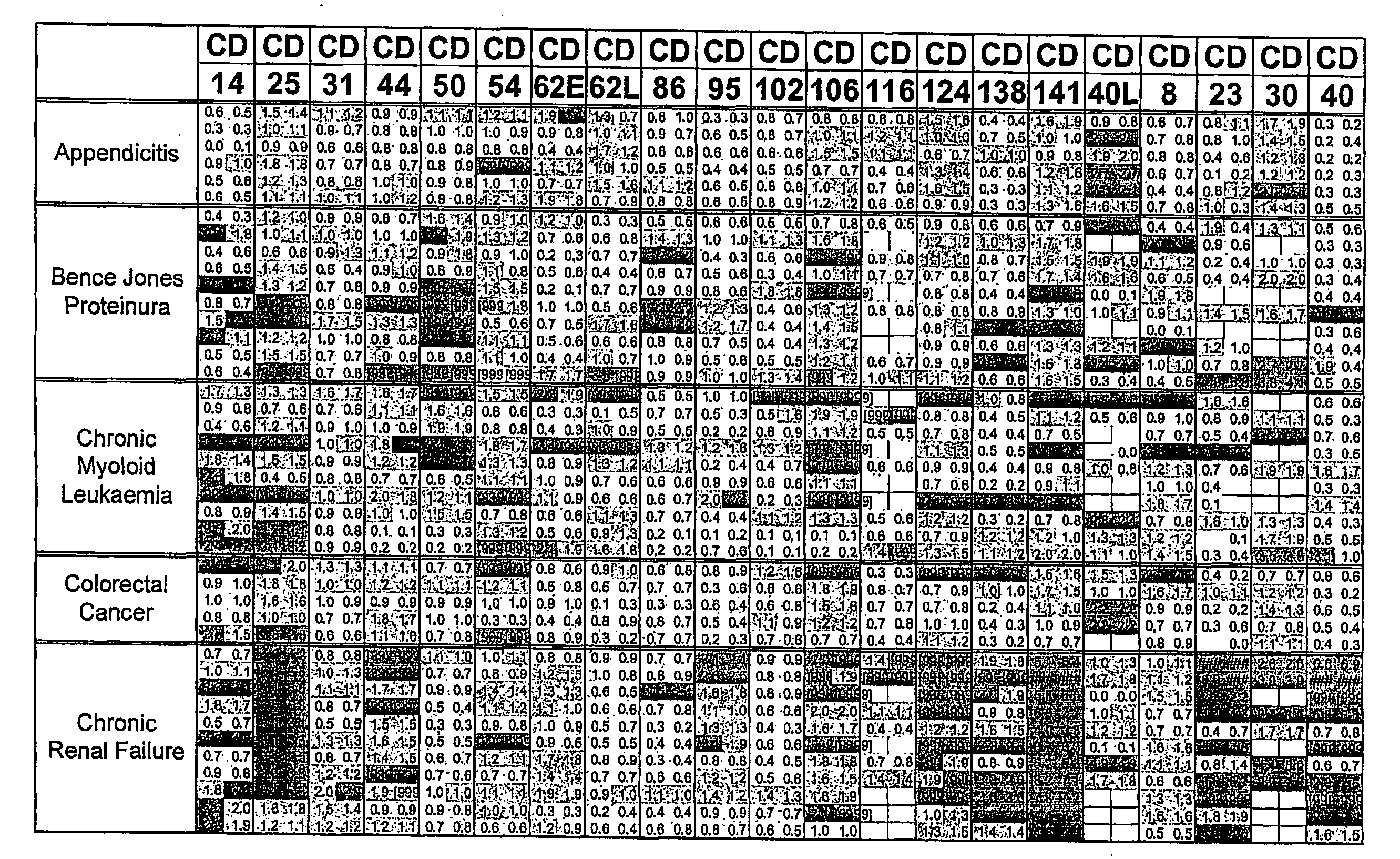
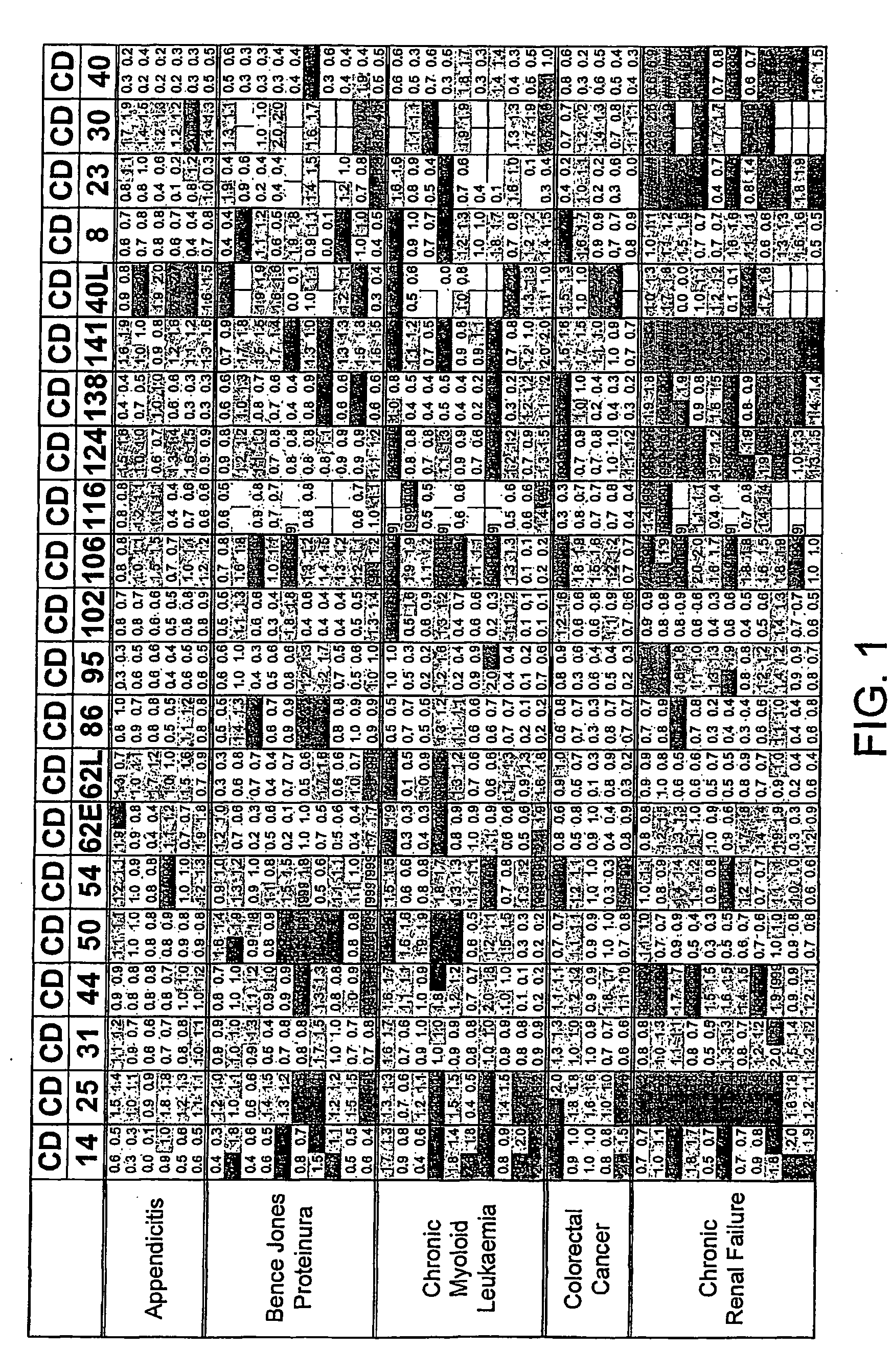
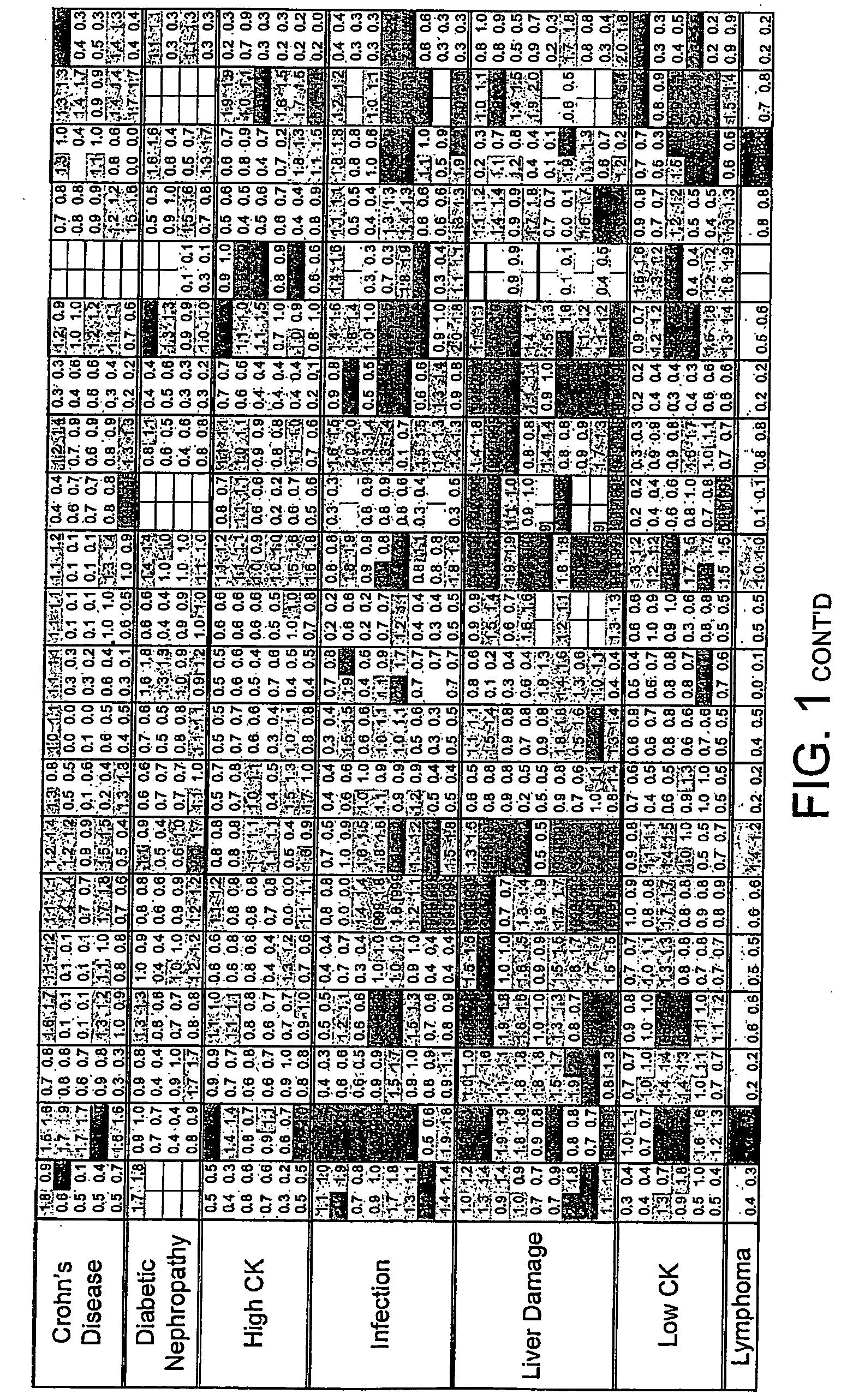
FIG. 1. Disease Groups. Multiples of ULN All sCD's included

[0215]Two values obtained (CD40L and CD30) for the individual, classified as normal, with a suspected drug overdose were omitted from the calculation of the upper limit of normal. The dilution factor for each sCD was fixed throughout the study. The results obtained are those of the diluted sample and have not been multiplied by the dilution factor. The absolute value of each data point was divided by the upper limit of normal (ULN) as defined above. Where the absolute value was greater than the dynamic range of the assay the result [9999] was recorded.

[0216]The limits indicated by each point are:[0217]Green ≦1×ULN[0218]Blue 1-2×ULN[0219]Red >2×ULN[0220]A white block indicates no data available.

example 2

FIG. 2 Remove all sCD's that Appear not to Discriminate from the Normals (sCD's 21; 102; 117; 126; 130; 26; 44v5; 44v6; 62P)

[0221]To simplify the diagram the above sCD plots were removed.

[0222]The data suggests (FIG. 1.) that the concentration of some of these sCD may actually be lowered in disease. As we initially worked on the premise that there would be over-expression of these molecules in disease, samples have been diluted optimally to focus on high, rather than low concentrations.

example 3

FIG. 3. Disease Groups. Mode of Response for Remaining 20 sCD's

[0223]To simplify the data further the modal response for each disease group has been plotted. As the lymphoma and “oligoclonal-banding positive” group contain only a single subject, they have been omitted. Where there is no clear mode, both responses have been shown.


PUM

PropertyMeasurementUnit
timeaaaaaaaaaa
timeaaaaaaaaaa
timeaaaaaaaaaa

Abstract

The present invention relates to the use of cluster of differentiation (CD) molecules in detecting the presence and progression of one or more disease states in an individual. In particular it relates to the use of profiles of shed CD (sCD) molecules in detecting an assessing the progress of one or more disease states in an individual. Further uses of sCD profiles according to the present invention are also described.

Description

[0001]This application is a continuation of Ser. No. 10 / 506,906, filed Jun. 27, 2006, which is a 371 national phase application of PCTGB03 / 00974 filed Mar. 7, 2003, which claims the benefit of GB0205394.0 filed Mar. 7, 2002; GB0207746.9 filed Apr. 3, 2002; and GB0228195.4, filed Dec. 3, 2002. Each of these applications in their entirety is incorporated by reference herein.[0002]The present invention relates to the use of cluster of differentiation (CD) molecules in detecting the presence and / or assessing the progression and / or assessing the response to therapeutic intervention of one or more disease states in an individual. In particular I relates to the use of profile / fingerprints of shed CD (sCD) molecules in body fluids in detecting and / or assessing the progression of one or more disease states in an individual. Further uses of sCD profiles according to the present invention are also described.BACKGROUND TO THE INVENTION[0003]Rapid and accurate diagnosis is essential in medicine ...

Claims


Application Information

Patent Timeline
Patent Type & Authority Applications(United States)
IPC IPC(8): A61K38/02G06F19/00C07K2/00A61P35/00G01N33/68G16H10/60
CPCG01N33/68G01N33/6803Y02A90/22G06Q50/24G01N33/54306G01N33/6845A61P35/00G16H40/63G16H70/60Y02A90/10
Inventor WOOLFSON, ADRIANHALES, CHARLES NICHOLASHALES, MARGARETMILSTEIN, CESARPRILLELTENSKY MILSTEIN, CELIA
Owner CAMBRIDGE ENTERPRISE LTD
Why Eureka
Try Eureka