System for closing a puncture in a vessel wall

a technology for a vessel wall and a puncture site, which is applied in the field of percutaneous closure of arterial and venous puncture sites, can solve the problems of increasing the time required before completion of compression techniques, relying on clot formation, and time-consuming, and achieves the effect of closing the puncture and constant tensioning for

Active Publication Date: 2008-12-25
ABBOTT LAB INC
View PDF99 Cites 52 Cited by
  • Summary
  • Abstract
  • Description
  • Claims
  • Application Information

AI Technical Summary

Benefits of technology

[0014]According to one example, a closure device can also be included that includes a body and guide lumens extending beyond the lumens that are in communication with the body. The body can have a clamping or securing mechanism coupled thereto that allows wires to be secured to the body. According to one example, the wires can be fed through the distal ends of the guide lumens to the body and secured thereto by the clamping mechanism. The closure device can be rotated to twist the suture wires and thereby close the puncture. The clamping mechanism may be configured to provide substantially constant tensioning force on the suture wires as the suture wires are twisted. Further, the clamping mechanism may be configured to separately and independently apply tensioning force to each wire. The force may be independently chosen to help ensure that the wires twist equally. In one example, the forces can be approximately equal. The closure device may also include a guide tip coupled to the distal ends of the guide lumens. The guide tip helps ensure that twisting of the wires begins at a desired location away from the vessel wall.

Problems solved by technology

It is time consuming, frequently requiring one-half hour or more of compression before hemostasis is assured.
Additionally, such compression techniques rely on clot formation, which can be delayed until anticoagulants used in vascular therapy procedures (such as for heart attacks, stent deployment, non-optical PTCA results, and the like) wear off.
This can take two to four hours, thereby increasing the time required before completion of the compression technique.
The compression procedure is further uncomfortable for the patient and frequently requires analgesics to be tolerable.
Moreover, the application of excessive pressure can at times totally occlude the underlying blood vessel, resulting in ischemia and / or thrombosis.
During this time renewed bleeding may occur, resulting in blood loss through the tract, hematoma and / or pseudo-aneurysm formation, as well as arteriovenous fistula formation.
The incidence of complications from compression induced hemostasis increases when the size of the introducer sheath grows larger, and / or when the patient is anti-coagulated.
It is clear that the compression technique for arterial closure can be risky and is expensive and onerous to the patient.
Although the risk of complications can be reduced by using highly trained individuals, dedicating such personnel to this task is both expensive and inefficient.
While potentially effective, this approach suffers from a number of problems.
It can be difficult to properly locate the interface of the overlying tissue and the adventitial surface of the blood vessel.
Locating the fastener too far from that interface can result in failure to provide hemostasis, and subsequent hematoma and / or pseudo-aneurysm formation.
Conversely, if the sealing body intrudes into the artificial lumen, intravascular clots and / or collagen pieces with thrombus attached can form and embolize downstream, causing vascular occlusion.
Also, thrombus formation on the surface of a sealing body protruding into the lumen can cause a stenosis, which can obstruct normal blood flow.
Other possible complications include infection, as well as adverse reaction to the collagen or other implant.

Method used


Image

  • System for closing a puncture in a vessel wall
  • System for closing a puncture in a vessel wall
  • System for closing a puncture in a vessel wall

Examples


Embodiment Construction

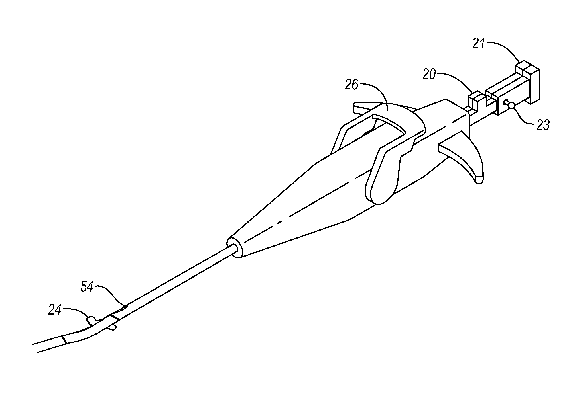
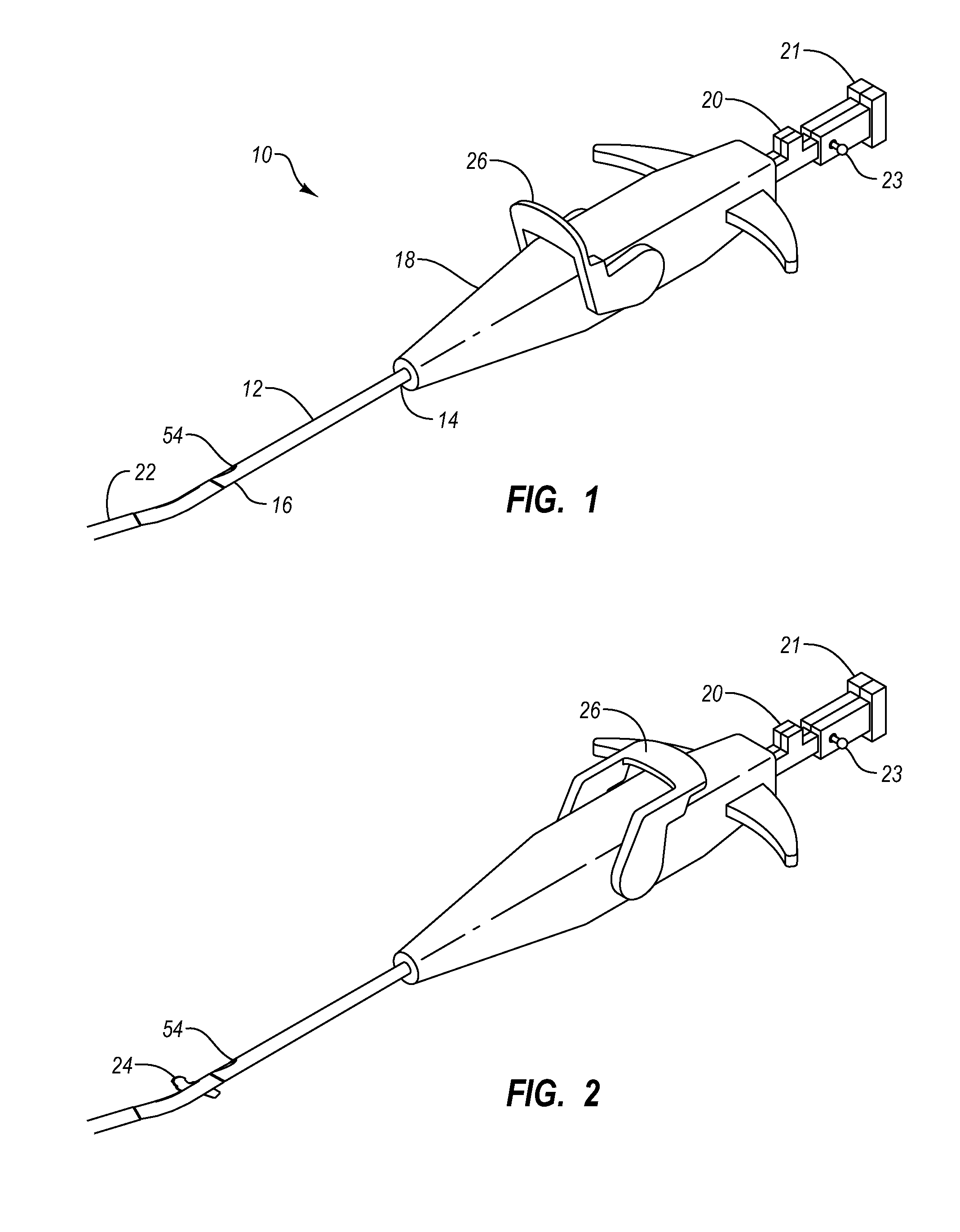
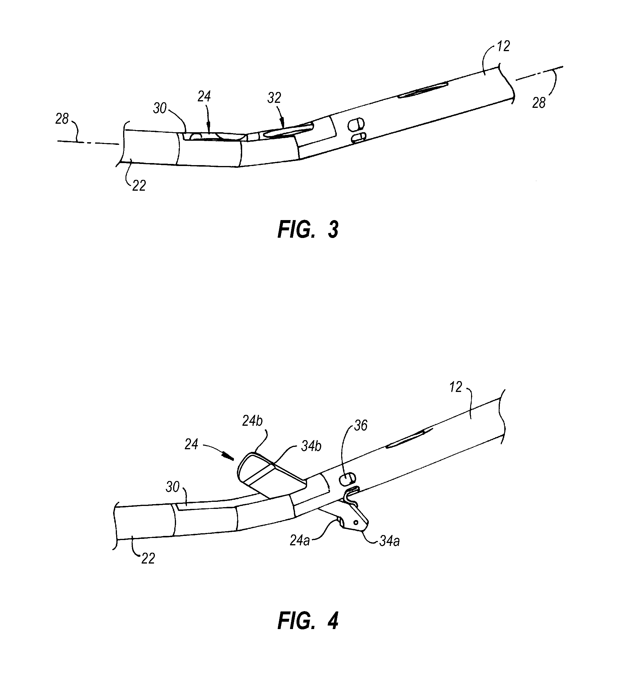
[0040]A closure system is provided herein that includes a deployment device and a closure device. According to one example, the deployment device provides for repeatable and reliable deployment of wires within a vessel having a puncture therein. The wires have securing member, such as hooks, on the distal ends thereof that allow the distal ends of the wires to be anchored to a proximal wall of the vessel to be closed. As used herein, a hook shall be broadly understood to mean any structure configured to secure the wires to tissue, such as to secure a wire from moving proximally from engagement with the tissue. According to one example, the wires are deployed by advancing needles from outside the vessel to a location within the vessel. The needles carry the hooks. In particular, the hooks may be stored completely within the needles prior to deployment. A practitioner deploys the hooks by advancing the hooks beyond the distal ends of the needles. Thereafter, the hooks may be anchored ...


PUM


Abstract

A device is provided herein for deploying a suture in a puncture through a vessel wall of a blood vessel, the puncture disposed within a tissue tract of a patient body. The device includes a shaft suitable for insertion along the tissue tract and into the vessel through a puncture, a plurality of needles, each needle having a proximal end and a distal end suitable for forming a needle path through the vessel wall, wires associated with each needle, each wire having a distal end and a proximal end, and hooks coupled to each of the distal ends of each wire, each hook being configured to anchor the wire to an interior portion of the vessel wall.

Description

CROSS-REFERENCE TO RELATED APPLICATIONS[0001]This application claims the benefit of U.S. Provisional Patent Application Ser. No. 60 / 946,063, entitled “System for Closing a Puncture in a Vessel Wall”, and filed Jun. 25, 2007, the disclosure of which is incorporated herein by this reference in its entirety.BACKGROUND[0002]1. Field of the Invention[0003]The present invention relates generally to apparatus and methods for the suturing of body lumens. More particularly, the present invention relates to techniques for percutaneous closure of arterial and venous puncture sites, which are usually accessed through a tissue tract.[0004]2. Relevant Technology[0005]A number of diagnostic and interventional vascular procedures are now performed translumenally. A catheter is introduced to the vascular system at a convenient access location and guided through the vascular system to a target location using established techniques. Such procedures require vascular access, which is usually established...

Claims


Application Information

Patent Timeline
Patent Type & Authority Applications(United States)
IPC IPC(8): A61B17/04
CPCA61B17/0057A61B17/0469A61B17/0482A61B17/0487A61B2017/00663A61B2017/0419A61B2017/0427A61B2017/047A61B2017/0472A61B2017/0496A61B2017/06052
Inventor REYNOLDS, TIMOTHY C.
Owner ABBOTT LAB INC
Why Eureka
Try Eureka