Apparatus and Method for Endovascular Device Guiding and Positioning Using Physiological Parameters

a technology of endovascular devices and physiological parameters, applied in the direction of catheters, instruments, angiography, etc., can solve the problems of inaccurate method, decreased effectiveness, perforation of the heart, etc., and achieve the effect of increasing the reliability of information and accuracy of location identification

Active Publication Date: 2009-01-01
TELEFLEX LIFE SCI LTD
View PDF104 Cites 213 Cited by
  • Summary
  • Abstract
  • Description
  • Claims
  • Application Information

AI Technical Summary

Benefits of technology

[0025]Another aspect of the invention includes a method for positioning an instrument in the vasculature of a body. The method comprises the steps of: accessing the vascular system of the body; positioning an instrument in the vascular system of the body; using the instrument to transmit an ultrasound signal into the vascular system of the body; using the instrument to receive a reflected ultrasound signal from the vasculature indicating flow rates between 2 and 20 cm/s; using the instrument to measure intravascular electrical activity of the heart, processing the reflected ultrasound and electrocardiogram signals to determine one or more parameters and their temporal behavior from a group consisting of: a blood flow direction, a blood flow velocity, e.g., the highest, the lowest, the mean or the average velocity, a blood flow signature pattern, a pressure signature pattern, A-mode information, a preferential non-random direction of flow, the shape of the different waveforms and complexes charactering the intravascular electrocardiogram, e.g., P-wave, QRS complex, T-wave, the peak-to-peak amplitudes, the absolute and relative amplitude changes and other distinctive elements of the intravascular ECG; and advancing the instrument within the vasculature using the one or more of the determined parameter or parameters within the vasculature. In yet other embodiments, processing the reflected ultrasound signal and the intravascular or intracardiac electrocardiogram signal and their correlation to determine the position of the instrument relative to the caval-atrial junction and other specific location within the body is performed. In other aspects of the invention, the specific target vasculature for positioning an instrument is included, for example, the specific structure is a valve of a heart, a blood vessel wall, a heart wall, a pacemaker node of the heart. In another aspect of the invention, the method can further comprise identifying a certain obstructing structure in a blood vessel or within the lumen of an endovascular device which can obstruct either the advancement of the endovascular device in the blood vessel or the delivery of a payload through a lumen of the devices, e.g., a blood clot In another aspect of the invention, the method can further comprise: using the instrument determine a location to secure a device within the vasculature of a body; and securing the device to the body to maintain the device in the location determined by the instrument. In still another aspect of the method, the method can further comprise: using the instrument to calculate the current position of the device; and determining if the devi

Problems solved by technology

One challenge common to all vascular-based therapies is health care provider access to the specific location or section of the vascular tree.
Even though it is routine, it is a very time and labor-intensive procedure for the hospital staff, which also makes it expensive.
If the catheter tip moves, not only its effectivenes

Method used


Image

  • Apparatus and Method for Endovascular Device Guiding and Positioning Using Physiological Parameters
  • Apparatus and Method for Endovascular Device Guiding and Positioning Using Physiological Parameters
  • Apparatus and Method for Endovascular Device Guiding and Positioning Using Physiological Parameters

Examples


Embodiment Construction

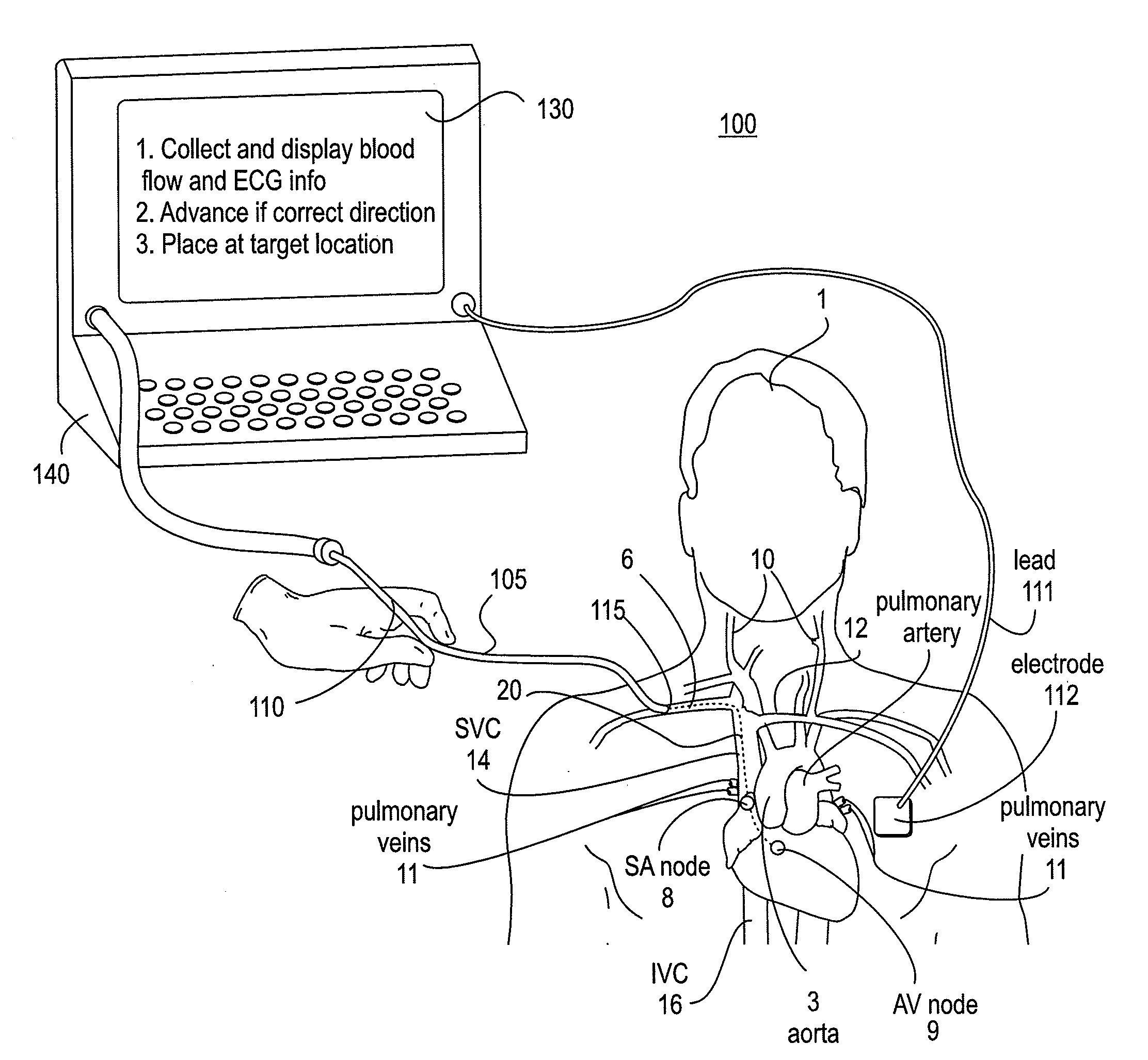
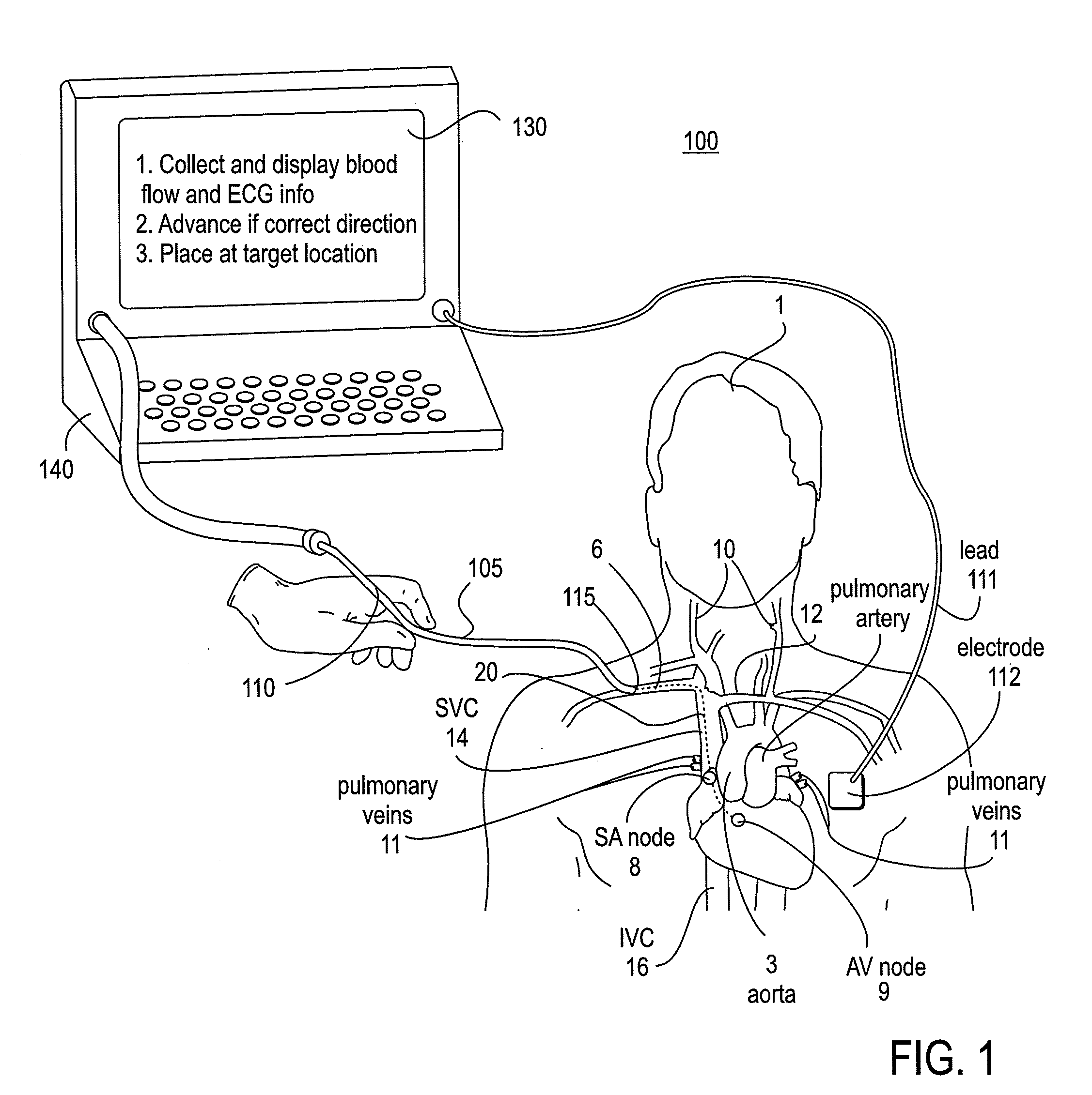
[0090]Embodiments of the present invention provide guided vascular access devices, systems for processing signals from the guided vascular access devices and user interface for providing information to a user based on outputs from the processing system. FIG. 1 illustrates one embodiment of an exemplary endovascular access and guidance system 100. The system 100 includes an elongate body 105 with a proximal end 110 and a distal end 115. The elongate body 105 is any of a variety of endovascular devices adapted to insertion into and navigation through the vasculature of the patient 1. FIG. 1 illustrates the distal end 115 inserted into the basilic vein 6. The expected path of travel (dashed line 20) in this illustrative example is into the a portion of the heart 20 or within the superior vena cava 14 in proximity to the sinoatrial node (SA node) 8. The aorta 3, the pulmonary arteries, pulmonary veins11, the jugular veins 10, the brachiocephalic vein 12, inferior vena cava 16 and atriov...


PUM


Abstract

An endovascular access and guidance system has an elongate body with a proximal end and a distal end; a non-imaging ultrasound transducer on the elongate body configured to provide in vivo non-image based ultrasound information of the vasculature of the patient; an endovascular electrogram lead on the elongate body in a position that, when the elongate body is in the vasculature, the endovascular electrogram lead electrical sensing segment provides an in vivo electrogram signal of the patient; a processor configured to receive and process a signal from the non-imaging ultrasound transducer and a signal from the endovascular electrogram lead; and an output device configured to display a result of information processed by the processor. An endovascular device has an elongate body with a proximal end and a distal end; a non-imaging ultrasound transducer on the elongate body; and an endovascular electrogram lead on the elongate body in a position that, when the endovascular device is in the vasculature, the endovascular electrogram lead is in contact with blood. The method of positioning an endovascular device in the vasculature of a body is performed by advancing the endovascular device into the vasculature; transmitting a non-imaging ultrasound signal into the vasculature using a non-imaging ultrasound transducer on the endovascular device; receiving a reflected ultrasound signal with the non-imaging ultrasound transducer; detecting an endovascular electrogram signal with a sensor on the endovascular device; processing the reflected ultrasound signal received by the non-imaging ultrasound transducer and the endovascular electrogram signal detected by the sensor; and positioning the endovascular device based on the processing step.

Description

CROSS-REFERENCE TO RELATED APPLICATIONS[0001]This application claims the benefit of U.S. Provisional Patent Application No. 60 / 937,280 filed on Jun. 26, 2007 by Sorin Grunwald et al., entitled “Apparatus and Method for Vascular Access”; U.S. Provisional Patent Application No. 60 / 957,316 filed on Aug. 22, 2007 by Sorin Grunwald et al., entitled “Apparatus and Method for Endovascular Guidance”; and U.S. Provisional Patent Application No. 61 / 023,183 filed on Jan. 24, 2008 by Sorin Grunwald et al., entitled “Configurations of Guided Endovascular Access devices”, each of which is incorporated herein by reference in its entirety.[0002]This application is also a continuation-in-part of U.S. Non-Provisional patent application Ser. No. 11 / 431,140 filed on May 8, 2006 by Sorin Grunwald et al., entitled “Endovenous Access and Guidance System Utilizing Non-Image Based Ultrasound”, now publication no. 2007-0016072-A1; U.S. Non-Provisional patent application Ser. No. 11 / 431,118 filed on May 8, 20...

Claims


Application Information

Patent Timeline
IPC IPC(8): A61B8/12A61B5/296A61B5/352A61B5/366
CPCA61B5/0215A61B8/565A61B5/029A61B5/042A61B5/0452A61B5/0472A61B5/0476A61B5/06A61B5/061A61B5/1459A61B8/06A61B8/08A61B8/0841A61B8/12A61B8/42A61B8/445A61B8/461A61B19/5244A61B19/56A61B2017/00044A61B2017/00106A61B2017/00115A61B2017/22067A61B2019/462A61B2019/464A61B2019/5251A61B2019/5255A61B2019/5276A61B2019/562G09B23/288A61B5/0205A61B5/065A61B7/04A61B5/026A61B8/465A61B2034/2055A61B34/20A61B34/25A61B2090/378A61B2034/2051A61B2034/252A61B2090/062A61B2090/064A61B8/463A61B5/283A61B5/366A61B5/349A61B5/369A61B5/352A61B5/389A61B5/742A61B5/6853A61B8/02A61B8/488A61B5/7405
Inventor GRUNWALD, SORINSANDER, FIONA MARIASAMSON, WILFRED J.HILL, BRADLEYCHENG, E. TINA
Owner TELEFLEX LIFE SCI LTD
Why Eureka
Try Eureka