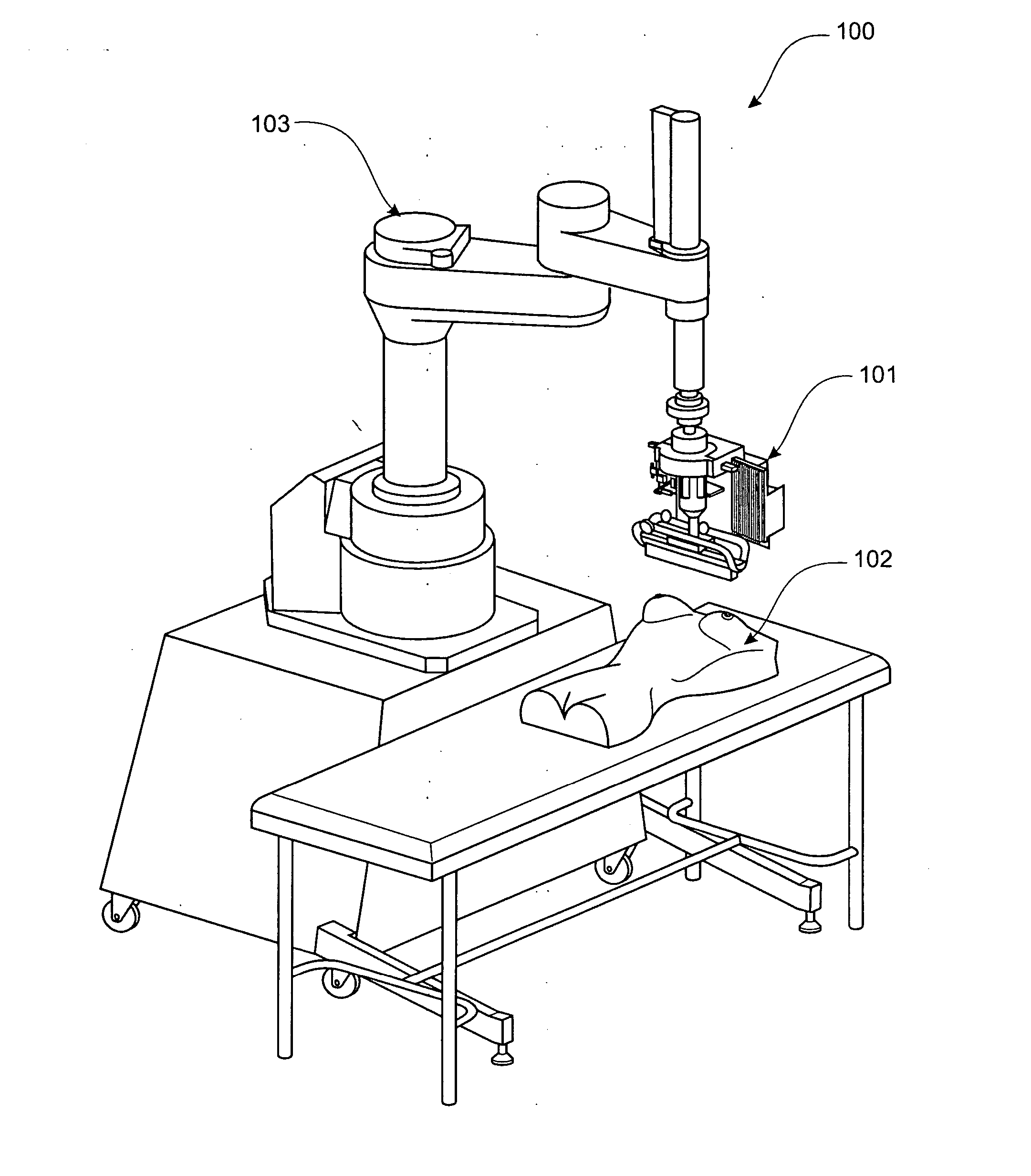
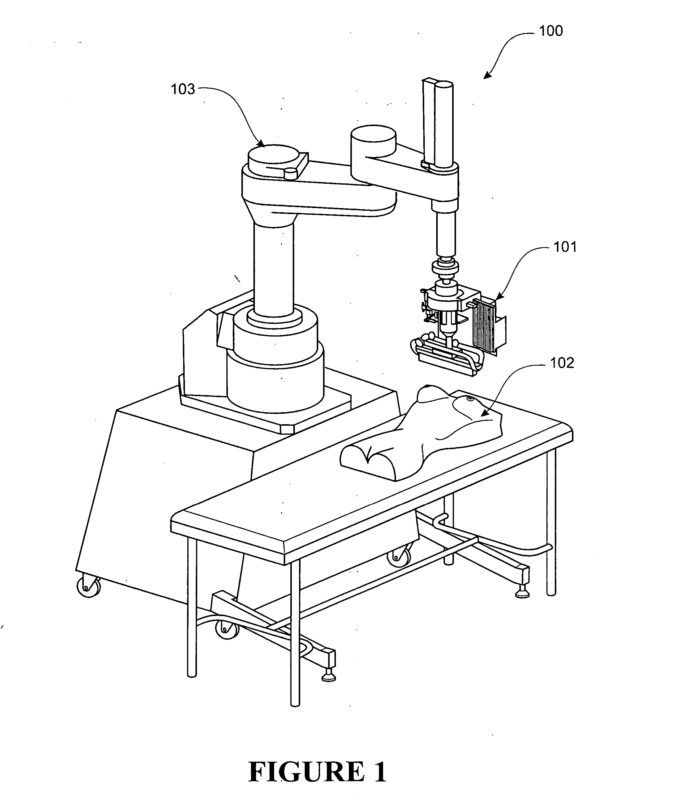
Imaging system

a technology of imaging system and image, applied in the field of imaging system, can solve the problems of x-ray mammography with a number of associated limitations and drawbacks, difficult to determine the true three-dimensional (3d) location of any suspicious features, and women with silicone breast implants are also at risk of implant ruptur

Inactive Publication Date: 2009-01-22
SIMPKIN RAY ANDREW
View PDF24 Cites 14 Cited by
  • Summary
  • Abstract
  • Description
  • Claims
  • Application Information

AI Technical Summary

Problems solved by technology

However, x-ray mammography has a number of associated limitations and drawbacks.
X-ray mammography requires the patient's breasts to be compressed between two plates which is uncomfortable for many women and makes it difficult to determine the true three-dimensional (3D) location of any suspicious features.
Furthermore, women with silicone breast implants are also at risk from implant rupture due to the compression process.
X-ray detection of suspicious features relies on differences in density within the breast tissue under test and the density contrast between healthy and malignant breast tissue is small, typically only about 2%, which can make detection of tumours difficult.

Method used


Image

  • Imaging system
  • Imaging system
  • Imaging system

Examples


Embodiment Construction

[0064] The preferred form imaging system of the invention is a breast cancer screening tool and is arranged to scan a patient's breasts with microwave radiation in order to generate 3D radar images of each breast which can be examined for suspicious features such as malignant tumours. There is a large difference in complex permittivity between healthy and malignant breast tissue and this leads to greater scattered field amplitudes from malignant tumours embedded in healthy tissue which show up readily in a microwave image of scattered field intensity. For example, the real part of complex permittivity (the dielectric constant) for a malignant tumour is of the order of 50 at a frequency of 10 GHz whereas healthy tissue has a value of about 9. Hence, radar images are suited to breast tumour detection since the high permittivity contrast between malignant and healthy tissue translates to high-contrast images.

[0065] The imaging system generates 3D radar images based on the intensity of...


PUM


Abstract

An imaging system (100) for generating a three-dimensional image of a body part of a patient (102). The imaging system (100) comprises a sensor head (101) that is moved relative to the patient by a robot (103) to conduct a scan of the body part. The sensor head (101) comprises a three-dimensional profiler that is arranged to obtain surface profile information and a radar device that is arranged to obtain radiation information at frequencies of at least approximately 10 GHz at multiple scan locations defining a synthetic aperture relative to the body part. The imaging system (100) has a control system that is arranged to operate the three-dimensional profiler and radar device. The control system also receives and processes the radiation information and surface profile information to generate a three-dimensional image of the body part that has multiple image points by synthetically focusing the radiation information.

Description

FIELD OF THE INVENTION [0001] The present invention relates to an imaging system for body parts that utilises non-ionizing electromagnetic radiation, for example microwaves. In particular, although not exclusively, the imaging system is suitable for breast cancer screening. BACKGROUND TO THE INVENTION [0002] Breast cancer is the most common cancer to affect women. The detection of malignancies at an early stage is deemed to offer the best prognosis for patients and this has lead to the establishment of screening programmes aimed at early detection. [0003] X-ray mammography is one commonly used breast cancer screening method due to its simplicity, high-resolution images and cost effective implementation. However, x-ray mammography has a number of associated limitations and drawbacks. X-rays are an example of ionizing electromagnetic radiation which can damage tissue and in some cases initiate malignant tumours. X-ray mammography requires the patient's breasts to be compressed between...

Claims


Application Information

Patent Timeline
Patent Type & Authority Applications(United States)
IPC IPC(8): A61B5/05
CPCA61B5/05A61B5/1077A61B5/0507
Inventor SIMPKIN, RAY ANDREW
Owner SIMPKIN RAY ANDREW
Why Eureka
Try Eureka