Computerized image analysis for acetic acid induced Cervical Intraepithelial Neoplasia

a technology of intraepithelial neoplasia and computerized image analysis, which is applied in the field of medical imaging and image processing, can solve the problems of false positives, difficult automated detection of acetowhite epithelial depicted on cervical images, and large false positives, and achieve high intensity variation, and controllable local contrast enhancement

Inactive Publication Date: 2009-02-05
STI MEDICAL SYST
View PDF8 Cites 46 Cited by
  • Summary
  • Abstract
  • Description
  • Claims
  • Application Information

AI Technical Summary

Benefits of technology

[0020]A textured region is identified in the original image by determining regions within the image with high variations in color and intensity (brightness). Texture analysis attempts to quantify rough, smooth, silky, or bumpy as a function of the spatial intensity variations in the image (the spatial extent of variations in brightness (or intensity) of the image, preferably measured by using variations in brightness in a black and white version of the image, or “grayscale”). Thus, roughness or bumpiness can be understood as the spatial extent of variations in intensity values, or gray levels, in a black and white version of the image. For the cervix, the texture region is referred to as the region which is rich in texture content or, in other words, has high variations in intensity (instead

Problems solved by technology

However, blood vessels do not turn white after application of acetic acid and therefore still appear red, even though they are in abnormal tissue that has become white, so that blood vessels may mask acetowhitening.
Further, some lesions do not have visible blood vessels, and some normal regions are naturally white, or turn white after application of acetic acid, so reliance solely on acetowhitening leads to substantial

Method used


Image

  • Computerized image analysis for acetic acid induced Cervical Intraepithelial Neoplasia
  • Computerized image analysis for acetic acid induced Cervical Intraepithelial Neoplasia
  • Computerized image analysis for acetic acid induced Cervical Intraepithelial Neoplasia

Examples


Embodiment Construction

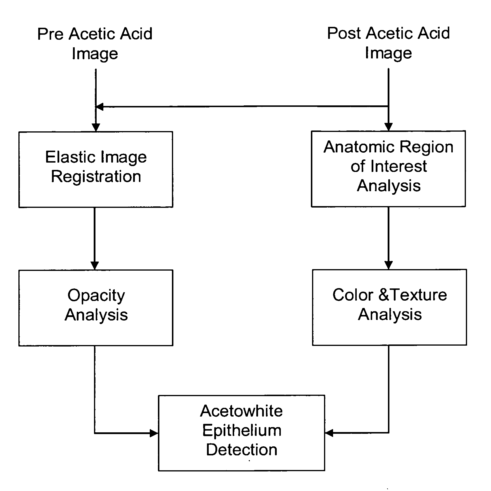
[0031]The present invention described herein and more fully below, comprises an automated image analysis system for quickly and efficiently detecting acetic acid induced intraepithelial lesions from surrounding tissue, and to characterize both color and opacity properties of acetowhite epithelium. FIG. 1 depicts a block diagram of the multi-step procedure of analyzing the acetowhite epithelium using a set of image processing algorithms.

1. Obtaining Pre Acetic Acid and Post Acetic Acid Images of the Cervix

[0032]Images of the cervix, before and after acetic acid application, are required. The images could, be images from a digital imager, such as a digital colposcope, or they could be digitized copies of film or other images. Prior to application of acetic acid (preferably 5% acetic acid), polarized and non-polarized high-resolution digital color images of the cervix are taken with a digital imager (colposcope). The post acetic acid image is considered to be the reference image.

[0033]...


PUM


Abstract

A method for differentiating cancerous lesions from surrounding tissue, which includes extracting an opacity parameter from acetowhite regions of pre acetic acid and post acetic acid images of a cervix.

Description

[0001]The present application claims the priority of U.S. provisional patent application 60 / 963351 filed Aug. 3, 2007, which is incorporated herein by reference.[0002]This invention was made with partial government support under US Army Medical Research and Material Command under Contract No. W81XWH-07-C-0006. The US government has certain rights in the invention.TECHNICAL FIELD[0003]This invention generally relates to medical imaging and image processing. The invention relates more specifically to the computer aided diagnosis of uterine cervical cancer through the evaluation of acetowhite regions and extraction of an opacity parameter.BACKGROUND ART [0004]Although this invention is being disclosed in connection with cervical cancer, it is applicable to many other areas of medicine. Uterine cervical cancer is the second most common cancer in women worldwide, with nearly 500,000 new cases and over 270,000 deaths annually (LARC, “Globocan 2002 database, “International agency for resea...

Claims


Application Information

Patent Timeline
IPC IPC(8): G06K9/00G06V10/56
CPCG06T7/0012G06T7/0081G06T7/0097G06K9/4652G06T2207/20041G06T2207/30096G06T2207/10024G06T7/11G06T7/174G06V10/56
Inventor LI, WENJINGWOLTERS, ROLF HOLGER
Owner STI MEDICAL SYST
Why Eureka
Try Eureka