Device, System and Method for In-Vivo Examination

a technology of in-vivo examination and computer-generated images, applied in the field of medical procedures, can solve the problems of discouraging patients from getting examined, and the details provided by the computer-generated image of a vc procedure may not be available, so as to facilitate patient compliance, effective timing, and effective timing

Inactive Publication Date: 2009-04-23
GIVEN IMAGING LTD
View PDF16 Cites 11 Cited by
  • Summary
  • Abstract
  • Description
  • Claims
  • Application Information

AI Technical Summary

Benefits of technology

[0007]Some embodiments of the invention provide procedures for preparing a patient for an in-vivo examination of the GI tract. According to some embodiments, a procedure is provided for examination of the large intestine (colon). Some embodiments may allow effective timing for colon cleansing and/or effective timing for propelling an in-vivo imaging device to the colon and in the colon.
[0008]Procedures according to some embodiments of the invention may be typically shorter than conventional procedures, may be performed in non hospital environments (e.g., in the patient's home), and/or may facilitat

Problems solved by technology

Bleeding and puncture of the colon are possible complications of colonoscopy, and it is usually necessary to give the patient pain medication and sedatives to keep him comfortable and relaxed during the colonoscopy.
This is an unpleasant preparation pr

Method used


Image

  • Device, System and Method for In-Vivo Examination
  • Device, System and Method for In-Vivo Examination
  • Device, System and Method for In-Vivo Examination

Examples


Embodiment Construction

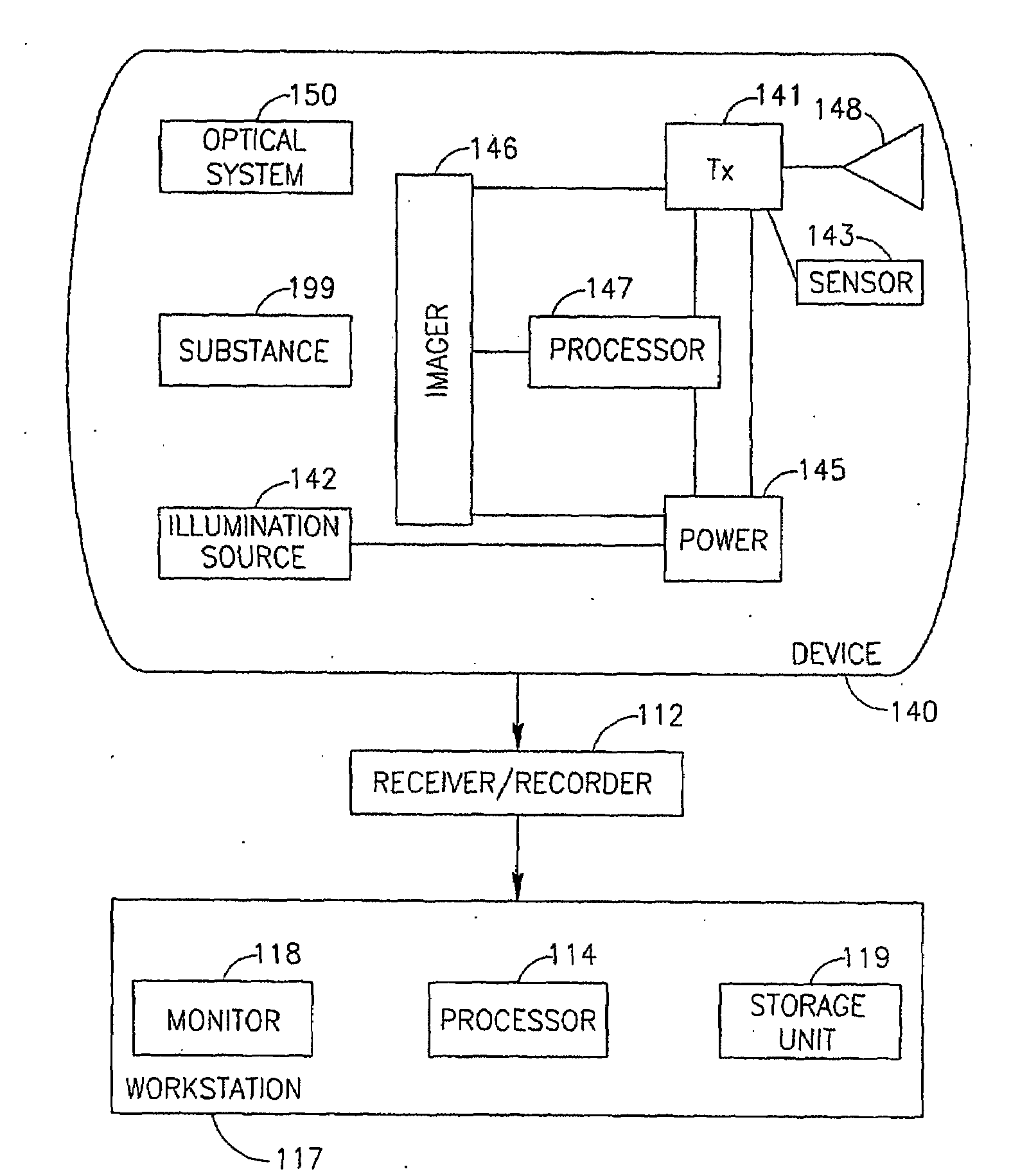
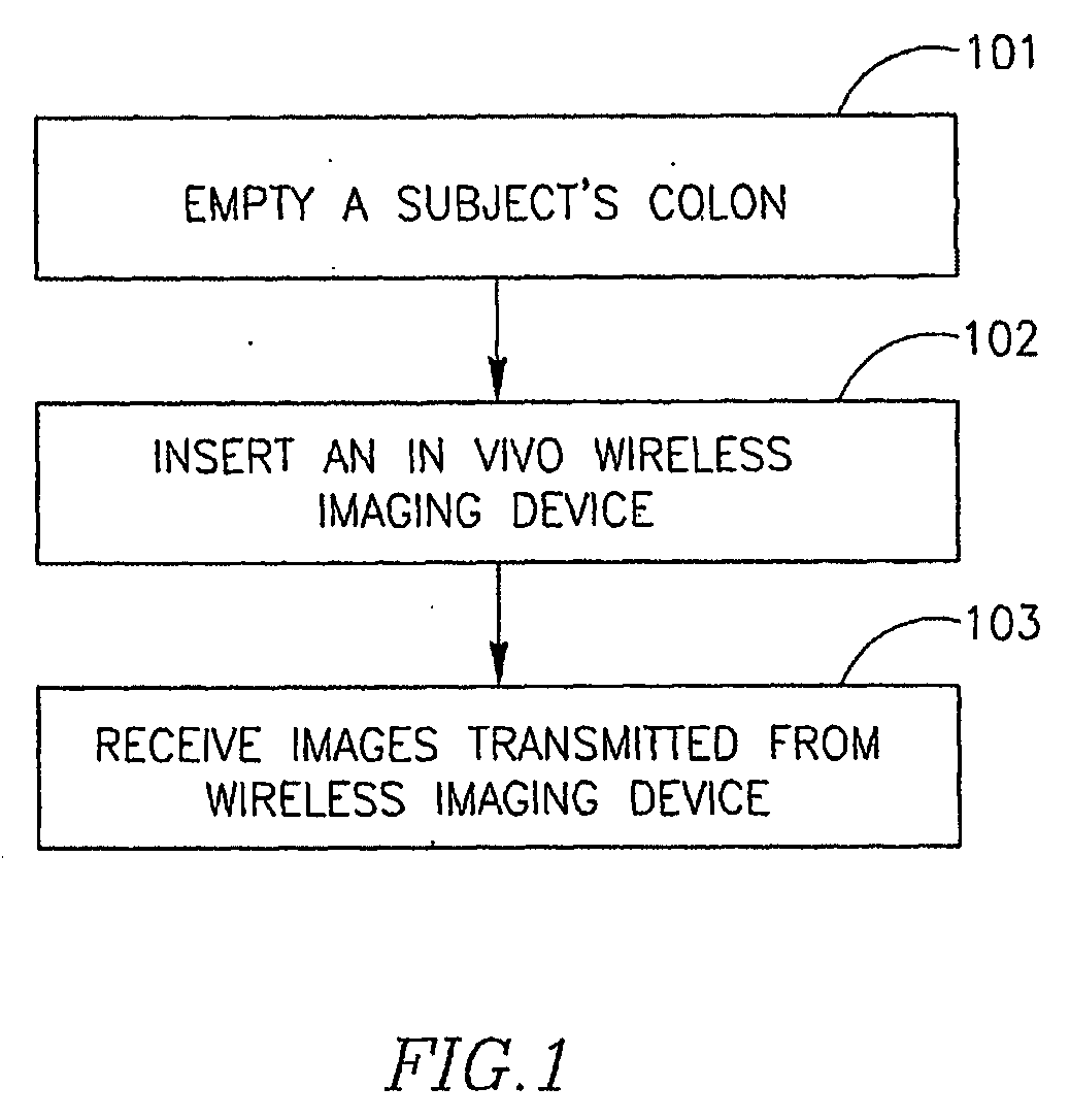
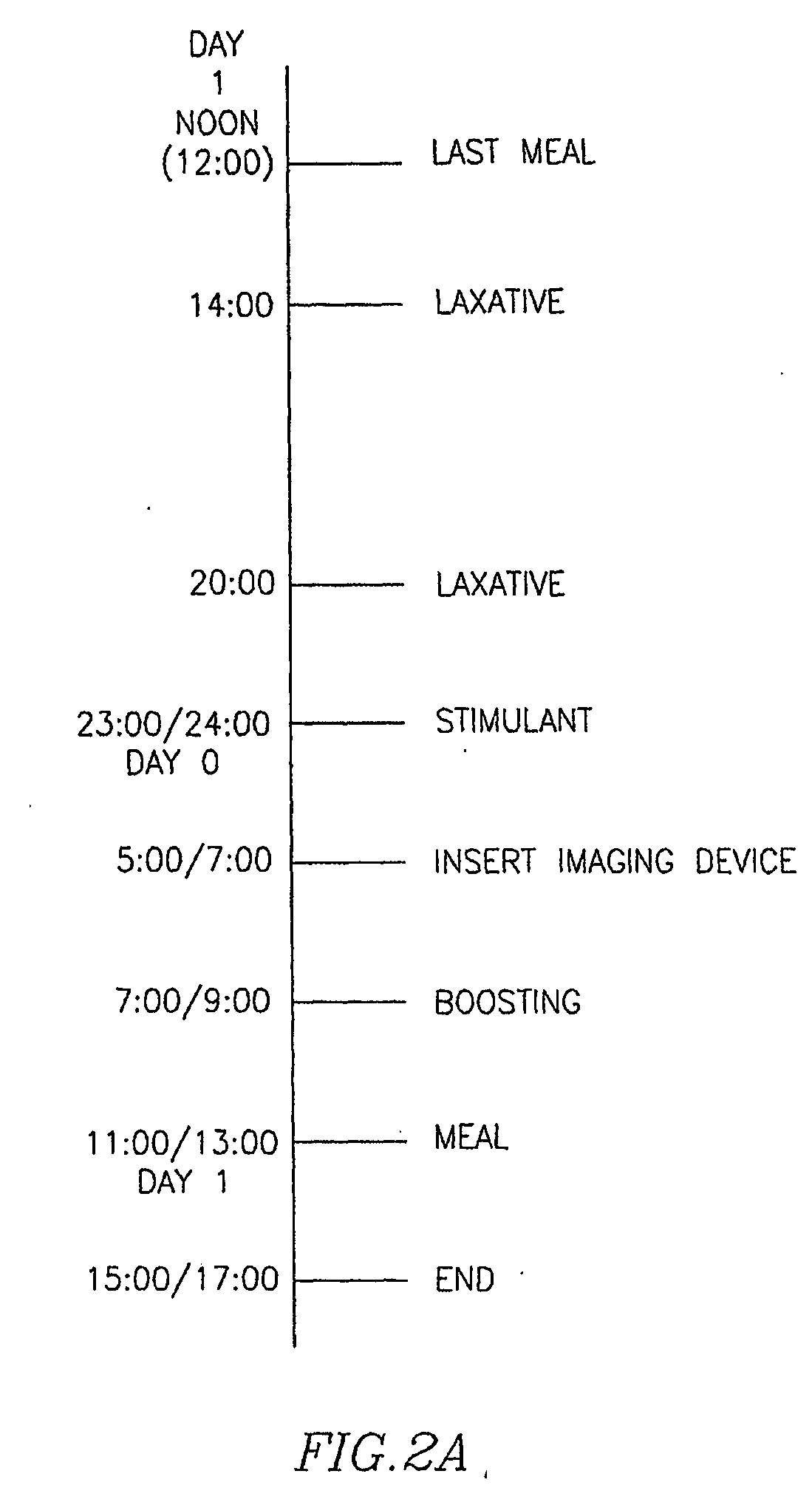
[0040]In the following description, various aspects of the invention are set forth. For purposes of explanation and in order to provide an understanding of the invention, specific configurations and details are set forth in order to provide a thorough understanding of the present invention. However, it will also be apparent to one skilled in the art that the invention may be practiced without the specific details presented herein. Furthermore, well-known features may be omitted or simplified in order not to obscure the invention. Some embodiments of the present invention are directed to a typically swallowable in-vivo sensing device, e.g., a typically swallowable in-vivo imaging device. Devices according to embodiments of the present invention may be similar to embodiments described in U.S. patent application Ser. No. 09 / 800,470, entitled “Device And System For In-vivo Imaging”, filed on 8 Mar. 2001, published on Nov. 1, 2001 as United States Patent Application Publication Number 20...


PUM

PropertyMeasurementUnit
Timeaaaaaaaaaa
Timeaaaaaaaaaa

Abstract

A device, a system and a method for in-vivo examination. A method for in-vivo examination includes substantially emptying a subject's colon from content, and inserting an autonomous in-vivo imaging device into the subject's gastrointestinal tract. An in-vivo examination kit includes an autonomous in-vivo imaging device, a wireless receiver, an antenna set, a laxative, and optionally a stimulant and an instructions leaflet.

Description

FIELD OF THE INVENTION[0001]The present invention relates to the field of medical procedures. More specifically, the present invention relates to methods for preparing a subject for an examination of the gastrointestinal tract.BACKGROUND OF THE INVENTION[0002]An endoscopic examination of the large intestine (colonoscopy) may be used to look for early signs of cancer in the colon. It may further be used to diagnose the causes of unexplained changes in bowel habits. Colonoscopy may allow a physician to see pathology in the colon, such as inflamed tissue, abnormal growths, polyps, cancers ulcers, and bleeding.[0003]Colonoscopy involves inserting a long, flexible, lighted tube through a patient's rectum and guiding it into the colon. An image of the inside of the colon is obtained, so the physician can examine the lining of the colon. Usually, air is blown into the colon, which inflates the colon and helps the physician see.[0004]Bleeding and puncture of the colon are possible complicat...

Claims


Application Information

Patent Timeline
IPC IPC(8): A61B1/04A61K33/42A61P1/10A61K31/765
CPCA61B1/041A61P1/10A61B1/00A61B5/05
Inventor GAT, DANIELRABINOVITZ, RAPHAELBONFIS, SHARON
Owner GIVEN IMAGING LTD
Why Eureka
Try Eureka