Method and apparatus for skin documentation and analysis

a skin and documentation technology, applied in the field of dermatological documentation and analysis, can solve the problems of limited high resolution images of the body to particular areas of interest, slow imaging process, etc., and achieve the effect of convenient scalable and adaptable, and more flexibility

Inactive Publication Date: 2009-05-07
ORTIZ JOSEPH L +1
View PDF63 Cites 131 Cited by
  • Summary
  • Abstract
  • Description
  • Claims
  • Application Information

AI Technical Summary

Benefits of technology

[0004]Accordingly, what is needed is an integrated and automated system for imaging total visible skin areas that: captures total visible skin mages in about the time it takes to perform a chest x-ray or mammogram; provides images in a zoomable, interactive format; reduces the total number of images taken overall while increasing skin image detail viewable within the global context of the skin detail; accommodates both ambulatory and non-ambulatory subjects; may be configured to be portable; and provides analysis that aids in documentation, assessment and treatment of the skin.
[0008]Exemplary embodiments of the disclosure may be used to capture high resolution total body digital photographs in a manner that is quick, automated, and consistent in quality due to reduction of human error. Accordingly, the disclosed embodiments may have applications across numerous medical areas of need as well as outside the field of medicine. For example, the systems and methods described herein may be suitable for non-invasive calculation of skin wound or burn size, shape, and depth, visualizations before and after cosmetic surgery, calculation of skin area affected by psoriasis or acne lesion counts, for following the effectiveness of certain drugs on skin disease, or for non-medical applications such as reverse engineering competitors products, ergonomic based design or made-to-fit apparel construction, among others.
[0009]Other advantages of the systems and methods described herein may be that the systems are readily scalable and adaptable to be used in a variety of locations and settings. The systems may be configured to be fixed or portable thereby providing more flexibility for use of the systems. Accordingly, the systems and methods may eliminate the need to have images produced by professional photographers remote from the physician or medical professional's office.

Problems solved by technology

Often, high resolution images of the body are limited to particular areas of interest and are not viewed in the anatomical context of an expansive total body image.
Such an imaging process is typically slow and requires positioning a subject in multiple predetermined positions, with multiple images being taken from each individual position.

Method used


Image

  • Method and apparatus for skin documentation and analysis
  • Method and apparatus for skin documentation and analysis
  • Method and apparatus for skin documentation and analysis

Examples


Embodiment Construction

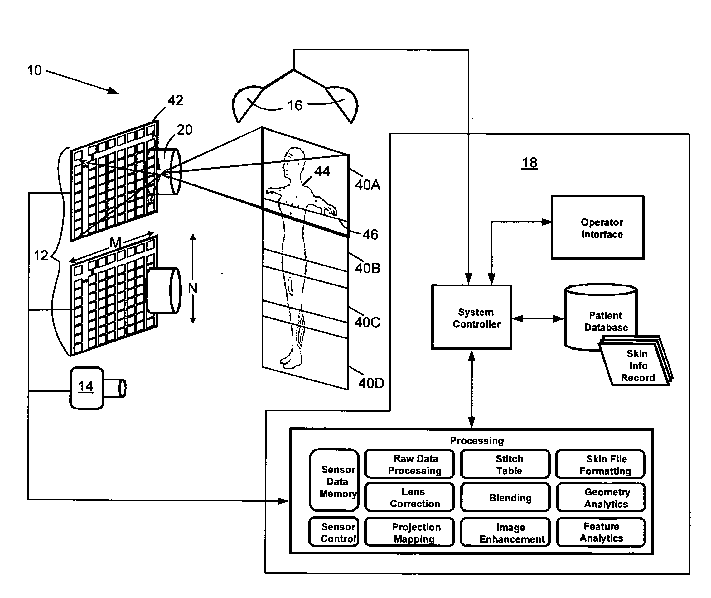
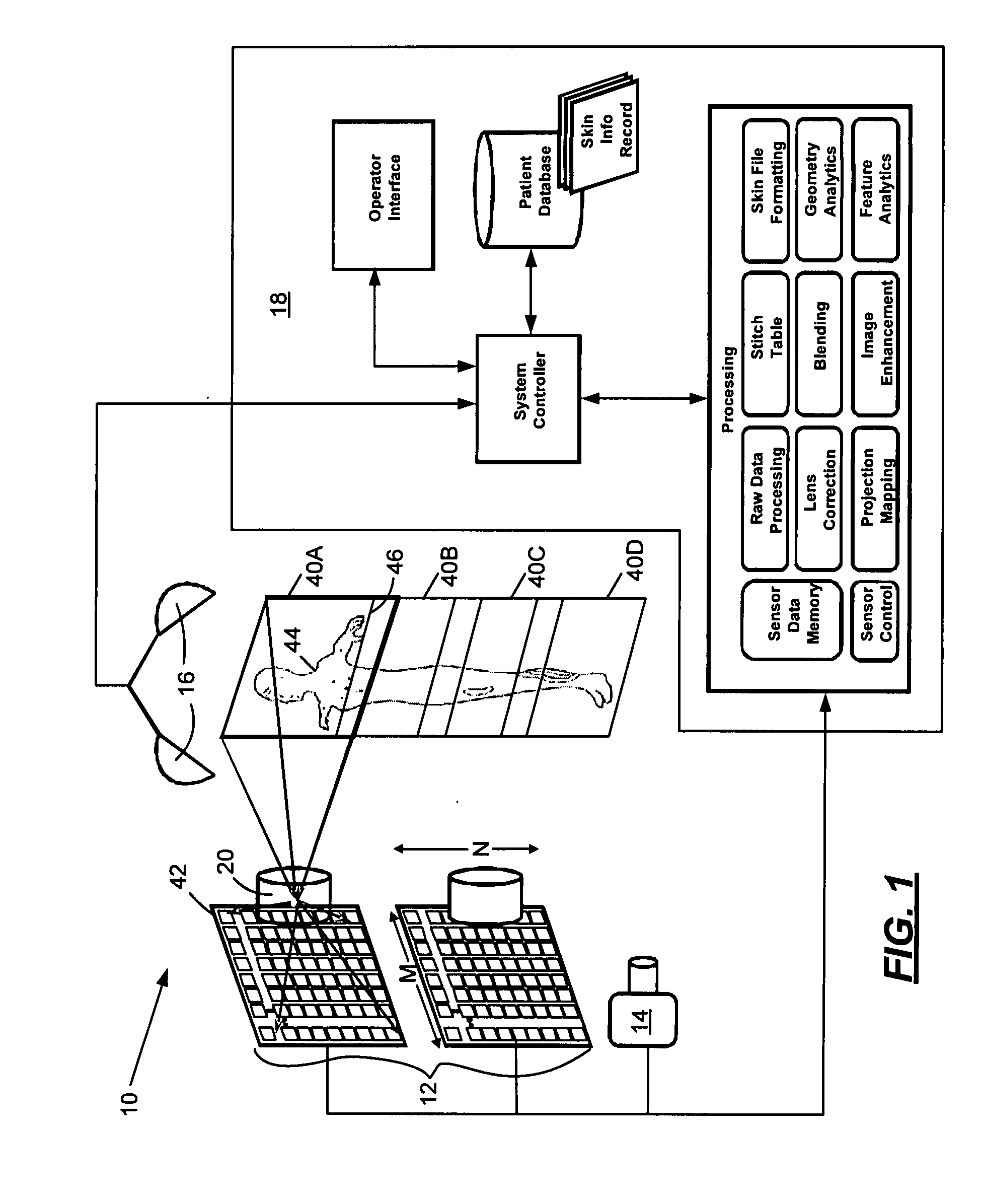
[0024]A schematic overview of a system according to an exemplary embodiment of the disclosure is illustrated in FIG. 1. The system 10 includes an imaging component 12, an optional geometry component 14, a lighting component 16, and a data processing component 18. The foregoing components are unified into a stand-alone system 10 that may be reconfigured or scaled to accommodate a variety of locations and purposes. Each of the components of the system 10 will be described in more detail below.

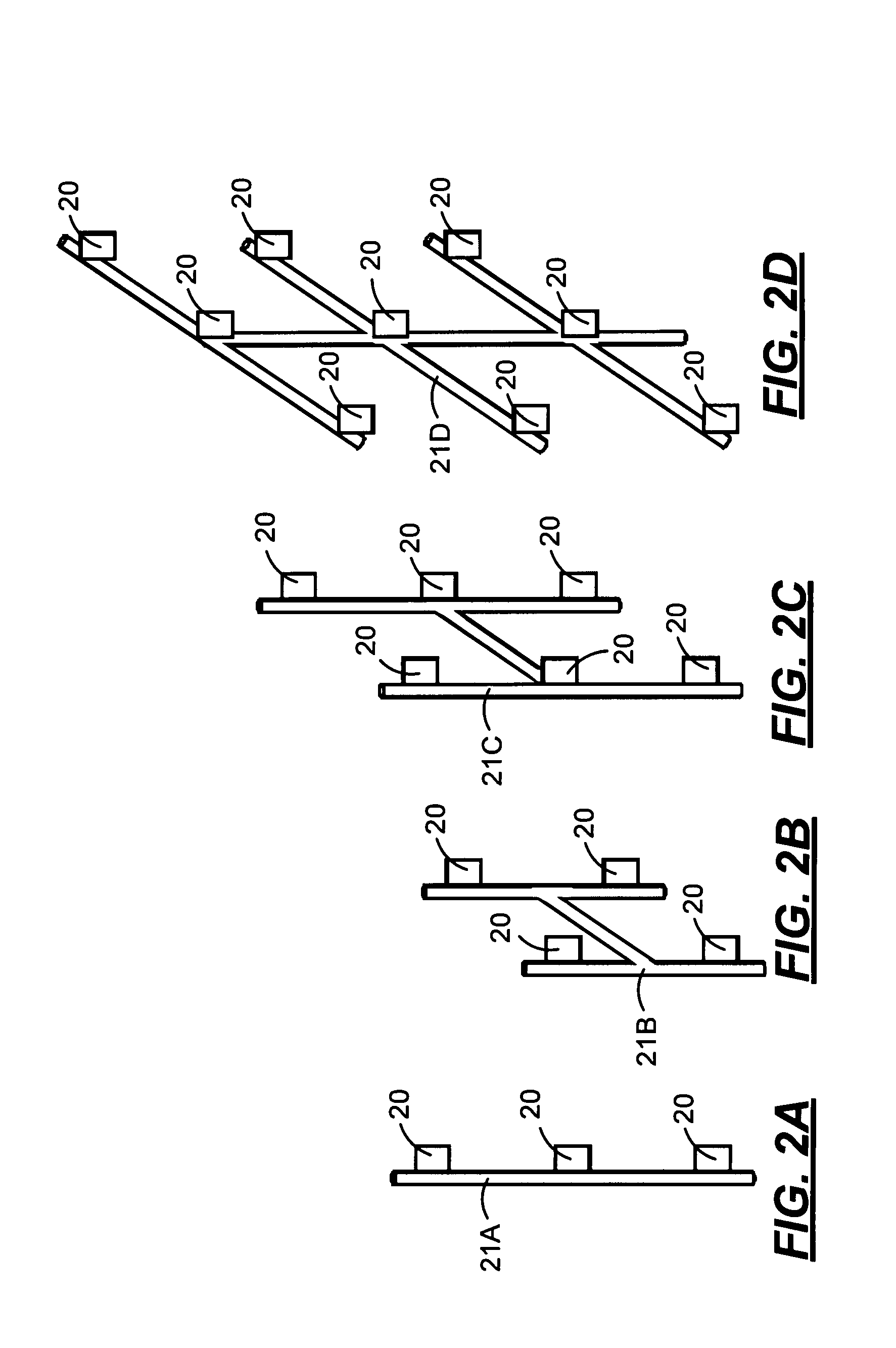
[0025]As shown in FIGS. 2 and 3, the imaging component 12 of the system 10 may be arranged in a variety of predetermined configurations. For the purposes of this disclosure, the imaging component 12 may include one or more image sensors 20 on support 21A (FIG. 2). At least three image sensors 20 are desirably used for the purposes of obtaining a full body image. The image sensors may be disposed in a single plane or in multiple planes as illustrated in FIGS. 1-3. In FIGS. 2A-2D, multiple image se...


PUM


Abstract

A system and method for documentation and analysis of dermatological aspects of a human body. The system has a predetermined arrangement of at least three image sensors selected from visible light sensors, ultraviolet (UV) light sensors, infrared (IR) sensors. Each of the image sensors has an effective normalized focal length of from about 8 to about 28 millimeters, an aperture stepped-down to at least f / 4 or higher F-stop, and a shutter exposure length of no longer than about 125 milliseconds. Output from the image sensors provide a single relatively high resolution image of a skin surface of the human body obtained from a distance of at least about 0.1 meters. A data collection and processing system is integrated with the image sensors to provide storage, analysis, and output of in-situ dermatological information to a system operator.

Description

TECHNICAL FIELD[0001]The disclosure relates to documentation and analysis of dermatological properties, and in particular to systems that provide improved capturing and analysis of images of skin surfaces for the purpose of aiding in the documentation, assessment, and treatment of the skin.BACKGROUND AND SUMMARY[0002]Imaging portions of a body for documenting and tracking physiological and pathological changes over time has been useful for the purposes of early detection and treatment of a variety of conditions including cancer, burns, and the like. Visible light and multi-spectral cameras have been used to capture digital images of partial regions of the body. Typically, handheld cameras and scanner devices are used for the manual collection of images, usually by a skilled professional. Once collected, the images are manually inspected by a medical professional to determine the appropriate treatment regimen if any. Often, high resolution images of the body are limited to particular...

Claims


Application Information

Patent Timeline
Patent Type & Authority Applications(United States)
IPC IPC(8): A61B5/00
CPCA61B5/0064A61B5/445A61B5/444A61B5/0077
Inventor ORTIZ, JOSEPH L.ORTIZ, NANCY A.
Owner ORTIZ JOSEPH L
Why Eureka
Try Eureka