

Combination Therapy
a combination therapy and cell technology, applied in the field of mammalian disease manifested by abnormal cell growth and/or abnormal cell proliferation, can solve the problems of mitotic arrest or apoptosis of cells, and achieve the effect of potentiating therapeutic activity
- Summary
- Abstract
- Description
- Claims
- Application Information
AI Technical Summary
Benefits of technology
Problems solved by technology
Method used
Image

Examples
example 1
Production of Recombinant HDAC Isotypes
[0238]cDNAs of human HDAC1-8 and 11 were generated by RT-PCR reactions using primers complementary to the 5′ and 3′ coding sequence of human HDAC gene sequences in GenBank. cDNAs corresponding to the full length human HDAC1, 2, 3 and 11 were cloned into pBlueBac4.5 vector (Invitrogen). The constructs were used to generate recombinant baculoviruses using the Bac-N-Blue™ DNA according to the manufacturer's instructions (Invitrogen). The recombinant HDAC1, 2, 3, 11 proteins produced harbor a FLAG tag at their C-termini. cDNAs encoding truncated versions of HDAC4, 5 and 7 encompassing their deacetylase domains were cloned into pDEST10 as an N-terminal hexahistidine fusion protein and recombinant baculoviruses were generated using the Bac-to-Bac™ Baculovirus expression system (Invitrogen). HDAC6 and 8 were cloned as full length N-terminally His-tagged protein. All HDAC proteins were expressed in insect Sf-9 cells (Spodoptera frugiperdai) upon infect...
example 2
Fluorescence-Based HDAC Enzyme Assay Using Recombinant HDAC Enzymes
[0239]Recombinant HDAC enzymes were incubated with diluted compounds in assay buffer (25 mM Hepes, pH 8.0, 137 mM NaCl, 1 mM MgCl2 and 2.7 mM KCl) for 10 minutes at ambient temperatures in black 96-well plate. Boc-Lys(Ac)-AMC (for HDAC1, 2, 3, 6, and 8 enzymes), which was purchased from Bachem Biosciences Inc., (King of Prussia, Philadelphia) were added into enzyme-compound mixture and incubated at 37° C. For HDAC4, 5, 7 assays, Boc-Lys(TFA)-AMC, which was synthesized in house, was used as substrate and 0.1% BSA was added to the buffer. The final concentration of substrates was 2 times over Ki of each isotype enzyme (between 70 uM to 200 uM). Reaction time was predetermined to ensure that reaction was linear for the incubation time. Reaction was stopped by adding a freshly prepared trypsin (1 mg / ml final concentration) with 1 μM TSA (Biomol) in assay buffer. After 30 minutes, fluorescence was measured using a fluorom...
example 3
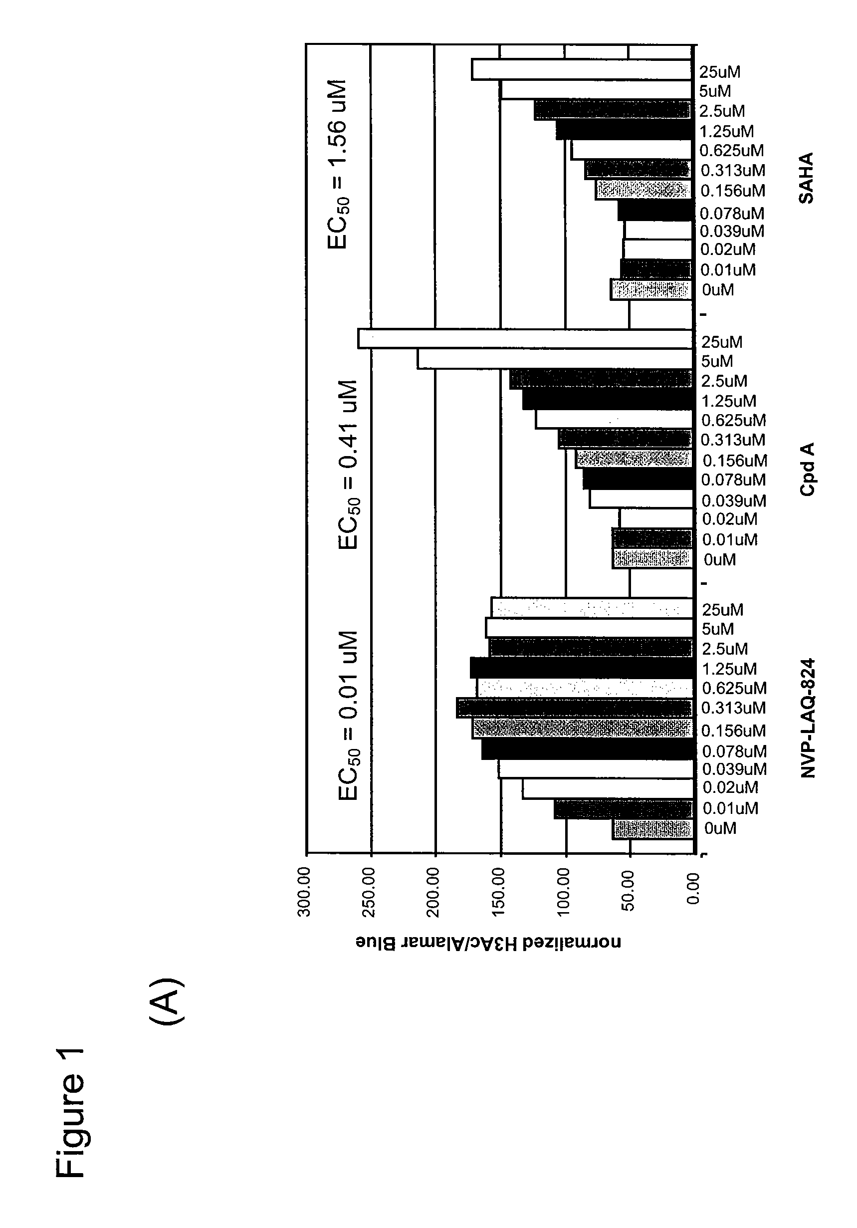
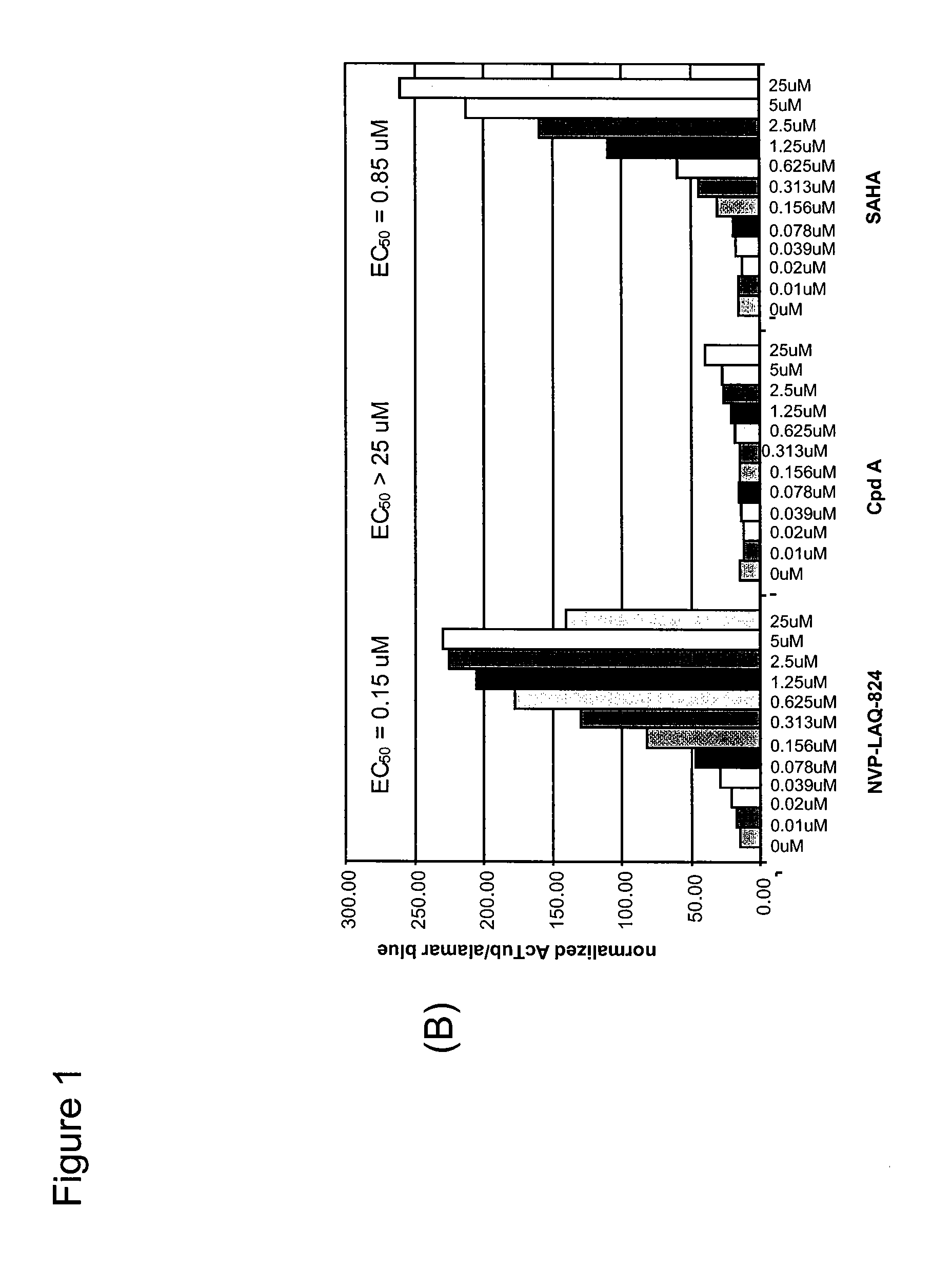
Cell-Based ELISA for H3 and Tubulin Acetylation
[0240]Bladder carcinoma T24 cells were seeded in black plates with clear bottoms (Costar #3603) at 1×10⁴ cells per well in a volume of 100 μl per well, and were allowed to settle for one day at 37° C. in a CO2 incubator. The cells were treated for 16 h with various concentrations of HDAC inhibitors. 3 h before the end of the treatment, Alamar Blue (BioSource) was added to monitor cell viability according to the manufacturer's instructions. At the end of the treatment time, Alamar Blue OD (at 570 nm and 600 nm) was recorded, then the cells were carefully washed in PBS, fixed in pre-chilled methanol for 10 min at −20° C., washed again twice in PBS, and blocked in PBS containing 0.1% Triton X-100 and 1% BSA for a minimum of 30 min. For H3 acetylation, rabbit-anti-acetyl-H3 (Upstate #06-599) was used as primary antibody at a dilution of 1:1000 for 45 min; the secondary antibody was HRP-coupled goat-anti-rabbit (Sigma #A-0545), used at 1:800...
PUM
| Property | Measurement | Unit |
|---|---|---|
| Structure | aaaaa | aaaaa |
| Cell proliferation rate | aaaaa | aaaaa |
| Cell growth | aaaaa | aaaaa |
Abstract
Description
Claims
Application Information