User interface system for mammographic imager

a user interface and imager technology, applied in the field of medical imaging/biopsy systems, can solve the problems of limited ability to utilize ultrasound technologists as opposed to experienced physician specialists to perform most breast ultrasound procedures, difficult to locate a potential lesion/suspicious mass, and difficult to mentally associate ultrasound images with x-ray images, etc., to facilitate efficient use of medical personnel time, avoid acquisition, storage and processing

Inactive Publication Date: 2009-06-04
NIELDS MORGAN W +2
View PDF7 Cites 72 Cited by
  • Summary
  • Abstract
  • Description
  • Claims
  • Application Information

AI Technical Summary

Benefits of technology

[0009]It is an object of the present invention to provide an enhanced imaging / biopsy system that can reduce trained medical personnel time requirements in diagnostic and biopsy procedures for tissue diagnosis. It is a related objective to provide such a system in a cost-effective manner; namely through the provision of a system having relatively expensive components that can be utilized for multiple medical procedures combinatively employed in a single system.
[0010]A further objective of the present invention is to provide an enhanced imaging / biopsy system for obtaining spatially correlated three-dimensional image information regarding a location of interest in the body, such system being apt for the obtainment of three-dimensional image information regarding a potential lesion or suspicious mass in a female patient's breast. It is a further objective to provide such information in a manner allowing for enhanced use of tissue removal systems used for obtaining tissue samples from the body, including specifically, tissue from a potential lesion or suspicious mass within a female patient's breast. Such information may also be used in conjunction with other targeted instruments such as guide wire placement devices and instruments for ablation, delivery, etc.
[0011]Yet another objective of the present invention is to provide an enhanced imaging / biopsy system for obtaining depth-related image information for diagnostic use and for otherwise yielding biopsy-related control and access advantages.

Problems solved by technology

Specifically, a free-hand procedure can be performed in which a hand-held ultrasound probe is manipulated on the breast while viewing a display to obtain depth-profile information As can be appreciated, location of a potential lesion / suspicious mass can be difficult, and the ultrasound images obtained are frequently difficult to mentally associate with the x-ray images.
As such, the ability to utilize ultrasound technologists as opposed to experienced physician specialists to perform most breast ultrasound procedures is limited.
Recent developments in tissue removal systems have resulted in larger, heavier devices that are difficult for a physician to use in conjunction with free-hand ultrasound guidance.

Method used


Image

  • User interface system for mammographic imager
  • User interface system for mammographic imager
  • User interface system for mammographic imager

Examples


Embodiment Construction

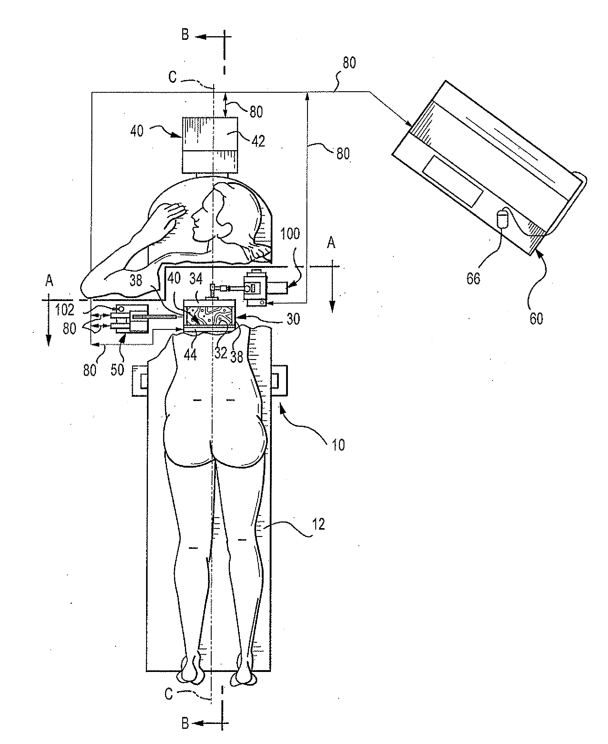
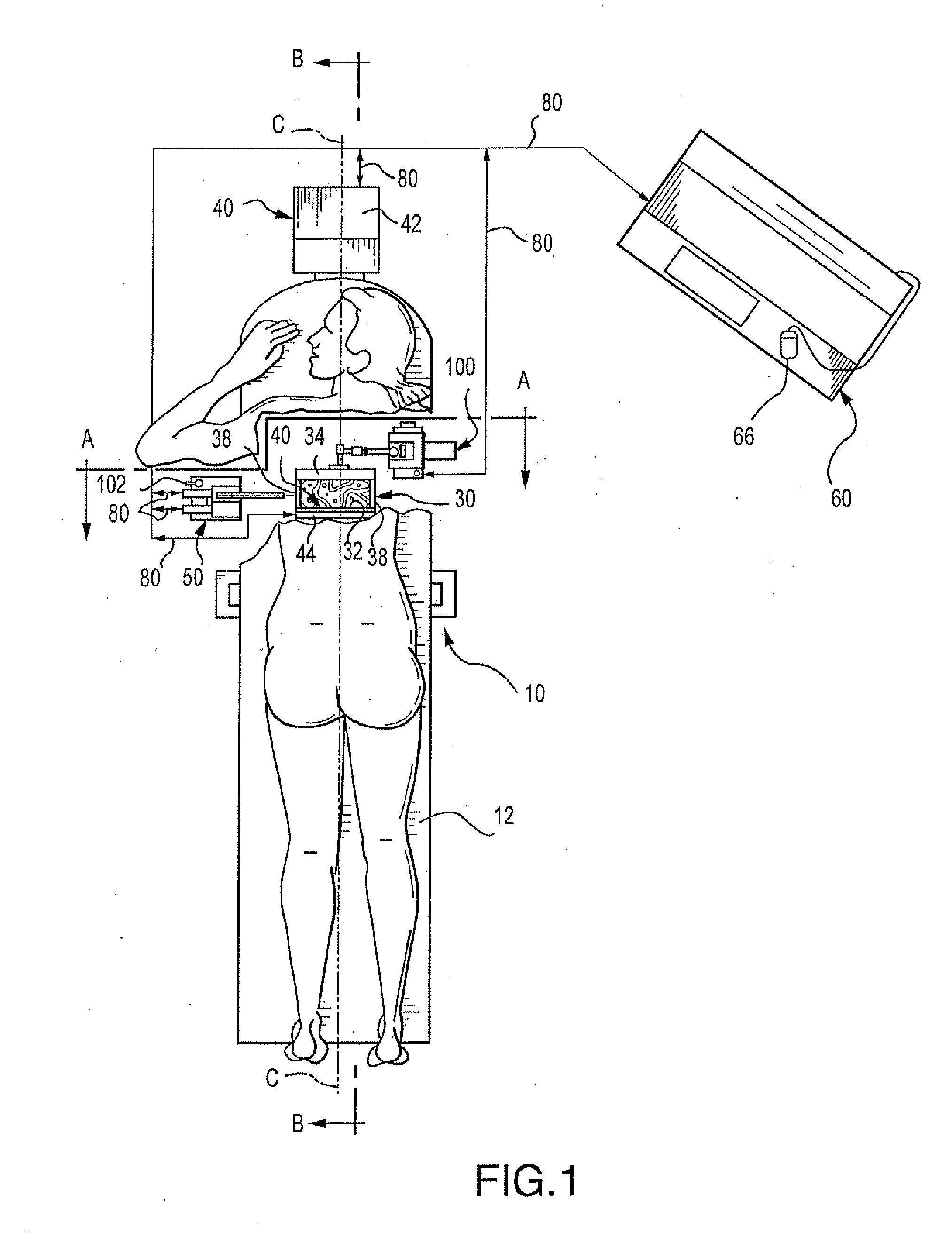
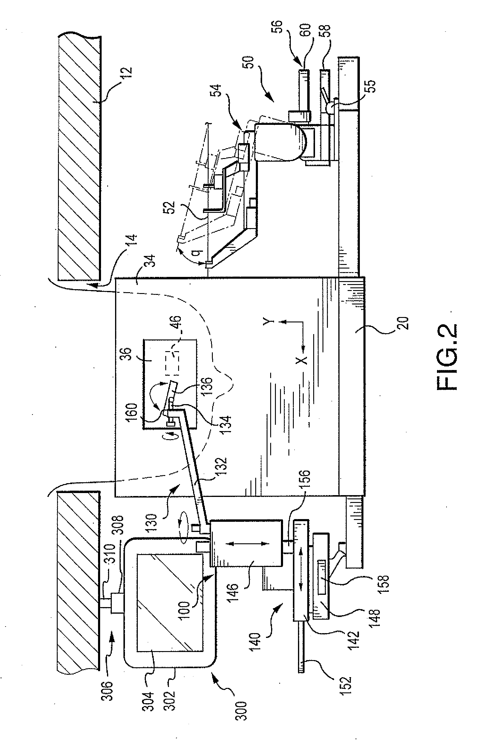
[0036]FIGS. 1-6 illustrate one embodiment of a diagnostic ultrasound / x-ray biopsy system comprising the present invention, as adapted for mammography / breast biopsy use.

[0037]Generally; the system comprises a support assembly 10 having a patient table 12 with breast-opening 14 therethrough, an immobilization assembly 30 for immobilizing a patient's breast within a predetermined XYZ frame of reference under the opening 14 of table 12, an x-ray imaging assembly 40 for providing two-dimensional x-ray images (e.g., X-Y images) of the patient's immobilized breast in correlated spatial relation to the predetermined XYZ frame of reference, and an ultrasound imaging assembly 100 for providing orthogonal depth-profile images (e.g., X-Z, Y-Z and / or X, Y-Z images) of the immobilized breast in correlated spatial relation to the predetermined XYZ frame of reference. A biopsy assembly 50 having puncture instrument 52 is also provided for obtaining samples from a patient's breast while the breast i...


PUM


Abstract

The present invention provides for x-ray imaging and ultrasound imaging of a body region of interest in a spatially correlatable manner. The resultant x-ray and ultrasound images may be combinatively employed to provide three-dimensional information regarding a location of interest within the body, and is particularly apt for use in the analysis / biopsy of potential lesions and suspicious masses in a female breast. The invention provides for direct body contact by an ultrasound imaging head, as well as targeted ultrasound imaging of a selected portion of the region from which x-ray images are obtained. A user interface system facilitates various procedures including ultrasound guided needle biopsy procedures.

Description

RELATED APPLICATION INFORMATION[0001]This application claims priority from U.S. Provisional Patent Application Ser. No. 09 / 449,267, filed Nov. 24, 1999, which is a continuation-in-part of prior U.S. patent application Ser. Nos. 60 / 109,881, filed on Nov. 25, 1998 and 09 / 111,094, filed on Jul. 6, 1998, both of which are incorporated herein by reference in their entireties. The latter application is a continuation-in-part of U.S. patent application Ser. No. 08 / 730,107, now issued as U.S. Pat. No. 5,776,062.FIELD OF THE INVENTION[0002]The present invention relates to medical imaging / biopsy systems, and more particularly, to an enhanced system that employs x-ray imaging and targeted ultrasound imaging in a combinative, spatially correlatable manner that is particularly apt for breast imaging / biopsy procedures. The invention further relates to targeted ultrasound features that yield plural modalities of operation as well as improved biopsy capabilities and a user interface system for faci...

Claims


Application Information

Patent Timeline
Patent Type & Authority Applications(United States)
IPC IPC(8): A61B8/00A61B6/00A61B8/08A61B10/00A61B10/02A61B17/34A61B19/00
CPCA61B6/0435A61B6/502A61B8/0825A61B8/406A61B8/4218A61B8/5238A61B6/5247A61B17/3403A61B2019/205Y10S128/915Y10S128/916A61B6/4417A61B10/0233A61B90/17
Inventor NIELDS, MORGAN W.CONNER, JOHNDALY, CURTIS
Owner NIELDS MORGAN W
Why Eureka
Try Eureka