Spatially correlated x-ray and ultrasound mammographic imaging systems and method

a mammography and ultrasound technology, applied in the field of medical imaging/biopsy systems, can solve the problems of limited ability to utilize ultrasound technologists as opposed to experienced physician specialists to perform most breast ultrasound procedures, difficult to mentally associate ultrasound images with x-ray images, and difficult to locate potential lesion/suspicious mass location, etc., to achieve convenient efficient use of medical personnel time, avoid acquisition, storage and processing

Inactive Publication Date: 2009-02-24
HOLOGIC INC
View PDF54 Cites 42 Cited by
  • Summary
  • Abstract
  • Description
  • Claims
  • Application Information

AI Technical Summary

Benefits of technology

[0017]As indicated above, x-ray images may be employed to select a limited, or targeted, portion of the x-rayed body region of interest to be imaged utilizing the ultrasound signal. Such targeted ultrasound imaging avoids the acquisition, storage and processing of unneeded imaging data, and otherwise facilitates efficient use of medical personnel time, and otherwise advantageously accommodates direct contact with the body portion to be imaged. Further, where necessary, the provision of a hand-held ultrasound imaging option provides practitioners with added flexibility as may be desirable in certain applications.
[0018]According to a further aspect of the present invention, an ultrasound imaging apparatus is provided that has an improved imaging focal depth. It has been noted that a linear array of transducer elements may have a focal depth that is only a portion of the thickness of a patient's immobilized breast. In this regard, in order to provide for more complete imaging for a range of patients, it is desirable to provide a greater focal depth. In particular, it would be desirable to provide a focal depth approaching to accommodate a range of patients and procedures. A corresponding apparatus with improved focal depth includes a probe structure supporting a transducer array that includes at least a first set of transducer elements disposed a first distance from the signal interface surface of the probe structure and a second set of transducer elements disposed a second distance from the signal interface surface. The first and second sets of elements thereby provide a combined focal depth that is greater than the focal depth that would be provided by either of the transducer sets considered alone. In a preferred implementation, a transducer array includes 7 or more columns of array elements where each column is disposed a different distance from the signal interface surface of the probe structure. Such a structure provides for improved imaging for a range of patients.
[0019]In accordance with a still further aspect of the present invention, a display is provided proximate to the patient's breast in order to facilitate real time monitoring of insertion of a medical instrument into the patient's breast. The associated apparatus includes: an immobilizer for immobilizing the patient's breast; a first graphical display for displaying one or more images of the patient's breast so as to permit identification of an area of interest within the patient's immobilized breast; a medical instrument operative for insertion to the identified area of interest within the patient's breast; and a second graphical display, separate from the first graphical display and located proximate to the patient's immobilized breast, for providing real time images of the patient's compressed breast so that a user can monitor insertion of the medical instrument to the identified location of interest using the second graphical display located proximate to the patient's immobilized breast. Preferably, the second graphical display can be translated and rotated to facilitate viewing during a medical procedure In one embodiment, the patient is supported in a prone position on a table with the breast under examination protruding through an opening in the table and the second display is disposed beneath the table for convenient viewing. Real time images such as ultrasound images can be monitored on the second display during insertion of a medical instrument such as a biopsy needle for improved guidance and confidence regarding sampling of suspicious masses.

Problems solved by technology

As can be appreciated, location of a potential lesion / suspicious mass can be difficult, and the ultrasound images obtained are frequently difficult to mentally associate with the x-ray images.
As such, the ability to utilize ultrasound technologists as opposed to experienced physician specialists to perform most breast ultrasound procedures is limited.
Recent developments in tissue removal systems have resulted in larger, heavier devices that are difficult for a physician to use in conjunction with free-hand ultrasound guidance.

Method used


Image

  • Spatially correlated x-ray and ultrasound mammographic imaging systems and method
  • Spatially correlated x-ray and ultrasound mammographic imaging systems and method
  • Spatially correlated x-ray and ultrasound mammographic imaging systems and method

Examples


Embodiment Construction

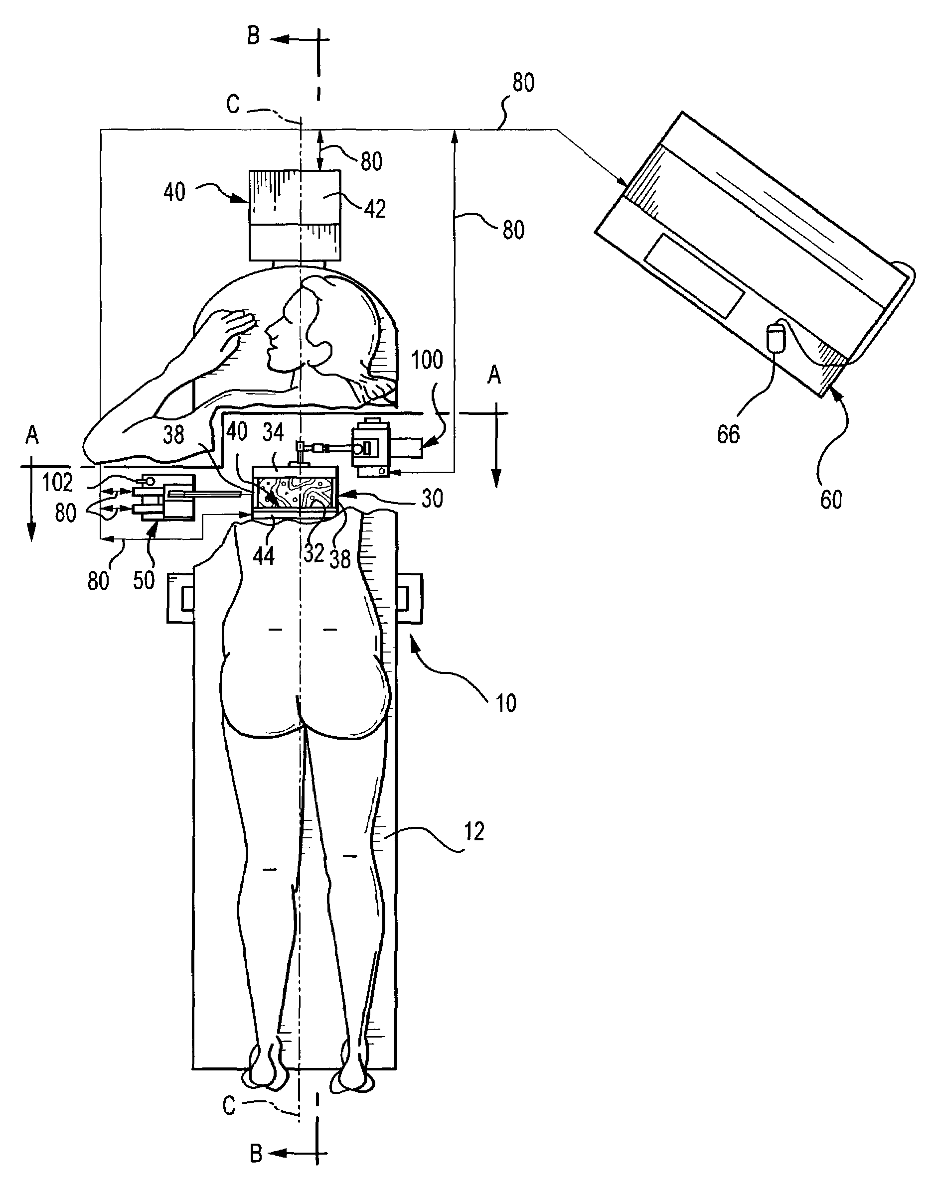
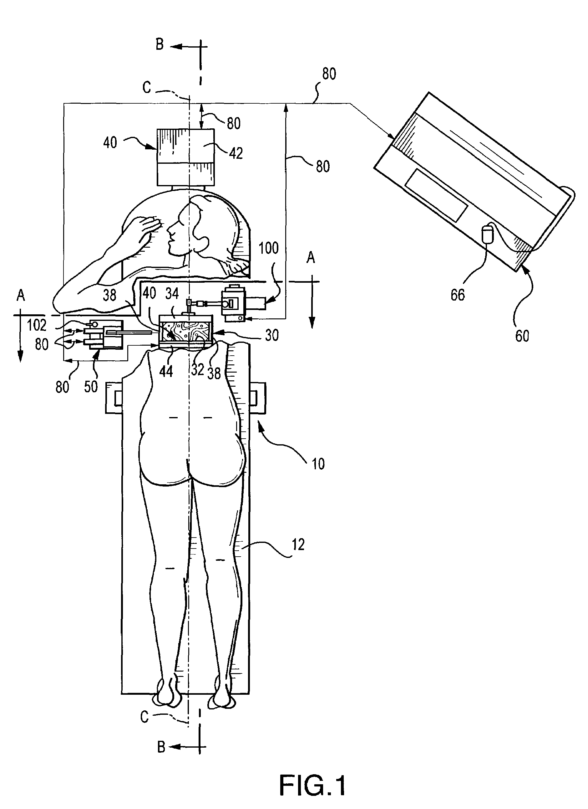
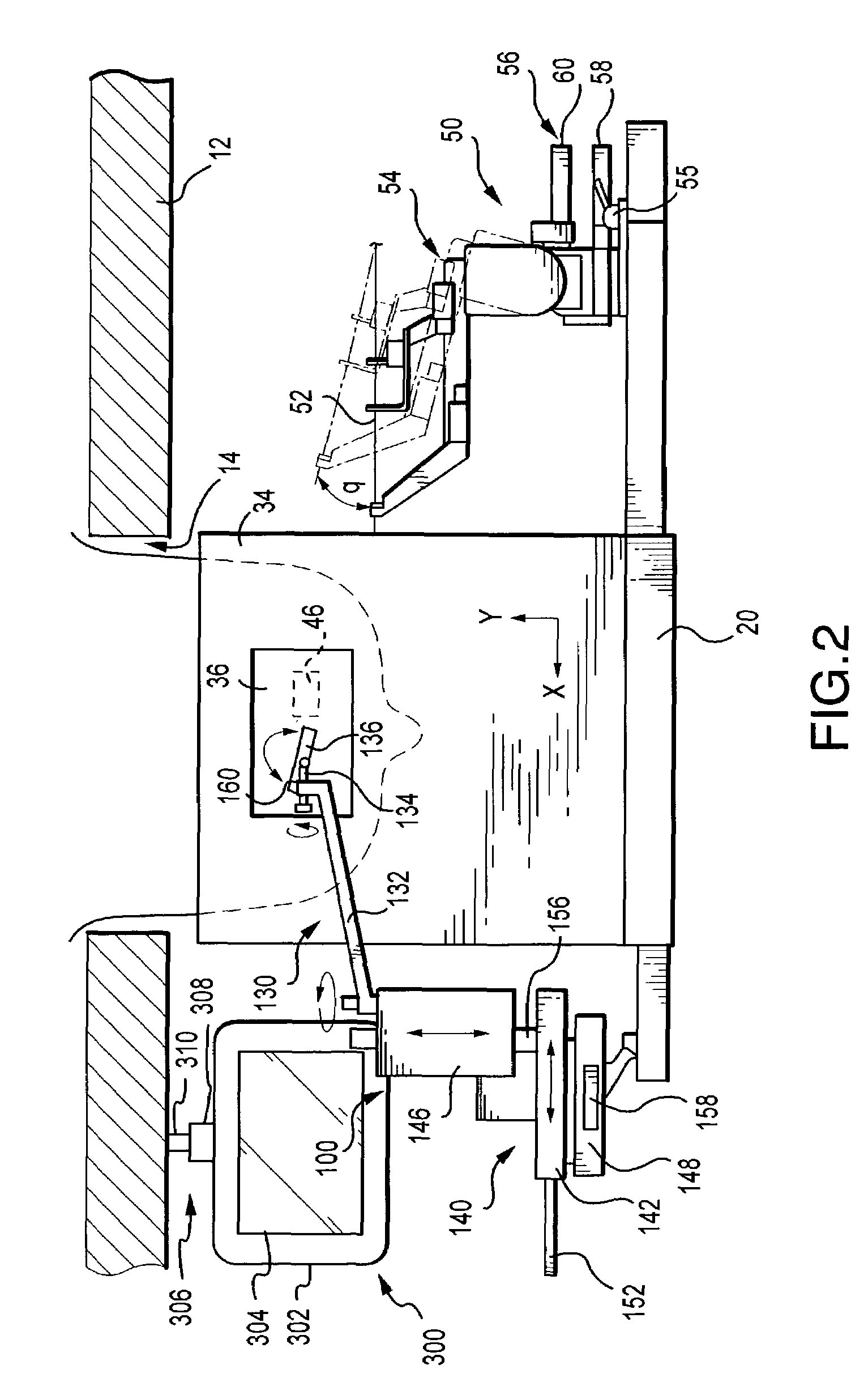
[0036]FIGS. 1-6 illustrate one embodiment of a diagnostic ultrasound / x-ray biopsy system comprising the present invention, as adapted for mammography / breast biopsy use.

[0037]Generally, the system comprises a support assembly 10 having a patient table 12 with breast-opening 14 therethrough, an immobilization assembly 30 for immobilizing a patient's breast within a predetermined XYZ frame of reference under the opening 14 of table 12, an x-ray imaging assembly 40 for providing two-dimensional x-ray images (e.g., X-Y images) of the patient's immobilized breast in correlated spatial relation to the predetermined XYZ frame of reference, and an ultrasound imaging assembly 100 for providing orthogonal depth-profile images (e.g., X-Z, Y-Z and / or X, Y-Z images) of the immobilized breast in correlated spatial relation to the predetermined XYZ frame of reference. A biopsy assembly 50 having puncture instrument 52 is also provided for obtaining samples from a patient's breast while the breast i...


PUM


Abstract

The present invention provides for x-ray imaging and ultrasound imaging of a body region of interest in a spatially correlatable manner. The resultant x-ray and ultrasound images may be combinatively employed to provide three-dimensional information regarding a location of interest within the body, and is particularly apt for use in the analysis/biopsy of potential lesions and suspicious masses in a female breast. The invention provides for direct body contact by an ultrasound imaging head, as well as targeted ultrasound imaging of a selected portion of the region from which x-ray images are obtained. A user interface system facilitates various procedures including ultrasound guided needle biopsy procedures.

Description

RELATED APPLICATION INFORMATION[0001]This application is a divisional of U.S. patent application Ser. No. 09 / 449,267, filed Nov. 24, 1999, now U.S. Pat. No. 6,459,925, issued Oct. 1, 2002, which claims the benefit of U.S. Provisional Application Ser. No. 60 / 109,881, filed on Nov. 25, 1998 and is a Continuation-in-Part of Ser. No. 09 / 111,094, filed on Jul. 6, 1998, now U.S. Pat. No. 6,102,866, issued Aug. 15, 2000, all of which are incorporated herein by reference in their entireties. The latter application is a continuation-in-part of U.S. patent application Ser. No. 08 / 730,107, filed Oct. 15, 1996 now issued as U.S. Pat. No. 5,776,062.FIELD OF THE INVENTION[0002]The present invention relates to medical imaging / biopsy systems, and more particularly, to an enhanced system that employs x-ray imaging and targeted ultrasound imaging in a combinative, spatially correlatable manner that is particularly apt for breast imaging / biopsy procedures. The invention further relates to targeted ult...

Claims


Application Information

Patent Timeline
Patent Type & Authority Patents(United States)
IPC IPC(8): A61B6/02A61B5/05A61B6/00A61B8/08
CPCA61B6/0435A61B6/463A61B6/465A61B6/469A61B6/502A61B6/5247A61B8/0825A61B8/406A61B8/463A61B8/465A61B8/467A61B8/5238A61B6/4417Y10S128/915Y10S128/916A61B8/4416A61B8/464A61B8/469
Inventor NIELDS, MORGAN W.CONNOR, JOHNDALY, CURTIS
Owner HOLOGIC INC
Why Eureka
Try Eureka