Radiotherapeutic system and radiotherapeutic dose distribution measuring method

Active Publication Date: 2009-06-25
TOSHIBA MEDICAL SYST CORP
View PDF6 Cites 44 Cited by
  • Summary
  • Abstract
  • Description
  • Claims
  • Application Information

AI Technical Summary

Benefits of technology

[0008]In view of the circumstances, an object of the present invention is to provide a radiotherapeutic system and a radiotherapeutic dose distribution measuring method, in external X-ray radiation therapy,

Problems solved by technology

However, at present, there is no means for confirming whether irradiation is actually performed to the patient with the planned position and dose or not.
However, it is difficult to place a patient in a position according to the irradiation plan, on a bed different from a phantom which is easily portable and whose position can be freely adjusted.
The confirmation prior to radiation does not completely assure the planned irradiation to the patient.
Since forward scattering is dominant in the sc

Method used


Image

  • Radiotherapeutic system and radiotherapeutic dose distribution measuring method
  • Radiotherapeutic system and radiotherapeutic dose distribution measuring method
  • Radiotherapeutic system and radiotherapeutic dose distribution measuring method

Examples


first embodiment

[0065]An embodiment of the present invention will be described below with reference to the drawings. In the following description, the same reference numerals are designated to components having almost the same function and configuration, and repetitive description will be given only when required.

[0066][Principle and Method]

[0067]A radiotherapeutic system of the first embodiment measures scattering radiation from a subject on the basis of radiation emitted to the subject and, on the basis of the scattering radiation, obtains information objectively showing an irradiated region in the subject and dose of the radiation. The principle and the method are as follows.

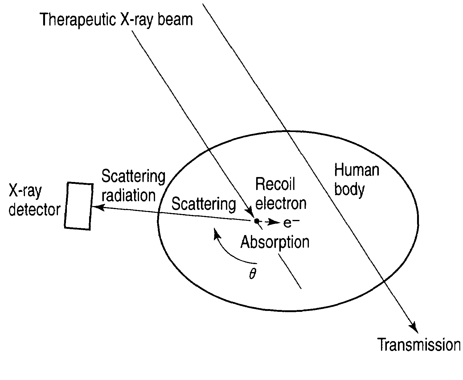
[0068]FIG. 1 is a diagram for explaining the principle and method of measuring scattering radiation from a subject on the basis of therapeutic radiation of the radiotherapeutic system of the invention.

[0069]The effect of treatment with the external X-ray radiation is produced mainly by X-ray scattered in the body of a patien...

second embodiment

[0159]A second embodiment of the present invention will now be described. A radiotherapeutic system in the second embodiment measures scattering radiation from a subject based on radiation applied to the subject and, on the basis of the measured scattering radiation, obtains, as absorbed dose, information objectively showing a region in the subject and a dose.

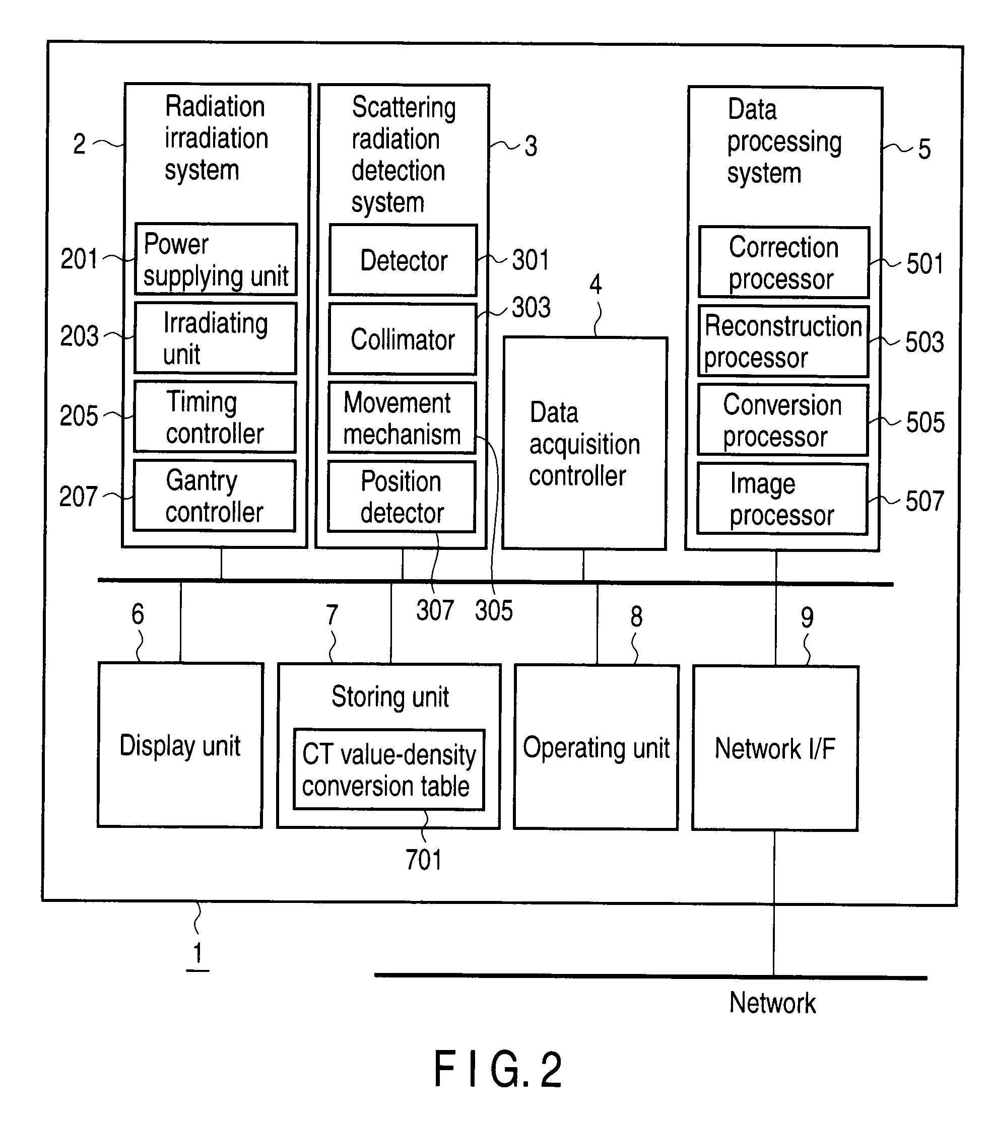
[0160]FIG. 19 is a block configuration diagram of a radiotherapeutic system 1 of the second embodiment. FIG. 19 is different from FIG. 2 with respect to the configuration of the data processing system 5 and the storing unit 7.

[0161]The data processing system 5 has a correction processor 501, a reconstruction processor 503, a conversion processor 505, and an image processor 507.

[0162]The correction processor 501 performs a data calibration process, a correcting process for eliminating noise, and the like as necessary. The correcting process executed by the correction processor 501 will be described in detail later.

[0163]The reco...

first example

[0174]An absorbed dose image data generating method using the radiotherapeutic system 1 of a first example will be described. In the radiotherapeutic system of the first example, a detector having a collimator is mounted in a position at a specific angle with respect to a therapeutic X-ray beam and selectively detects only scattering radiation in the direction. Further, to three-dimensionally obtain a distribution of places where scattering occurs in the patient body, the detector is rotated during irradiation and scattering radiation is measured from all of directions (refer to, for example, FIG. 3). After that, a reconstructing process is performed, and a distribution of occurrence of scattering radiation in the subject is three-dimensionally imaged.

[0175]FIG. 20 is a flowchart showing the flow of processes in radiation treatment including the absorbed dose image data generating process of the example. The processes in the steps will be described below.

[0176][Disposition of Subjec...


PUM


Abstract

A detector having a collimator in a position at a specific angle with respect to a therapeutic X-ray beam is mounted to selectively detect only scattering radiation in the direction. To three-dimensionally obtain a distribution of places where scattering occurs in a patient body, a detector is rotated during irradiation and scattering radiation is measured from all of directions. After that, a reconstructing process is performed, and a distribution of occurrence of scattering radiation in the subject is three-dimensionally imaged. Since angles and amounts of X-rays scattered by Compton scattering are known theoretically, if scattering radiation at a certain angle can be detected, the number of scattering radiation at other angles can be also estimated. On the basis of the theory, images of distribution of scattering radiation sources are converted to images of distribution of absorption of radiation.

Description

CROSS-REFERENCE TO RELATED APPLICATIONS[0001]This application is based upon and claims the benefit of priority from prior Japanese Patent Applications No. 2007-331108, filed Dec. 21, 2007; No. 2008-026733, filed Feb. 6, 2008; No. 2008-033361, filed Feb. 14, 2008; and No. 2008-033362, filed Feb. 14, 2008, the entire contents of all of which are incorporated herein by reference.BACKGROUND OF THE INVENTION[0002]1. Field of the Invention[0003]The present invention relates to a radiotherapeutic system and a radiotherapeutic dose distribution measuring method, in radiation treatment performed by emitting radiation from the outside of the body of a patient to an affected region (treatment for applying X-rays from the outside of the body of a patient to an affected region), capable of preventing excessive irradiation to an affected area and a normal tissue by measuring an actually irradiated region in the patient and actual dose and providing the result in a predetermined form.[0004]2. Desc...

Claims


Application Information

Patent Timeline
IPC IPC(8): A61B6/00A61N5/10
CPCA61B6/027A61B6/4291A61N5/1048
Inventor SAKURAI, YASUOOHYU, SHIGEHARUNAMBU, KYOJIROHARAGASHIRA, MOTOJIKUMAKURA, MASAHIROSHIBATA, MARIKOFUJIMOTO, KATSUHIKOTAKADA, YOICHI
Owner TOSHIBA MEDICAL SYST CORP
Why Eureka
Try Eureka