Nanoparticles that facilitate imaging of biological tissue and methods of forming the same

Inactive Publication Date: 2009-09-03
FLORIDA STATE UNIV RES FOUND INC
View PDF3 Cites 63 Cited by
  • Summary
  • Abstract
  • Description
  • Claims
  • Application Information

AI Technical Summary

Benefits of technology

[0004]Accordingly, there is a need for particles and/or nanoparticles that facilitate imaging of biological tissue. Further, there is a need for methods and/or techniques for forming particles and/or nanoparticles that facilitate imaging of biological tissue.
[0005]Some or all of the above needs and/or problems may be addressed by certain embodiments of the invention. Embodiments of the invention may include nanoparticles that facilitate imaging of biological tissue and methods for formulating the same. According to one embodiment of the invention, a method for forming polymeric nanoparticles is provided. An anionic surfactant may be applied to superparamagnetic nanoparticles to form modified nanoparticles. The modified nanoparticles may be mixed with a polym

Problems solved by technology

However, conventional contrast agents, such as iron oxide particles, may not adequately attach to tissue in which the agents are injected.
This failure to adequately attach to tissue m

Method used


Image

  • Nanoparticles that facilitate imaging of biological tissue and methods of forming the same
  • Nanoparticles that facilitate imaging of biological tissue and methods of forming the same
  • Nanoparticles that facilitate imaging of biological tissue and methods of forming the same

Examples


Embodiment Construction

[0023]Example embodiments of the invention now will be described more fully hereinafter with reference to the accompanying drawings, in which some, but not all embodiments of the invention are shown. Indeed, these inventions may be embodied in many different forms and should not be construed as limited to the embodiments set forth herein; rather, these embodiments are provided so that this disclosure will satisfy applicable legal requirements. Like numbers refer to like elements throughout.

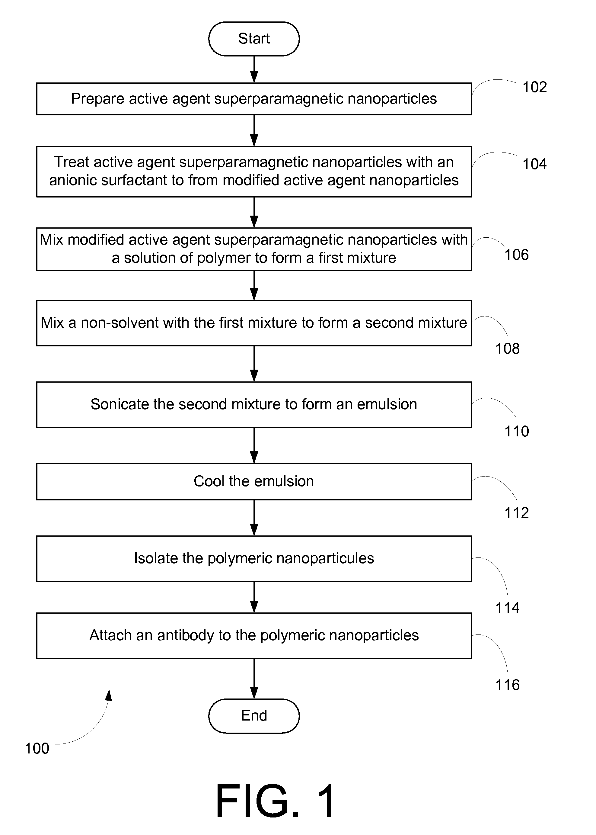
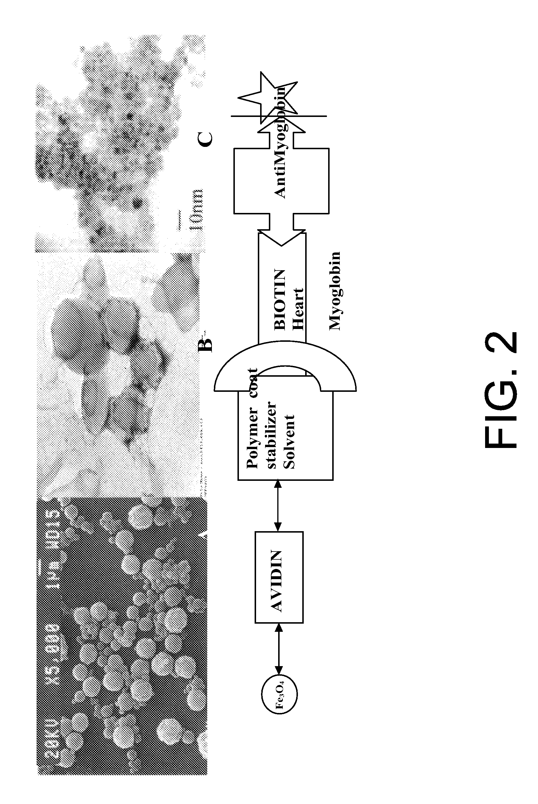
[0024]Example embodiments of the invention may provide for the formation, preparation and / or synthesis of nanoparticles that may facilitate the imaging of biological tissue. In various embodiments, the nanoparticles may facilitate the preparation of high resolution images and rapid imaging of various biological matter, such as cardiac tissue. According to an example embodiment of the invention, nanoparticles as described herein may be injected or supplied to the biological tissues to be imaged acc...


PUM

PropertyMeasurementUnit
Diameteraaaaaaaaaa
Diameteraaaaaaaaaa
Superparamagnetismaaaaaaaaaa

Abstract

Nanoparticles that facilitate imaging of biological tissue and methods for formulating the nanoparticles are provided. In order to form suitable nanoparticles for imaging, an anionic surfactant may be applied to superparamagnetic nanoparticles to form modified nanoparticles. The modified nanoparticles may be mixed with a polymer in a solvent to form a first mixture, and a non-solvent may be mixed with the first mixture to form a second mixture. An emulsion may be formed from the second mixture and the polymeric nanoparticles may be isolated from the emulsion. In certain embodiments of the invention, an antibody may be attached to the polymeric nanoparticles to facilitate attachment of the nanoparticles to biological tissue.

Description

CROSS REFERENCE TO RELATED APPLICATION[0001]The present application claims the benefit of U.S. Provisional Application No. 61 / 032,716, filed Feb. 29, 2008, and entitled “Systems and Methods for Biological Magnetic Resonance Imaging.” The priority application is incorporated by reference herein in its entirety.FIELD OF THE INVENTION[0002]Embodiments of the invention relate generally to nanoparticles and more particularly to nanoparticles that may facilitate imaging of biological tissue.BACKGROUND OF THE INVENTION[0003]When imaging tissue, such as biological tissue, a wide variety of imaging techniques are typically utilized, such as, magnetic resonance imaging (MRI). In some instances contrast agents are injected in tissue to enhance the appearance of the tissue. However, conventional contrast agents, such as iron oxide particles, may not adequately attach to tissue in which the agents are injected. This failure to adequately attach to tissue may lead to lower quality or lower resolu...

Claims


Application Information

Patent Timeline
IPC IPC(8): A61K49/16B05D7/00
CPCB82Y5/00A61K49/1824
Inventor SHARMA, RAKESH
Owner FLORIDA STATE UNIV RES FOUND INC
Why Eureka
Try Eureka