Method and device for optical detection of the eye

a technology of optical detection and eye, applied in the field of optical detection of eyes, can solve the problems of only visible disease-relevant changes in an oct image, many phenomena of icg angiography are still not quite understood, and achieve the effect of improving optical contrast and high transparency of optical system

Inactive Publication Date: 2009-12-10
CARL ZEISS MEDITEC AG
View PDF12 Cites 18 Cited by
  • Summary
  • Abstract
  • Description
  • Claims
  • Application Information

AI Technical Summary

Benefits of technology

[0048]The use of optical methods is strongly preferred for the diagnosis of the eye as a result of the high transparency of the optical system of the eye compared to other body parts. On the other hand, the added molecular markers, which selectively improve the optical contrast for the diagnosis, also influence the normal vision of the patient.

Problems solved by technology

However, for the integration of in-vitro techniques and methods in in-vivo environments, a number of problems must be overcome (e.g., toxicity, targeted transport to target cell, anatomical transport barriers).
This method is only effective for a very limited number of molecules.
However, the disadvantage is that disease-relevant changes in an OCT image are only visible once the disease has broken out.
Furthermore, anomalies detected in an OCT image do not necessarily have pathological causes (problem of structure and function).
While fluorescein remains the standard dye for diabetic retina changes, retinal vessel occlusions or macular edemas, ICG is increasingly used for age-related macular degeneration and other subretinal diseases, due to the limited diagnostic information from fluorescein angiography for technical reasons.
Despite extensive studies, many phenomena of the ICG angiography are still not quite understood.
Therefore, when compared to FA angiography, there is still no uniform terminology for the findings of an ICG angiography.
The described known methods for increasing contrast in ophthalmology (ICG or fluorescence angiography) are limited to contrasting blood vessels through attachment of fluorescence dyes to blood components, such as hemoglobin and albumin.
Even though this allows for detection of changes in the blood vessels, e.g., neovascularization, provided they are already in an advanced stage, a detection of disease-relevant molecules and cells as well as morphological changes in tissues and membranes, as would be necessary for early detection, is not possible.

Method used


Image

  • Method and device for optical detection of the eye
  • Method and device for optical detection of the eye
  • Method and device for optical detection of the eye

Examples


Embodiment Construction

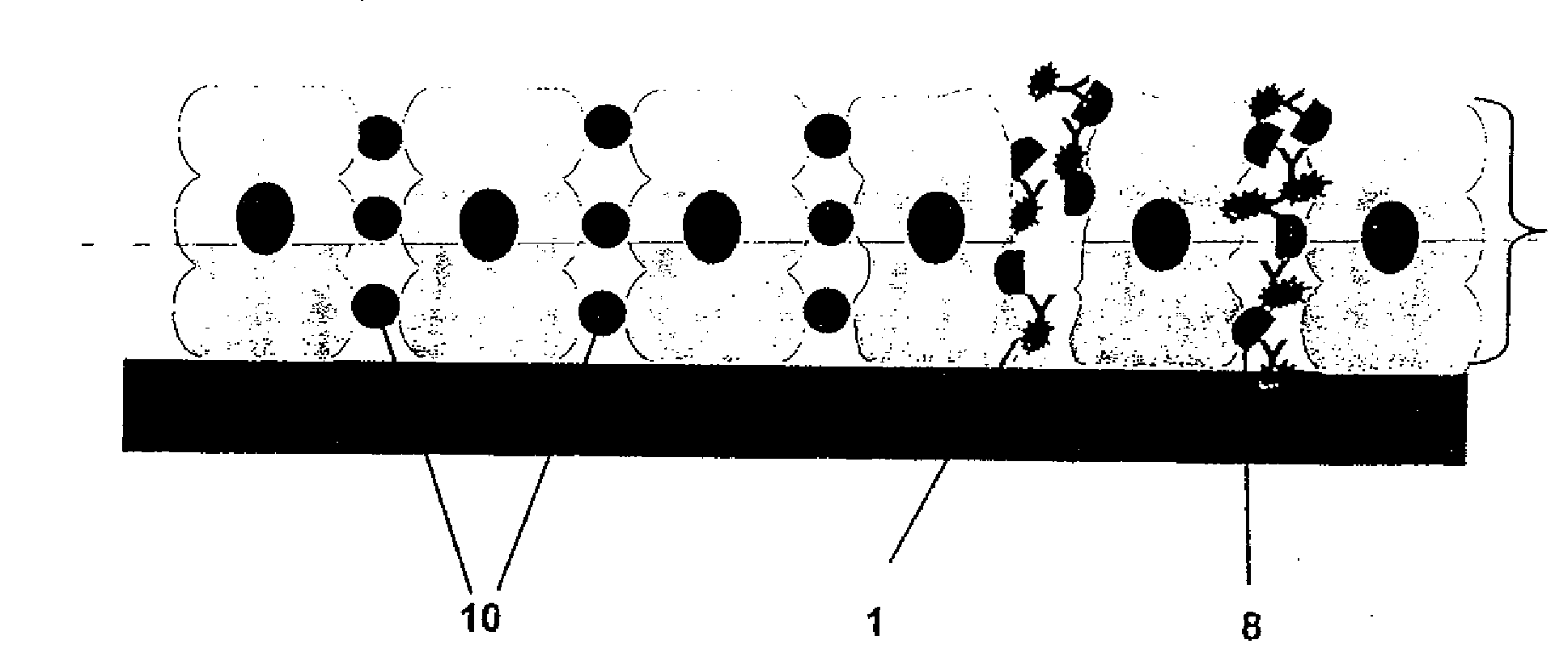
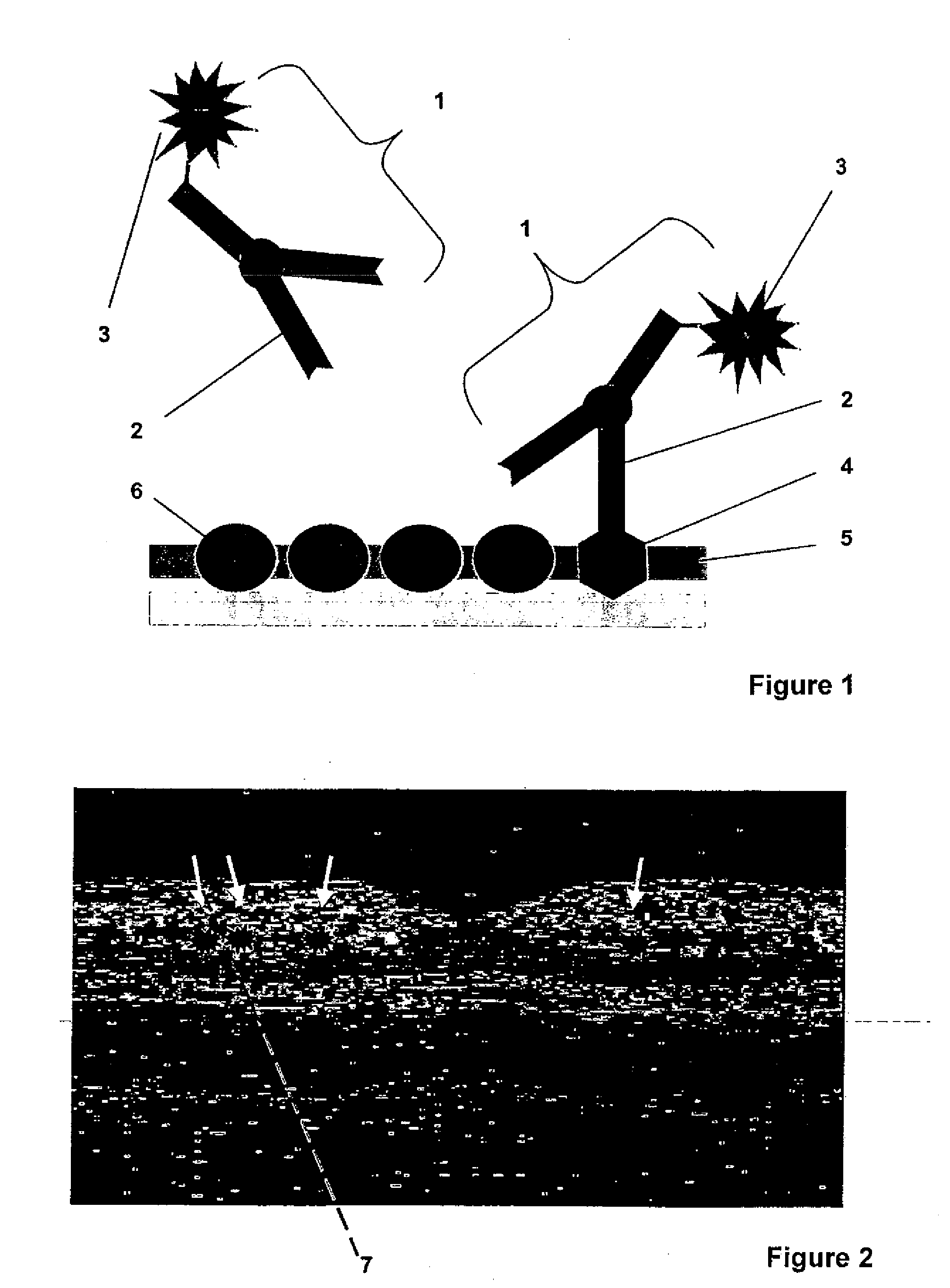
[0061]In the method for optical detection of changes of the eye, according to the invention, a molecular marker with spectral characteristics of absorption and / or dispersion in the visual and infrared spectral region or of fluorescence or luminescence is introduced into the eye and bound to a specific target. The interaction between the molecular marker and the target is detected by means of optical imaging methods. Since the molecular, physiologically compatible marker exhibits the characteristics of a temporally limited, selective binding to the targets in the eye with subsequent internal degradation in the body without noticeable impairment of the vision of the patient, only a slight strain to the patient and, particularly, the eye is achieved, which is adequate for diagnostic purposes.

[0062]The molecular marker, functioning as diagnostic reagent, can be injected in the patient, applied orally, or administered as eye drops. After the time period T0, when the molecular marker has ...


PUM

PropertyMeasurementUnit
wavelengthsaaaaaaaaaa
wavelengthaaaaaaaaaa

Abstract

A solution for optical detection of the eye. Molecular markers are used for high-contrast diagnosis of eye diseases, other diseases, and other vital parameters which can be diagnosed in the eye. For optical detection of the eye, a molecular marker with spectral characteristics of absorption and/or scattering in the visual and infrared spectral region is introduced into the eye and bound to a specific target. The interaction of the molecular marker with the target is detected by means of optical imaging methods, such as fundus photography, confocal laser microscopy, polarisation-optical imaging methods, holographic methods or especially OCT methods. The use of optical methods is strongly preferred for the diagnosis of the eye as a result of the high transparency of the optical system of the eye compared to other body parts.

Description

[0001]The present application is a National Phase entry of PCT Application No. PCT / EP2007 / 005555, filed Jun. 23, 2007, which claims priority from German Application Number 102006030382.2, filed Jun. 29, 2006, the disclosures of which are hereby incorporated by reference herein in their entirety.FIELD OF THE INVENTION[0002]The invention relates to a solution for optical detection of the eyes.[0003]Pathologically altered cells have altered metabolisms and gene activities, which, for example, manifest themselves in a change of the surface structure of the cells (so-called disease-correlated molecular markers). For quite some time, respective findings from molecular biological basic research have already been part of in-vitro diagnostics.[0004]However, for the integration of in-vitro techniques and methods in in-vivo environments, a number of problems must be overcome (e.g., toxicity, targeted transport to target cell, anatomical transport barriers).[0005]For the detection of cellular p...

Claims


Application Information

Patent Timeline
Patent Type & Authority Applications(United States)
IPC IPC(8): A61K49/00A61P27/02A61B6/00
CPCA61B5/0059A61K49/0058A61B5/0066A61B5/4088A61P27/02
Inventor RUSSMANN, CHRISTOPHDICK, MANFRED
Owner CARL ZEISS MEDITEC AG
Why Eureka
Try Eureka