In situ detection of early stages and late stages HPV einfection

a technology of hpv and in situ detection, which is applied in the field of in situ detection of early and late stages hpv einfection, can solve the problems of poor inter- and intra-observer agreement, high risk of progression toward invasive cervical cancer, and difficulty in obtaining samples

Active Publication Date: 2009-12-17
HEER MEDICAL TECH DEV CO LTD
View PDF88 Cites 26 Cited by
  • Summary
  • Abstract
  • Description
  • Claims
  • Application Information

AI Technical Summary

Benefits of technology

[0009]Embodiments of the invention provide various immunoassays for in situ detection of HPV proteins using various monoclonal antibodies against recombinant HPV proteins such that infection by high risk and / or low risk HPV types can be detected by a single specific monoclonal antibody and / or a general pan antibody. The invention also provides HPV immunocytochemistry (ICC) assay, HPV flow cytometry assay, HPV immunohistochmistry (IHC) assay to detect the presence of HPV proteins in cervical cells or cervical tissues. In addition, monoclonal antibodies highly specific for HPV viral proteins are also provided to be used in the HPV ICC or HPV flow cytometry assays.

Problems solved by technology

The presence of these lesions, preferentially observed in women aged 35-40 yrs, are at high risk of progression toward invasive cervical cancer.
However, due to subjective test criteria, there are various drawbacks for pap smear tests: difficulty in obtaining samples, poor inter- and intra-observer agreement, high rates of false negatives nd false positives, requiring specialized labs staffed with highly trained personnel, and inability to identify the majority of HPV-infected human subjects.
Detecting HPV infection by nucleic acid tests, such as “DNA Hybrid Capture”, has been developed with high assay sensitivity, but are still not ideal, due to not only its high cost, assay operation procedures, the requirements for facility, equipment, and highly trained personnel, but also its very low positive predictive value (PPV) in cervical intraepithelial neoplasia (CIN) testing samples.
In addition, DNA testing could not differentiate disease stages after HPV infection nor the diagnosis of different cell lesions (e.g., cannot diffrentiate LSIL from HSIL, nor CIN lesions from non-transforming latent or remissive viral infection).
Known protocols for producing monoclonal antibodies are generally unsuitable for the production of anti-HPV monoclonal antibodies and cannot be used in immunocytochemical diagnostic tests performed on human subjects of general population.
In addition, three problems exist in clinical HPV detection.
Secondly, there are too many HPV types and most HPV types present in clinical samples are not known or systemically identified due to the lack of available antibodies.
Third, HPV virus can not be cultured in labs by standard tissue culture techniques.
Thus, there is no available HPV proteins purified to large quantities as immunogens for generating anti-HPV antibodies, and there is no available HPV proteins or purified anti-HPV antibodies to recognize anti-viral antibodies or viral proteins present in clinical samples for clinical HPV detection.
Infections by only about 15 HPV types (out of more than 100 available HPV types) are at high risk of developing into cervical intraepithelial neoplasia (CIN) or cervical cancer.
However, some progressive cervical cancer cases are reported to be infected by low risk HPV types, while infection of some high risk HPV types will never progress into cervical cancer.

Method used


Image

  • In situ detection of early stages and late stages HPV einfection
  • In situ detection of early stages and late stages HPV einfection
  • In situ detection of early stages and late stages HPV einfection

Examples


examples

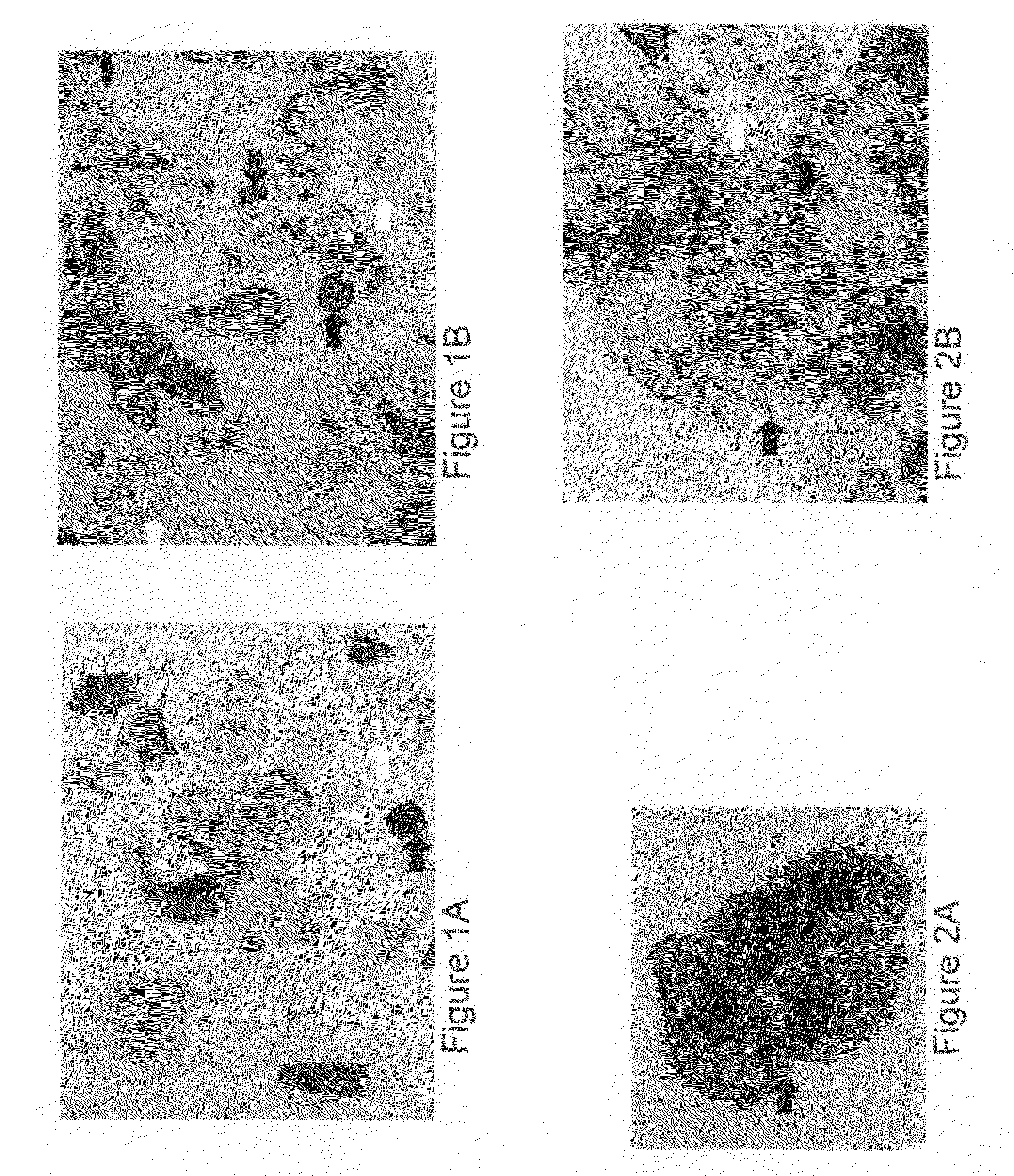
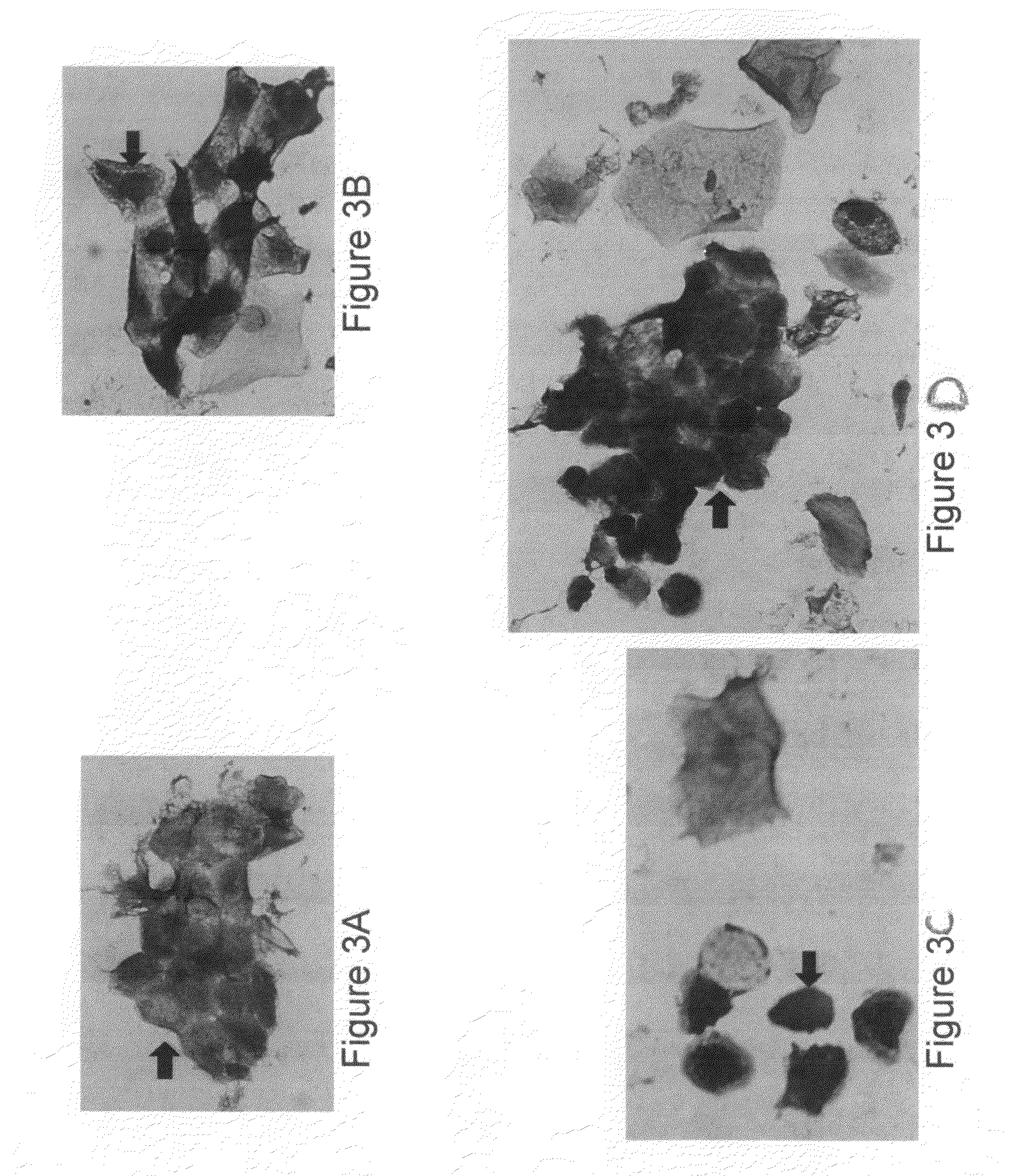
[0068]1. Expression, Purification, and Preparation of HPV Recombinant Proteins to be Used as Immunogens for Generating Antiserum and Anti-HPV Antibodies, and Screening Hybridoma Cell Lines for Monoclonal Antibodies

[0069]HPV recombinant proteins can be any kinds of HPV proteins, HPV proteins of early genes and / or late genes, including, but not limited to, E2, E6, E7, L1, L2 and can be from various HPV types. Full-length E6, E7, and / or L1 polypeptide sequence, which have been found very difficult to obtain and purify due to undesirable aggregation during protein purification, protein instability, low levels of expression, low immunogenic responses of purified proteins. For example, many early E6 oncoproteins contain many cysteine amino acids and thus the correct topography of the E6 oncoproteins requires formation of many disulfide bonds properly. In addition, it was known that certain immunological assays using small peptides of early E6 and E7 proteins results in extremely low assay...


PUM

PropertyMeasurementUnit
concentrationaaaaaaaaaa
morphologyaaaaaaaaaa
nucleic acid testsaaaaaaaaaa

Abstract

Embodiments of the invention provide methods, monoclonal antibodies, polyclonal antibodies, assays, and kits for detecting HPV infection and HPV related cancer diagnosis, including infection by various HPV genotypes, early and/or late stage HPV-associated or HPV-specific cancers. The anti-HPV antibodies are used in performing immunological assays on clinical samples. Various immunological assays and kits for detecting HPV infection, cervical cancer, other HPV related cancers, early stage precancerous lesions as well as late stage cancer progression are also provided.

Description

CROSS-REFERENCE TO RELATED APPLICATIONS[0001]This application claims benefit of U.S. provisional patent application Ser. No. 61 / 131,991, filed Jun. 13, 2008, and U.S. provisional patent application Ser. No. 61 / 192,912, filed Sep. 22, 2008. Each of the aforementioned related patent applications is herein incorporated by reference.BACKGROUND OF THE INVENTION[0002]Infection by human papillomaviruses (HPV) at specific epithelium cells to induce epithelial proliferations plays an important role for cervical carcinogenesis. About 99 percent of confirmed cervical cancer cases are found to be associated with HPV infection with biopsy-confirmed squamous intraepithelial lesions (SIL) or cervical intraepithelial neoplasia (CIN). The incidence of HPV infection, primarily transmitted through sexual contact, is highest among young women and about 20 millions of sexually active men and women worldwide are currently infected. Approximately 1% of the population has genital warts and 4% of women have...

Claims


Application Information

Patent Timeline
Patent Type & Authority Applications(United States)
IPC IPC(8): C12Q1/70
CPCC07K16/084C12Q1/708G01N2469/10G01N33/571G01N2333/025G01N33/56983
Inventor CHENG, SHULING
Owner HEER MEDICAL TECH DEV CO LTD
Why Eureka
Try Eureka