

Recombinant deamidated gliadin antigen
a technology of gliadin and antigen, which is applied in the field of severe gastrointestinal disease, can solve the problems of false positives or incomplete epitope repertoire of current assays
- Summary
- Abstract
- Description
- Claims
- Application Information
AI Technical Summary
Benefits of technology
Problems solved by technology
Method used
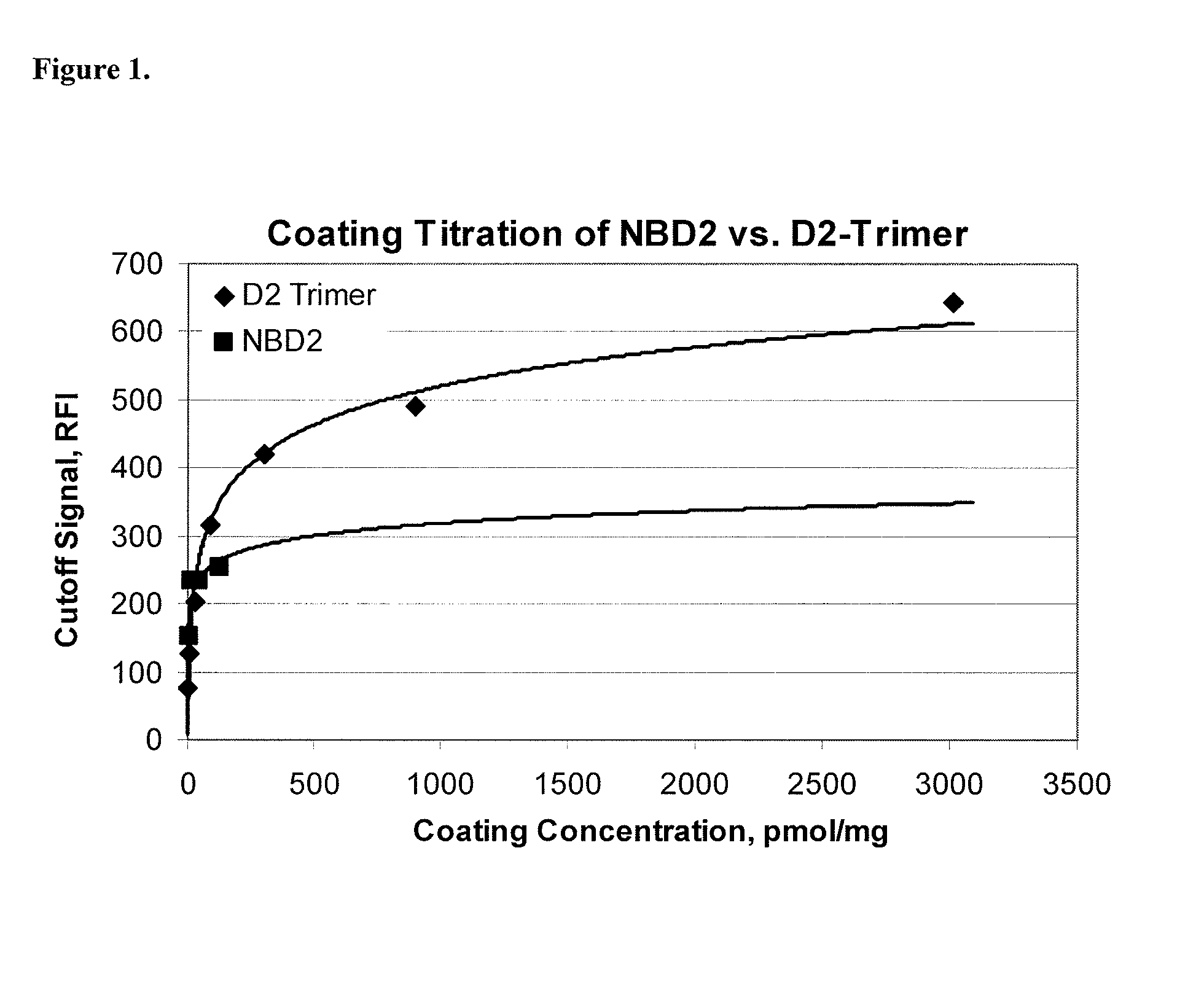
Image

Examples
example 1
Preparation of the Gliadin Fusion Protein Using the D2 Trimer
[0102]This example provides a method for preparing the gliadin fusion protein of the present invention using the D2 Trimer.
[0103]A DNA sequence encoding the D2 trimer, SEQ ID NO:2, was prepared, digested with a restriction enzyme and inserted into an expression vector containing a DNA fragment encoding GST, at the C-terminal position of GST, for expression of the gliadin fusion protein, SEQ ID NO:4.
example 2
Preparation of the Immobilized-Antigen without tTG
[0104]This example provides a method for preparing the antigen of the present invention in the absence of tTG that generally involves immobilization of a gliadin fusion protein (GST-D2 trimer) on a solid support.
Immobilization of Gliadin Fusion Protein
[0105]Into a microfuge tube is placed 8 mg of carboxyl modified magnetic beads. To the tube is added 800 μL of 50 mM 2-(N-morpholino)ethanesulfonic acid (MES) pH 6.1 in 70% EtOH (ethanol). Mix and magnetically separate. Pipet off and discard the supernatant. Repeat one more time.
[0106]Add 400 μL of 120 mM N-hydroxysuccinimide (NHS) in 50 mM MES pH 6.1 in 70% EtOH into the tube and mix. Add 400 μL of 100 mM N-Cyclohexyl-N′-(2-morpholinoethyl)carbodiimide metho-p-toluenesulfonate (CMC) in 50 mM MES pH 6.1 in 70% EtOH into the tube and mix. Mix for 30 minutes at room temperature.
[0107]Separate the beads from the supernatant and add 800 μL of 5 mM MES pH 6.1. Mix, magnetically separate, pip...
example 3
Preparation of the Immobilized-Antigen with tTG
[0113]This example provides a method for preparing the antigen of the present invention using tTG that involves immobilization of the gliadin fusion protein (GST-D2 trimer) and tTG onto the solid support such that the tTG and gliadin fusion protein become complexed together through transamidation reactions. The tTG and gliadin fusion protein are then cross-linked.
Immobilization of Gliadin Fusion Protein-tTG Complex
[0114]Into a microfuge tube is placed 8 mg of carboxyl modified magnetic beads. To the tube is added 800 μL of 50 mM 2-(N-morpholino)ethanesulfonic acid (MES) pH 6.1 in 70% EtOH (ethanol). Mix and magnetically separate. Pipet off and discard the supernatant. Repeat one more time.
[0115]Add 400 μL of 120 mM N-hydroxysuccinimide (NHS) in 50 mM MES pH 6.1 in 70% EtOH into the tube and mix. Add 400 μL of 100 mM N-Cyclohexyl-N′-(2-morpholinoethyl)carbodiimide metho-p-toluenesulfonate (CMC) in 50 mM MES pH 6.1 in 70% EtOH into the tu...
PUM
| Property | Measurement | Unit |
|---|---|---|
| pH | aaaaa | aaaaa |
| nucleic acid | aaaaa | aaaaa |
| signal-to-noise ratio | aaaaa | aaaaa |
Abstract
Description
Claims
Application Information