
Cryotherapy Planning and Control System

a control system and cryotherapy technology, applied in the field of systems and methods for planning and supervising ablative cryosurgery, can solve the problems of prior art methods that fail to provide adequate means for visualizing therapeutic probes in operative situations, fail to provide precise positions of cryoprobes, and prior art methods that fail to provide adequate means for visualizing surgical target environments

Inactive Publication Date: 2009-12-24
GALIL MEDICAL
View PDF38 Cites 48 Cited by
  • Summary
  • Abstract
  • Description
  • Claims
  • Application Information

AI Technical Summary

Benefits of technology

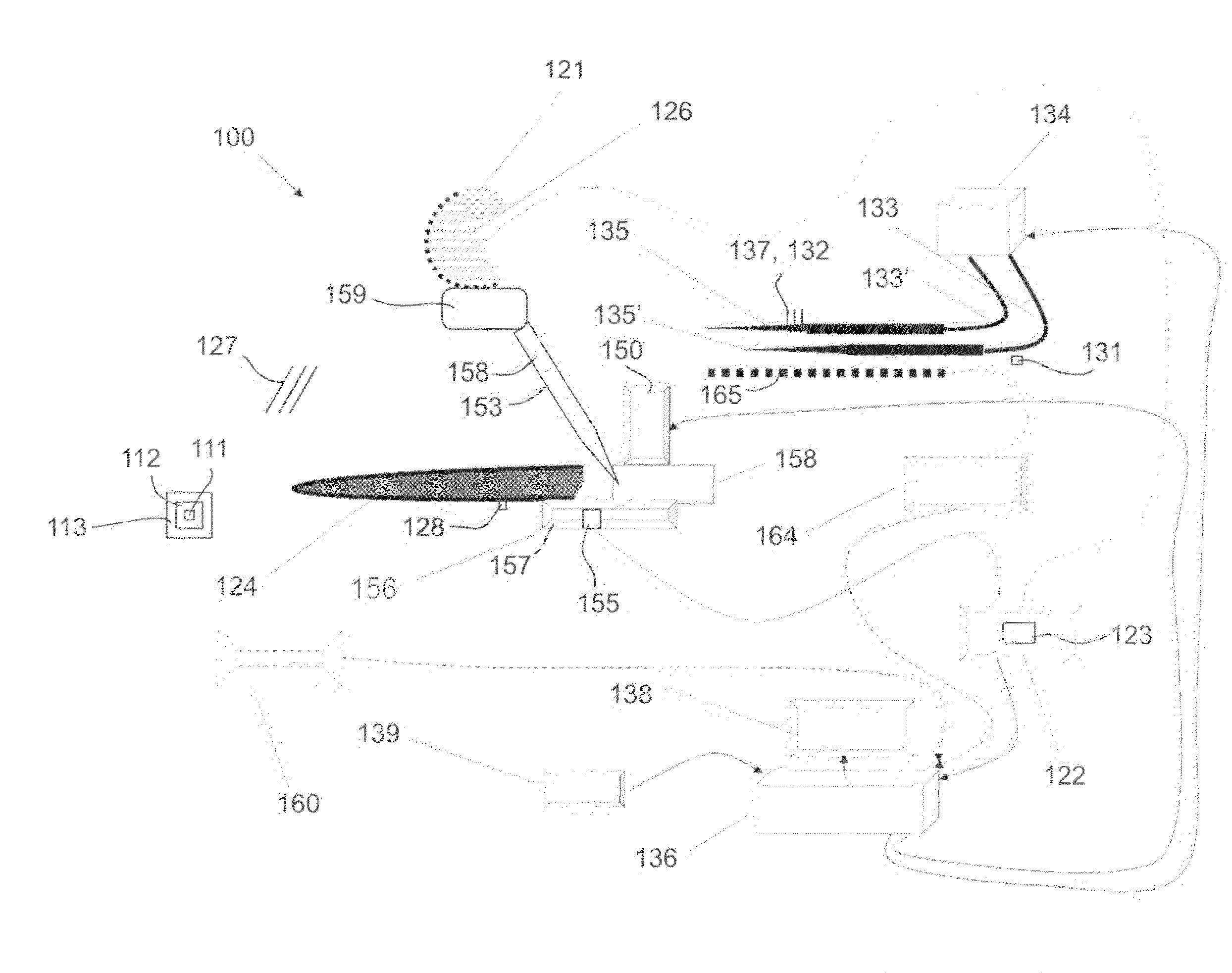
[0009]The present invention relates in particular to improved technologies for pre-operative user-input characterization of surgical target sites, for pre-operative and operative use of ultrasound and other imaging modalities to characterize surgical sites before and during surgery and more particularly during cryosurgery, to production and display of predictive evaluations of planned and real-time situations in terms of probabilities of tissue survival, and to improved methods for relating planned surgical procedures to actual surgical contexts.
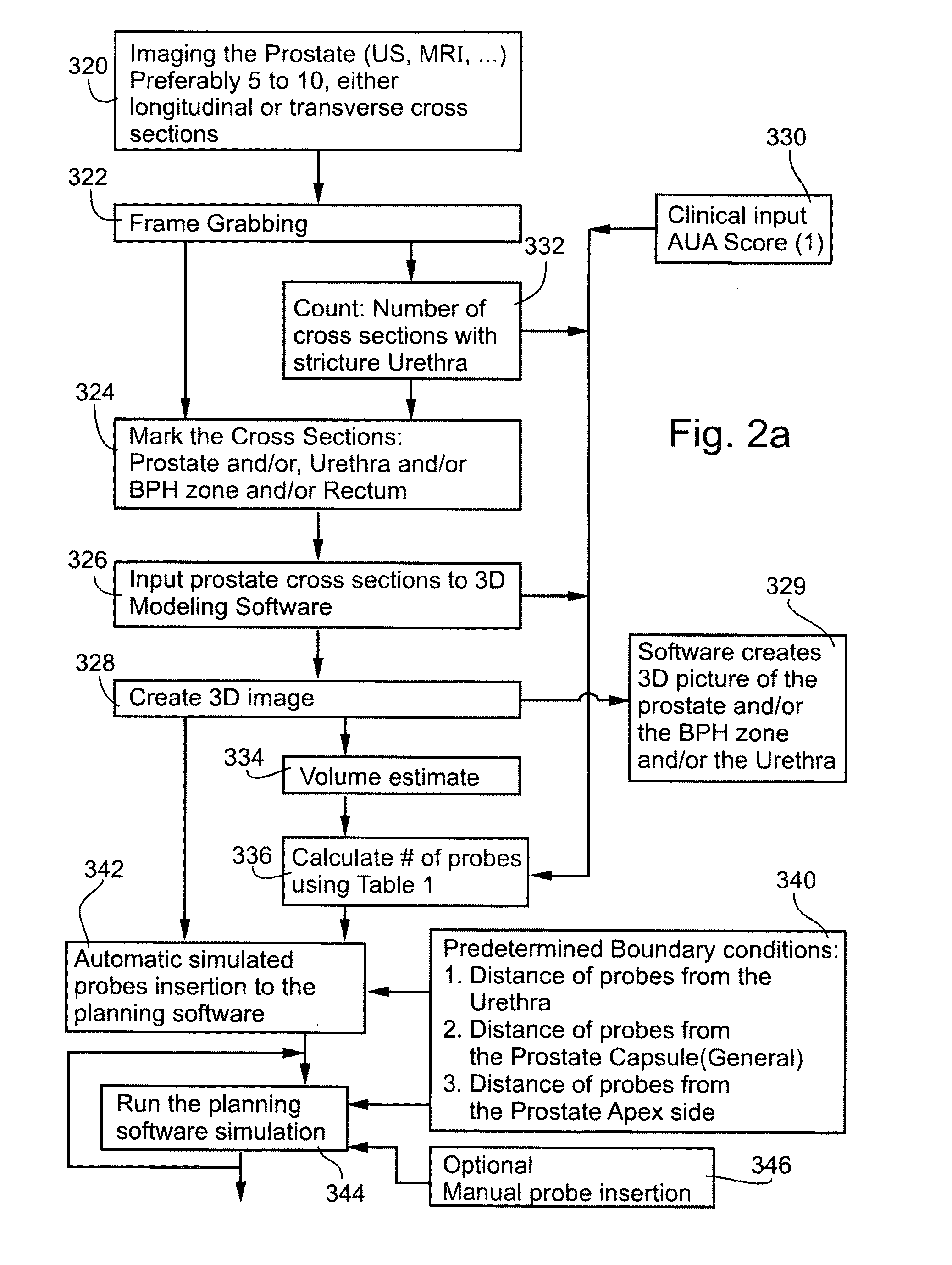
[0014]Methods of prior art provide for calculation and display of estimated surgical outcomes in terms of temperature gradients in tissues. Yet, there is a widely recognized need for, and it would be highly desirable to have, devices and methods for calculating and displaying therapy predictions in terms of assessed probabilities of tissue survival, as compared to user evaluations or automated evaluations of desirability of tissue survival. The present invention successfully addresses the shortcomings of the presently known configurations by providing means for inputting graduated tissue survival desirability scores for various tissues in a surgical context, and by providing means for calculating and displaying probabilities of tissues survival according in simulated or actual clinical contexts in a manner which facilitates comparing tissue survival desirability with tissue survival probability.
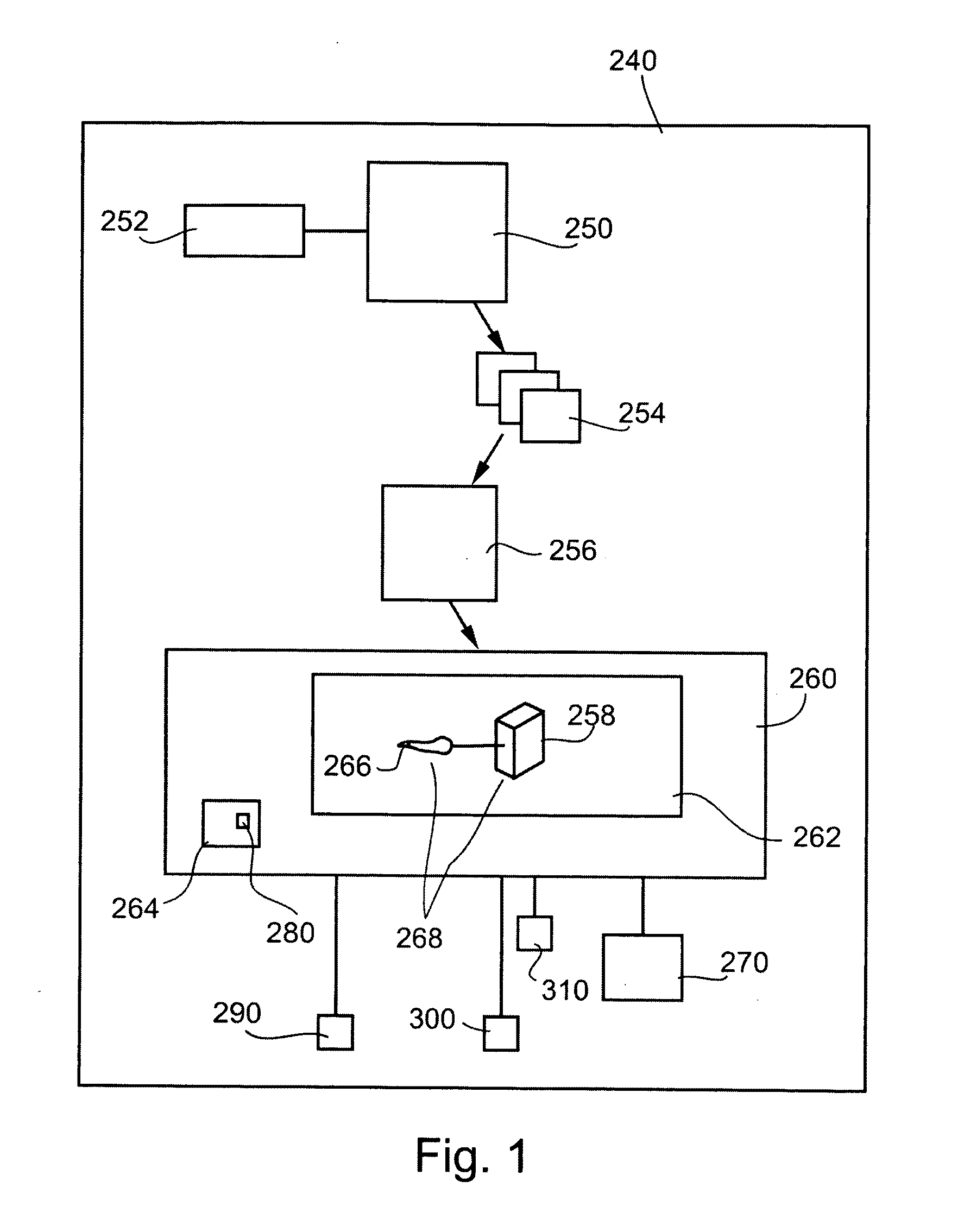
[0015]Surgical planning systems generally request user input in response to patient images created by imaging modalities such as CT scans, MRI and ultrasound images, in order to identify, differentiate and characterize tissues in the vicinity of a lesion. Thus, there is a widely recognized need for, and it would be highly desirable to have, devices and methods for facilitating the process of user evaluation of such images. The present invention successfully addresses the shortcomings of the presently known configurations by providing methods and devices for facilitating user input serving to characterize tissues presented in pre-operative patient imaging.
[0031]In an exemplary embodiment of the invention, the method comprises alternating operation of said first and second ultrasound probes, thereby avoiding signal interference between said first and second probes.
[0037](b) creating an image which comprises information received from said first ultrasound probe and information received from said second ultrasound probe. Optionally, the method comprises alternating operation of said first and second ultrasound probes, thereby avoiding acoustical interference between said first and second probes. Alternatively or additionally, the method comprises displaying said created image.
[0083]Optionally, the method comprises accepting said input from a user with respect to a first image, reproducing said user input from said first image on a second image, and enabling said user to identifying an anatomical feature present in said second image by modifying said reproduced input with respect to said second image.

Problems solved by technology

Methods of prior art fail to provide adequate means for visualizing therapeutic probes in operative situations under certain popular imaging modalities, and in particular fail to provide means for visualizing exact positions of cryoprobes before and most particularly during cryosurgery.
Methods of prior art fail to provide adequate means for visualizing surgical target environments under various standard clinical situations, such as for example during cryotherapy of a prostate under guidance of rectal ultrasound probe imaging.
Methods of prior art fail to provide means for detailed control of contours of an ablation volume created by cooling of a given set of inserted cryoprobes.

Method used


Image

  • Cryotherapy Planning and Control System
  • Cryotherapy Planning and Control System
  • Cryotherapy Planning and Control System

Examples


Embodiment Construction

[0127]The present invention relates to devices and methods for planning and supervising minimally invasive surgery. Specifically, the present invention can be used to enhance various imaging modalities used before and during cryosurgery, to enhance and facilitate user-input characterization of body tissues based on images provided by imaging modalities, to output predictions based on simulated and actual surgical situations in a form well suited to guiding a surgeon in decision-making processes, and to enhance controlled contouring of a cryoablation volume produced by a plurality of cryoprobes.

[0128]Before explaining at least one embodiment of the invention in detail, it is to be understood that the invention is not limited in its application to the details of construction and the arrangement of the components set forth in the following description or illustrated in the drawings. The invention is capable of other embodiments or of being practiced or carried out in various ways. Also...


PUM


Abstract

The present invention relates to devices and methods for planning and supervising minimally invasive surgery. Included are enhancements to systems for planning, monitoring, and controlling cryosurgery.

Description

RELATED APPLICATIONS[0001]The present application is related to U.S. patent application Ser. No. 11 / 219,648, filed on Sep. 7, 2005, which is a Continuation of U.S. patent application Ser. No. 11 / 066,294, filed on Feb. 28, 2005, which is a Divisional of U.S. patent application Ser. No. 09 / 917,811, filed on Jul. 31, 2001, now U.S. Pat. No. 6,905,492, issued on Jun. 14, 2005, which claims priority from U.S. Provisional Patent Application No. 60 / 221,891, filed on Jul. 31, 2000.[0002]The present application further claims priority U.S. Provisional Patent Application 60 / 796,519, filed May 2, 2006. The contents of all of the above-mentioned applications are incorporated herein by reference.[0003]This application is related to two other PCT applications being filed on even date with this application in the Israel Receiving Office having the titles and PROBE INSERTION GUIDE WITH USER-DIRECTING FEATURES and CRYOTHERAPY INSERTION SYSTEM AND METHOD, and Attorney docket Nos. 39261 and 39262, and...

Claims


Application Information

Patent Timeline
Patent Type & Authority Applications(United States)
IPC IPC(8): A61B8/14A61B18/02
CPCA61B18/02A61B19/201A61B19/2203A61B19/50A61B19/56A61B2019/504A61B2018/0262A61B2018/1861A61B2019/501A61B2019/5276A61B2017/00482A61B2034/101A61B2034/104A61B34/25A61B34/30A61B90/11A61B34/10A61B2090/378
Inventor AVITAL, OFERKOCHIVI, EYALPANSKY, AMIRHEFETZ, YARONPIANKA, PAZIT
Owner GALIL MEDICAL
Learn More