Ph sensitive liposome compositions for controlling surface topography and binding reactivity in functionalized liposomes
a liposome and functionalized technology, applied in the field of ph sensitive liposome compositions for controlling surface topography and binding reactivity in functionalized liposomes, can solve the problems of changing the effective reactivity and remodeling of the cell surface topography, and achieve the effects of reducing toxicities, minimizing toxicities, and maximizing tumor penetration
- Summary
- Abstract
- Description
- Claims
- Application Information
AI Technical Summary
Benefits of technology
Problems solved by technology
Method used
Image
Examples
example 1
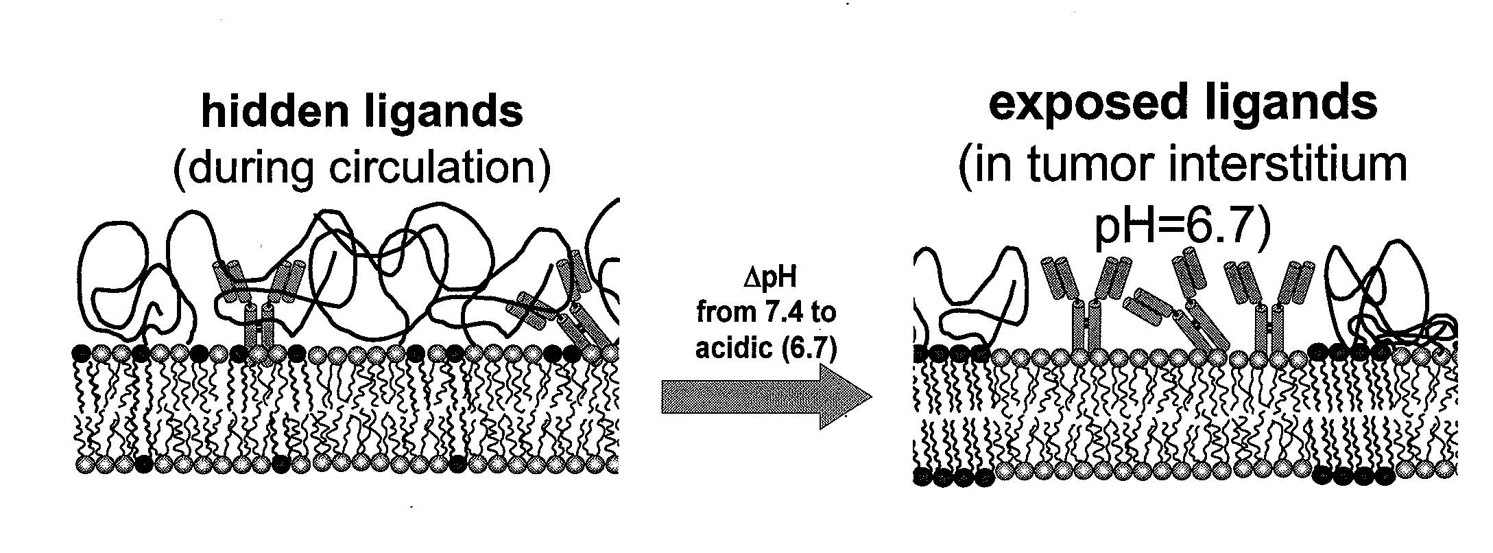
[0150]Biotinylated liposomes (1% mole DPPE-biotin) were developed containing PEGylated lipids that contain domain-forming lipids, which were tuned to become activated at conditions similar to those of tumor interstitial pH. Rigid liposomes consisting of DPPC (16:0), DSPS (18:0) (at 1:1 mole ratios), and 5% cholesterol and 0.1% to 1.5% mole DSPE-PEG (2000 MW) were incubated in PSB at 37° C. at various pH values.
example 2
[0151]Binding of rigid biotinylated liposomes (FIGS. 16, 17, 18, 19, 20, 21, filled symbols) to streptavidin-covered-magnetic microbeads was evaluated at various pH values ranging from pH 7.4, approximating the pH of the blood during circulation of liposomes to pH 6.5 that corresponds to the pH of the tumor interstitium after extravasation of liposomes into the tumor. The extent of liposomes bound was evaluated for different amounts of PEG-linked lipids in the liposome composition ranging from 0.1% to 1.5% mole (of total lipid) and was also compared to identical liposomes without biotin (plain liposomes) indicated by the open symbols in FIGS. 16, 17, 18, 19, 20, and 21. In biotinylated liposomes the amount of biotin-linked lipids was retained constant at 1% mole of total lipid (FIG. 16 shows liposomes containing 0.1% mole PEG-linked lipid, FIG. 17 0.25% mole, FIG. 18 0.5% mole, FIG. 19 0.75% mole, FIG. 20 1.0% mole, and FIG. 21 1.5% mole). The liposomal membrane was labeled with rho...
example 3
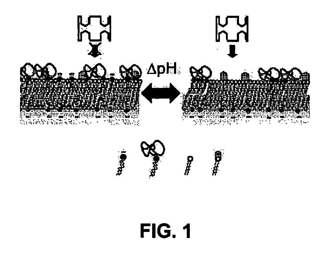
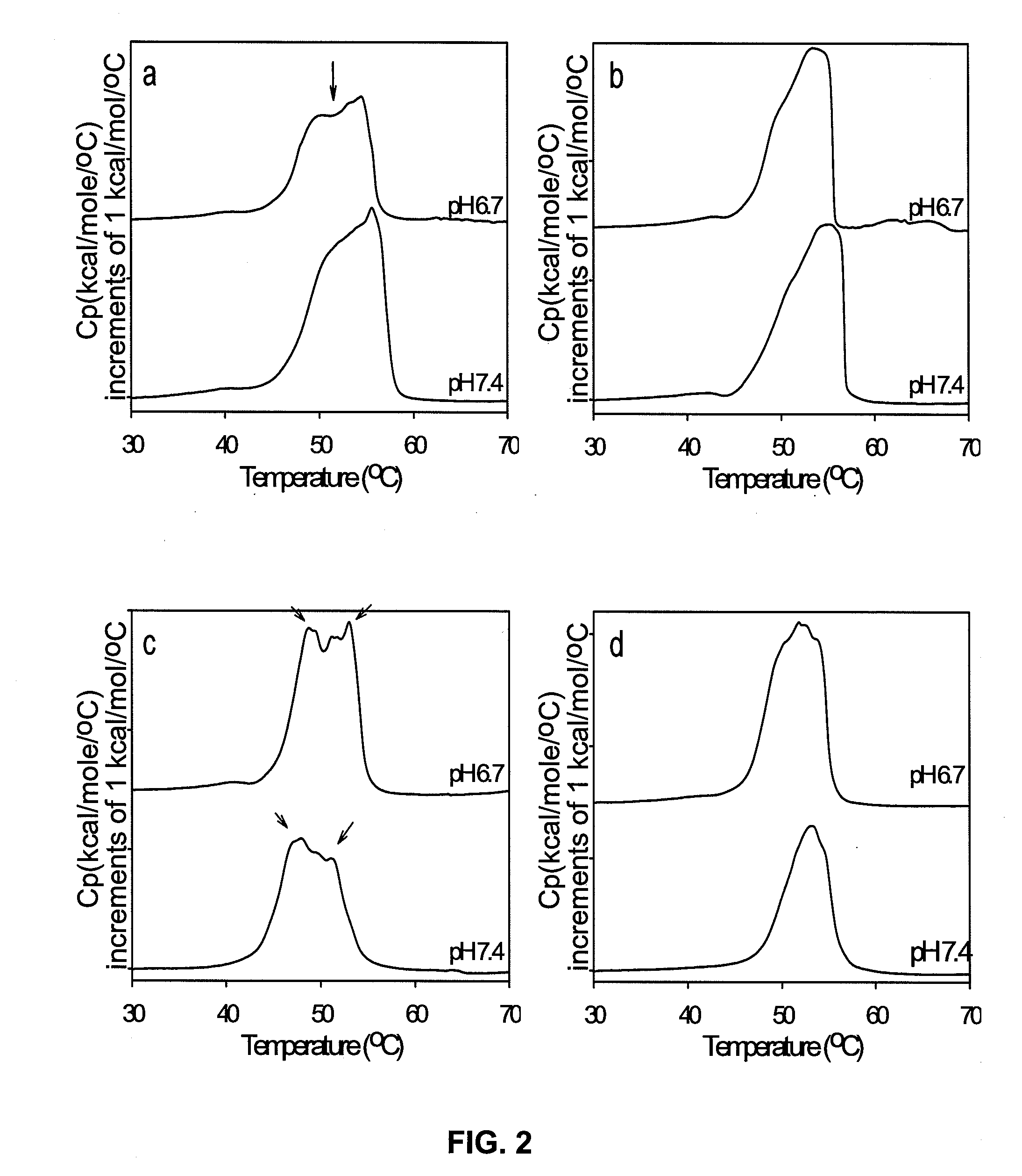
[0153]Differential Scanning Calorimetry (DSC) was used because it can provide direct evidence of phase separation of lipid membranes. FIG. 22 shows the thermal scans of the same liposome composition (equimolar DPPC and DSPS with 5% mole cholesterol and 2% mole DSPE-PEG), performed at a rate of 60° C. / h. As the pH was decreased from 7.4 to 4.0, an enhancement was observed on the contributions from thermal transitions at higher temperatures. Higher thermal transitions at lower pH values suggest increasing formation of lipid phases that are rich in clustered (protonated) DSPS lipids (that has higher Tg) and phases poor in DSPS lipids (or richer in DPPC lipids, FIG. 22). These results demonstrate that in membranes containing lipids with different hydrocarbon chain lengths (with one lipid type bearing charged headgroups), lipid mixing or domain formation is controlled by the pH that affects the extent of electrostatic repulsion among the titratable lipids.
PUM
 Abstract
Description
Claims
Application Information
 - R&D
- Intellectual Property
- Life Sciences
- Materials
- Tech Scout
- Unparalleled Data Quality
- Higher Quality Content
- 60% Fewer Hallucinations
Browse by: Latest US Patents, China's latest patents, Technical Efficacy Thesaurus, Application Domain, Technology Topic, Popular Technical Reports.
© 2025 PatSnap. All rights reserved.Legal|Privacy policy|Modern Slavery Act Transparency Statement|Sitemap|About US| Contact US: help@patsnap.com


