Process for preserving three dimensional orientation to allow registering histopathological diagnoses of tissue to images of that tissue

a three-dimensional orientation and image processing technology, applied in the field of histopathology analysis and reconstruction, can solve the problems of increased infection risk, bleeding, physical and emotional discomfort, cost, etc., and achieve the effect of reducing the risk of infection, reducing the risk of bleeding, and reducing the accuracy of the diagnosis

a three-dimensional orientation and image processing technology, applied in the field of histopathology analysis and reconstruction, can solve the problems of increased infection risk, bleeding, physical and emotional discomfort, cost, etc., and achieve the effect of reducing the risk of infection, reducing the risk of bleeding, and reducing the accuracy of the diagnosis

US20100093023A1Inactive Publication Date: 2010-04-15CADES SCHUTTE A LIMITED LIABILITY LAW PARTNERSHIP

Image

  • Process for preserving three dimensional orientation to allow registering histopathological diagnoses of tissue to images of that tissue
  • Process for preserving three dimensional orientation to allow registering histopathological diagnoses of tissue to images of that tissue
  • Process for preserving three dimensional orientation to allow registering histopathological diagnoses of tissue to images of that tissue

Examples


Embodiment Construction

Overview

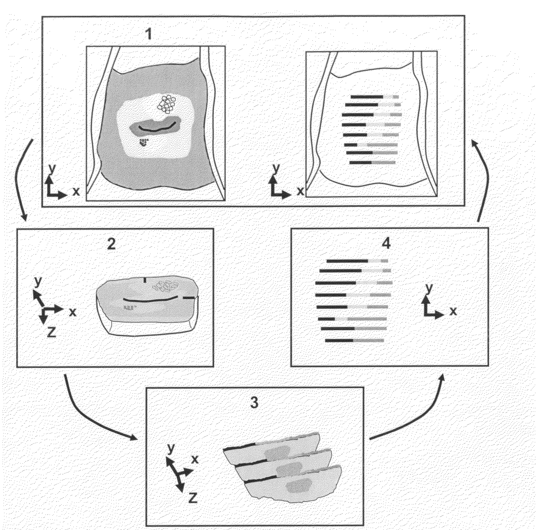
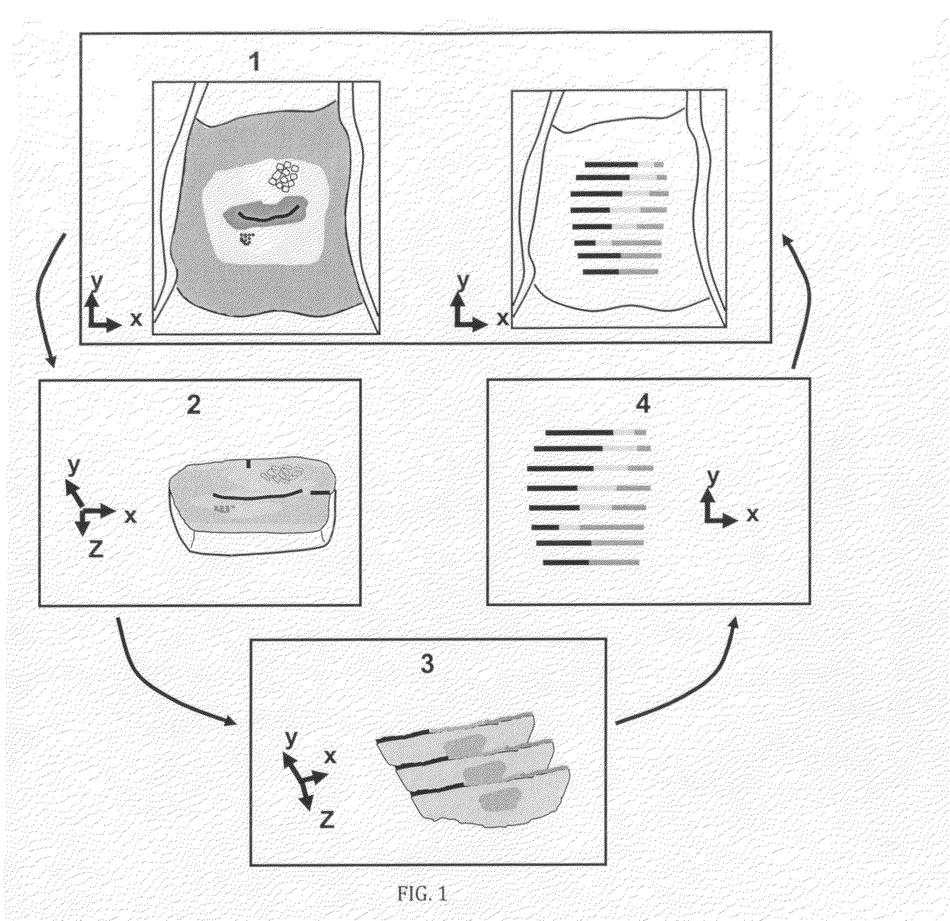
[0111]The presently preferred embodiments of the invention described herein disclose a systematic framework to maintain the spatial orientation at each level of tissue processing of a three-dimensional tissue specimen removed from an area of investigation, perform detailed histopathology analysis, and precisely map the location of the histopathology back to a digital image of the area of investigation acquired prior to removing the tissue specimen.

[0112]By utilizing tissue image acquisition, processing, and registration techniques in combination with mechanical and image processing tools and novel tissue processing procedures, the present invention accurately determines the location of the histopathology analysis (the ‘z-axis’) of the tissue specimen as it relates to the overlying tissue structure (the ‘x-y-axes’). The procedural steps of the presently preferred embodiments, as schematically illustrated in FIG. 1, and which will be described in detail in the following sectio...


PUM


Abstract

A process for maintaining 3 dimensional orientation between a tissue specimen and images of the area of investigation, to register histopathologic diagnoses of multiple locations within the specimen with corresponding locations on the surface of said area of investigation, by marking at least two fiduciary lines on the area of investigation; acquiring a fiduciary image of the tissue with the fiduciary lines; excising the tissue to form a tissue specimen; inserting at least two parallel needles through said specimen; acquiring a specimen image of the specimen with inserted needles over an alignment grid; fixing the specimen by immersing the specimen; acquiring a fixed image of the fixed specimen with the inserted needles over the alignment grid; forming a paraffin mold containing the fixed specimen and inserted needles; injecting different colored inks through the needles while withdrawing them from the fixed specimen, so that different colored needle tracks are formed in the specimen; sectioning the specimen to create specimen blocks having different colored needle tracks; further sectioning the specimen to cut the specimen blocks into specimen slices having different colored ink dots corresponding to the different colored needle tracks; forming pathology images from the specimen slices; performing histopathology analyses on the pathology images; annotating the pathology images with histopathology annotations; aligning the annotations with the fixed image using the colored ink dots; determining shrinkage between the fixed image and the annotations by using the grid to compare the distance between the needles in the fixed image with the distance between the ink dots on the specimen slices; registering the fixed image to the specimen image to account for shrinkage caused by fixation, using locations of the needles in both of the images as landmarks; registering the specimen image to the fiduciary image to account for tissue translation and soft tissue movement using the fiduciary lines and geographical features of said area of investigation as landmarks; registering the fiduciary image to the reference image to account for tissue translation and soft tissue movement using said geographical features; whereby annotations of histopathologic diagnoses are provided for multiple locations on or under the surface of the specimen that are registered to images of the specimen.

Description

[0001]This application claims the benefit of U.S. Provisional Patent Application No. 61 / 195,771 filed Oct. 9, 2008.TECHNICAL FIELD[0002]This invention relates to histopathology analysis and reconstruction and, more specifically, to maintaining the spatial orientation of three-dimensional tissue specimens removed from an area of investigation as an aid in the diagnosis of cancer and pre-cancerous lesions.BACKGROUND ART[0003]Although this invention is being disclosed in connection with cervical cancer, it is applicable to many other areas of medicine and other fields. Diagnosing cervical cancer in women is a multi-step procedure involving examination of the cervix, possible biopsy and follow-up (D. G. Ferris, J. T. Cox, D. M. O'Connor, V. C. Wright, and J. Foerster, Modern Colposcopy. Textbook and Atlas, pp. 1-699, American Society for Colposcopy and Cervical Pathology, 2004; 2. E. Burghardt, H. Pickel, and F. Girardi, Colposcopy—Cervical Pathology, Textbook and Atlas, Thieme, New Yor...

Claims


Application Information

Patent Timeline
15 Apr 2010
Publication
US20100093023A1
IPC
G01N1/28
CPC
G01N1/286
Inventors
GUSTAFSSON, ULF PETER; OYAMA, JODY C.