System and Method for Quantitative Molecular Breast Imaging

Inactive Publication Date: 2010-04-29
MAYO FOUND FOR MEDICAL EDUCATION & RES
View PDF13 Cites 31 Cited by
  • Summary
  • Abstract
  • Description
  • Claims
  • Application Information

AI Technical Summary

Benefits of technology

[0011]The present invention overcomes the aforementioned drawbacks by providing systems and methods for performing quantitative tumor analysis using information acquired with a dual-headed molecular breast imaging system. Specifically, the present invention provides systems and methods to utilize the info

Problems solved by technology

However, since bilateral breast MRI is currently approximately 20 times more expensive than mammography, it is not in widespread use as a screening technique.
The PEM technology offers excellent resolution; however, the currently available radiotracer (F-18 Fluoro deoxyglucose) requires that a patient fast overnight, the patient must have low blood levels (this is often a problem for diabetics), and after injection, the patient must wait 1-2 hours for optimum uptake of F-18FDG in the tumor.
The high cost of these PET procedures coupled with the long patient preparation time reduces the usefulness of this procedu

Method used


Image

  • System and Method for Quantitative Molecular Breast Imaging
  • System and Method for Quantitative Molecular Breast Imaging
  • System and Method for Quantitative Molecular Breast Imaging

Examples


Embodiment Construction

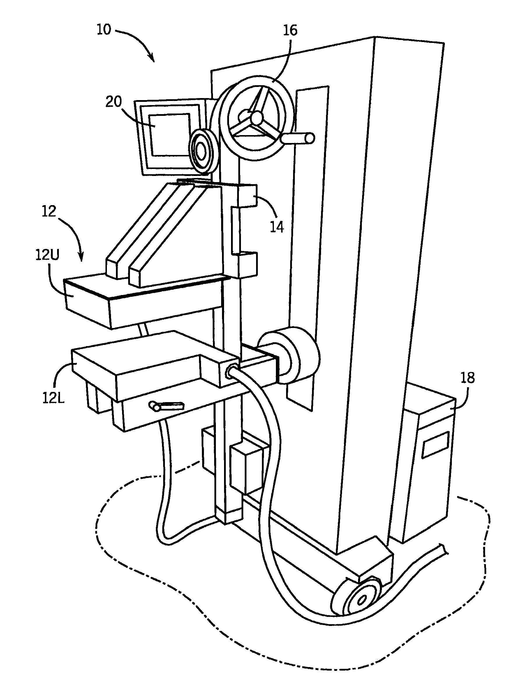
[0029]Referring to the Figs., and in particular FIG. 1, a molecular breast imaging (“MBI”) system 10 includes two opposing cadmium zinc telluride (“CZT”) detectors (detector heads) 12. In particular, the detector heads 12 include an upper detector head 12U and a lower detector head 12L. Each detector head 12U, 12L is, for example, 20 cm by 16 cm in size and mounted on a modified upright type mammographic gantry 14. In accordance with one embodiment, the detector heads 12 are LumaGEM 3200S high-performance, solid-state cameras from Gamma Medica having a pixel size of 1.6 mm. LumaGEM is a trademark of Gamma Medica, Inc. Corporation of California.

[0030]The relative position of the detector heads 12 can be adjusted using a user control 16. Specifically, the detector head assemblies 12 are, preferably, designed to serve as a compression mechanism. Accordingly, this system configuration reduces the maximum distance between any lesion in the breast and either detector head 12 to one-half o...


PUM


Abstract

A system and method for performing quantitative lesion analysis in molecular breast imaging (MBI) using the opposing images of a slightly compressed breast that are obtained from the dual-head gamma camera. The method uses the shape of the pixel intensity profiles through each tumor to determine tumor diameter. Also, the method uses a thickness of the compressed breast and the attenuation of gamma rays in soft tissue to determine the depth of the tumor from the collimator face of the detector head. Further still, the method uses the measured tumor diameter and measurements of counts in the tumor and background breast region to determine relative radiotracer uptake or tumor-to-background ratio (T/B ratio).

Description

CROSS-REFERENCE TO RELATED APPLICATIONS[0001]This application is a continuation-in-part of, claims the benefit of, and hereby incorporates by reference in its entirety U.S. patent application Ser. No. 12 / 515,369 filed on May 18, 2009, and entitled “System and Method for Quantitative Molecular Breast Imaging,” which claims the benefit of PCT Application No. US2007 / 086991 filed on Dec. 10, 2007, which claims the benefit of U.S. Provisional Patent Application Ser. No. 60 / 869,419 filed on Dec. 11, 2006. This application also claims the benefit of and hereby incorporates by reference in its entirety U.S. Provisional Patent Application Ser. No. 61 / 121,217 filed on Dc. 10, 2008, and entitled “Method and Apparatus for X-Ray Mammography / Tomosynthesis and Emission Mammography of the Breast.”STATEMENT REGARDING FEDERALLY SPONSORED RESEARCH[0002]Not Applicable.BACKGROUND OF THE INVENTION[0003]The invention relates to a system and method for obtaining quantitative information regarding breast im...

Claims


Application Information

Patent Timeline
IPC IPC(8): A61K51/00A61B6/00
CPCA61B6/0414A61B6/4258A61B6/502
Inventor O'CONNOR, MICHAEL K.
Owner MAYO FOUND FOR MEDICAL EDUCATION & RES
Why Eureka
Try Eureka