Ultrasound Imaging with Targeted Microbubbles

a microbubble and ultrasonic imaging technology, applied in the field of ultrasonic imaging with targeted microbubbles, can solve the problem of relatively low ultrasonic signal from these microbubbles

Inactive Publication Date: 2010-08-05
LINDNER JONATHAN R +2
View PDF3 Cites 17 Cited by
  • Summary
  • Abstract
  • Description
  • Claims
  • Application Information

AI Technical Summary

Benefits of technology

[0025]FIG. 16A provides a contrast image of a collagen-coated string within the left ventricle of a rat. This is a baseline image, confirming the assumed clot location. The imaging power is 10 MHz, with a 19 Hz frame rate. FIG. 16B provides a contrast enhance ultrasound image of targeted, GPIbα-conjugated microbubbles attached to the clot. The imaging power is 7 MHz with an 18 Hz frame rate and a mechanical index of 0.14.

Problems solved by technology

The ultrasound signal from these microbubbles, however, is relatively low because of the small proportion of microbubbles that are retained and viscoelastic damping of microbubbles once phagocytosed.

Method used


Image

  • Ultrasound Imaging with Targeted Microbubbles
  • Ultrasound Imaging with Targeted Microbubbles
  • Ultrasound Imaging with Targeted Microbubbles

Examples


example 1

Microbubbles Comprising rPGSL-Ig

[0077]The targeted microbubble contrast agent was prepared as follows. Biotinylated microbubbles were prepared by high-power sonication of a decafluorobutane bas-saturated aqueous suspension of distearoylphosphatidylcholine, polyoxyethylene-40-stearate, and distearoyl-phosphatidylethanolamine-PEG(2000)biotin. Microbubbles were washed by flotation centrifugation, exposed to streptavidin (30 μg per 108 microbubbles), and washed. A recombinant P-selectin ligand composed of the amino terminal region of PSGL-1 in a selectin-binding glycoform fused to the Fc portion of human IgG1 (rPSGL-Ig) was conjugated to the microbubble (Y's Therapeutics, Burlingame Calif.). For this process, the Ig portion of the ligand was biotinylated. Microbubbles were then exposed to the biotinylated rPSGL-Ig (50 μg per 108 microbubbles), then washed. Microbubble size and concentration were measured by electrozone sensing (Multisizer III, Beckman-Coulter, Fullerton, Calif.). Select...

example 2

Comparative Studies

Materials and Methods

Preparation of Microbubbles

[0083]Microbubbles with monoclonal antibodies against P-selectin (MBAB); isotype control antibodies (MBc); or rPSGL-Ig (Y's Therapeutics; Tokyo, Japan) (MBPGSL) conjugated to their surfaces were created. Biotinylated microbubbles containing decafluorobutane gas were prepared as previously described (Klibanov et al. (1999) Proc. 26th Intl. Symp. Controlled Rel. Bioact. Mat. 124-125). Approximately 3×10⁸ biotinylated microbubbles were incubated for 30 minutes with 90 μg streptavidin (Sigma) and washed. Aliquots of the suspension (1×10⁸ microbubbles) were incubated for 30 minutes with 75 μg of biotinylated (EZ-Link, Pierce, Rockford, Ill.) rat anti-mouse monoclonal IgG1 against P-selectin (RB40.34) or isotype control antibody (R3-34, Pharmingen Inc., San Diego, Calif.). The antibody concentration used was determined by flow cytometry experiments.

Flow Chamber Studies

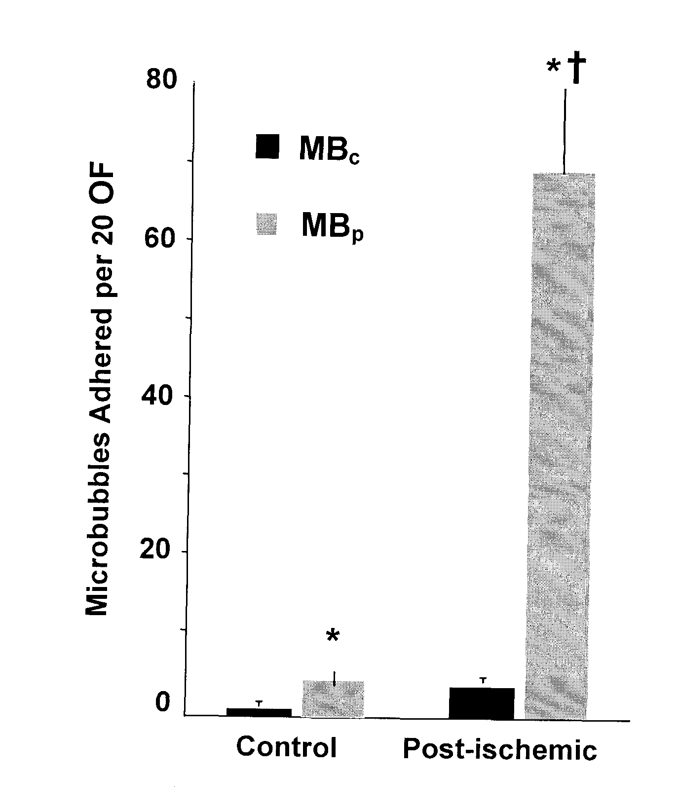
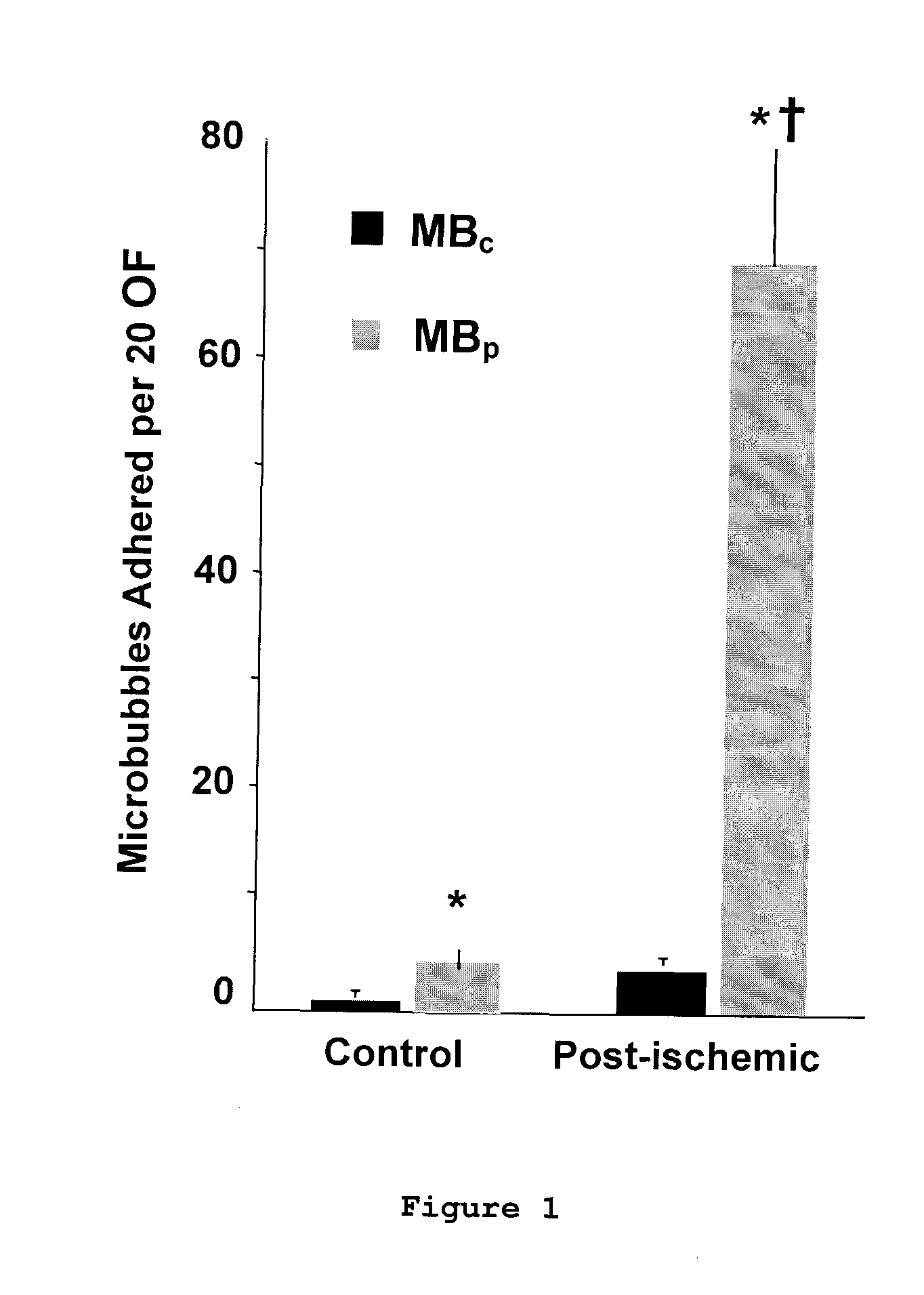
[0084]The in vitro binding capability of MBPSGL and MBA...

example 3

Microbubbles with VCAM-1

[0092]Atherosclerosis is a chronic inflammatory disorder that often progresses silently for decades before becoming clinically evident (Ross R.(1999) N Engl J Med 340:115-26). In current clinical practice, C-reactive peptide is the only inflammatory marker routinely used for risk assessment in patients. Non-invasive imaging of vascular changes such as coronary calcification, carotid intimal-medial thickening and plaque morphology have recently been used to assess patient risk (Arad et al. (2000) J. Am. Coll. Cardiol., 36:1253-60; Greenland et al. (2004) JAMA 291:210-5; Chambless et al. (2000) Am. J. Epidemiol., 151:478-87; O'Leary et al. (1999) N. Engl. J. Med., 340:14-22; Leber et al. (2006) J. Am. Coll. Cardiol., 47:672-7). However, these methods detect changes that occur relatively late in the disease process and do not directly assess inflammatory status. Since inflammation participates in plaque initiation and progression, a method capable of imaging the...


PUM


Abstract

Compositions and methods for detecting various disorders with targeted microbubbles are disclosed. Specifically, microbubbles comprising glycoprotein Ib (GPIb), ligands for VCAM-1, and ligands for P-selectin such as PSGL-1 are disclosed. Also disclosed are methods for using targeted microbubbles to detect cardiovascular disease comprising administering the disclosed microbubbles to a subject and detecting the microbubbles in the vasculature using ultrasound.

Description

[0001]This application claims priority under 35 U.S.C. §119(e) to U.S. Provisional Patent Application No. 60 / 913,086, filed on Apr. 20, 2007, and U.S. Provisional Patent Application No. 60 / 947,844, filed on Jul. 3, 2007. The foregoing applications are incorporated by reference herein.[0002]Pursuant to 35 U.S.C. Section 202(c), it is acknowledged that the United States Government has certain rights in the invention described herein, which was made in part with funds from the National Institutes of Health / National Heart, Lung, and Blood Institute Grant Nos. R01-HL074443 and R01-HL078610.FIELD OF THE INVENTION[0003]The present invention relates to the fields of imaging. Specifically, compositions and methods for detecting various disorders with targeted microbubbles are disclosed.BACKGROUND OF THE INVENTION[0004]Several publications and patent documents are cited throughout the specification in order to describe the state of the art to which this invention pertains. Each of these citat...

Claims


Application Information

Patent Timeline
Patent Type & Authority Applications(United States)
IPC IPC(8): A61K49/22A61P9/10
CPCA61K9/0009A61K9/127A61K9/10A61K9/0019A61P9/10
Inventor LINDNER, JONATHAN R.KAUFMANN, BEATMCCARTY, OWEN
Owner LINDNER JONATHAN R
Why Eureka
Try Eureka