
Cell Suspension Preparation Technique and Device

a cell suspension and preparation technique technology, applied in the field of cell suspension preparation technique and device, can solve the problems of requiring considerable skill, time and expensive equipment to perform, and limiting the area able to be covered, so as to reduce the complexity, reduce the complexity, and improve the effect of grafting efficiency

Inactive Publication Date: 2010-08-05
AVITA MEDICAL LTD
View PDF35 Cites 12 Cited by
  • Summary
  • Abstract
  • Description
  • Claims
  • Application Information

AI Technical Summary

Benefits of technology

[0010]The subject invention relates to a unique cell suspension and method for its preparation that is rapid, efficient and simple to prepare and apply. It also relates to a method for treating a patient using the unique cell suspension and to an apparatus suitable for use in the method preparation. Use of the described device while not essential for practicing the method of the invention has been found to significantly reduce the complexity associated with the use of conventional grafting technology such as CEA.
[0016]According to a second aspect of the invention there is provided a cell suspension produced according to the above method. Preferably the cells in the suspension are autologous cells (i.e. they are isolated from the patient requiring an autograft). The inventors have observed that by removing xenogenic serum from the cell suspension there is less likelihood of transmission of infection and xenogenic reactions between a patient and the serum are eliminated. Another feature of the cell suspension produced according to the above method is that the tissue sample used to isolate the cells in the suspension is removed from the enzyme solution before the cells are harvested. When cells are exposed to enzymes capable of damaging inter-cellular adhesion the viability of the cellular suspension decreases over time thereby reducing the efficiency of the grafting when applied to a patient. The cell suspension produced according to the above method has been observed to possess greater cellular viability compared to comparative methods that harvest the cells at regular intervals whilst the tissue is immersed in the presence of enzymes like trypsin.
[0019](b) administering the suspension directly to a region on the patient that requires a cell graft in a manner that facilitates spreading of the cell suspension in a relatively even distribution over the graft region.
[0034](a) harvesting cells suitable for grafting to the patient from a tissue sample of the patient in a nutrient solution that is (i) free of serum xenogenic to the patient, (ii) capable of maintaining viability of the harvested cells, and (iii) suitable for direct application to a second tissue region of the patient;

Problems solved by technology

Split-thickness grafts often require considerable skill, time and expensive equipment to perform.
Further, donor sites are painful, result in scarring and limit the area able to be covered.
Although they may be more successful than full-thickness grafts, they are usually less cosmetically attractive.
However, full-thickness grafts do not “take” as well as split-thickness grafts.
However, their cosmetic appearance is not as good as other techniques, as the resulting scarring is unacceptable.
There are several disadvantages of this method such as the amount of time required to prepare the graft is too long and the graft may not result in re-pigmentation of the area; or uneven pigmentation is common around the edges of the area of treatment.
However, these autografts are slow growing and require time for culturing of the grafts, which often exceeds the time of preparation of the recipient's sites.

Method used


Image

  • Cell Suspension Preparation Technique and Device
  • Cell Suspension Preparation Technique and Device
  • Cell Suspension Preparation Technique and Device

Examples


example 1

Preparation of Recipient Site

[0116]To optimize the success of the skin graft, the wound was cleaned and assessed to be of the appropriate depth. Further, blood haemostasis was established and the wound checked for evidence of surrounding cellulitis or infection. Techniques for preparing the area included sharp dissection, dermabrasion or laser-resurfacing.

Donor Site Biopsy

[0117]The donor site was chosen to appropriately match the recipient site. The donor site was infiltrated with local anaesthetic and adrenaline underneath the skin near the subcutaneous tissue. This allowed the donor site to be firm and aided in the taking a thin split-thickness biopsy. The dimensions of the biopsy were determined by the size of the surface area of the recipient site to be covered. Typically, the biopsy size has an expansion ratio of 1:10-1:80. In this case, a biopsy size of 2 cm×2 cm was taken from the donor site giving an expansion ratio of 1:60.

Cell Resurfacing Using the Rapid Technique

[0118]Tre...

example 2

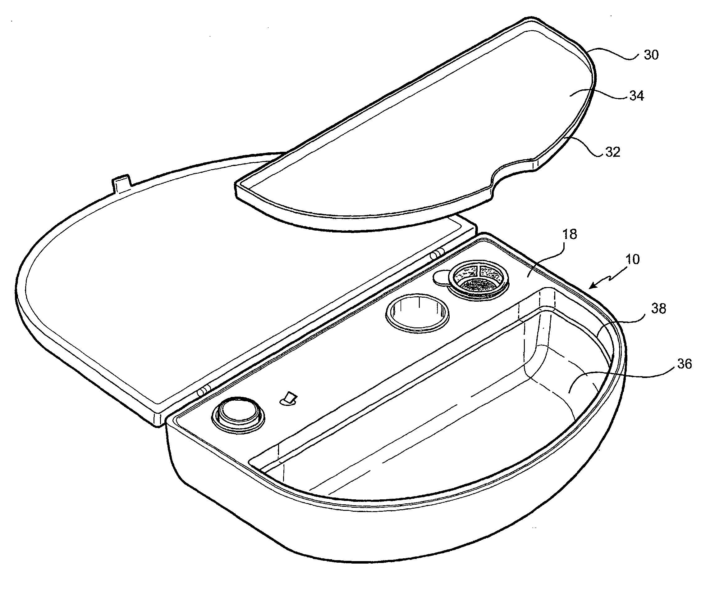
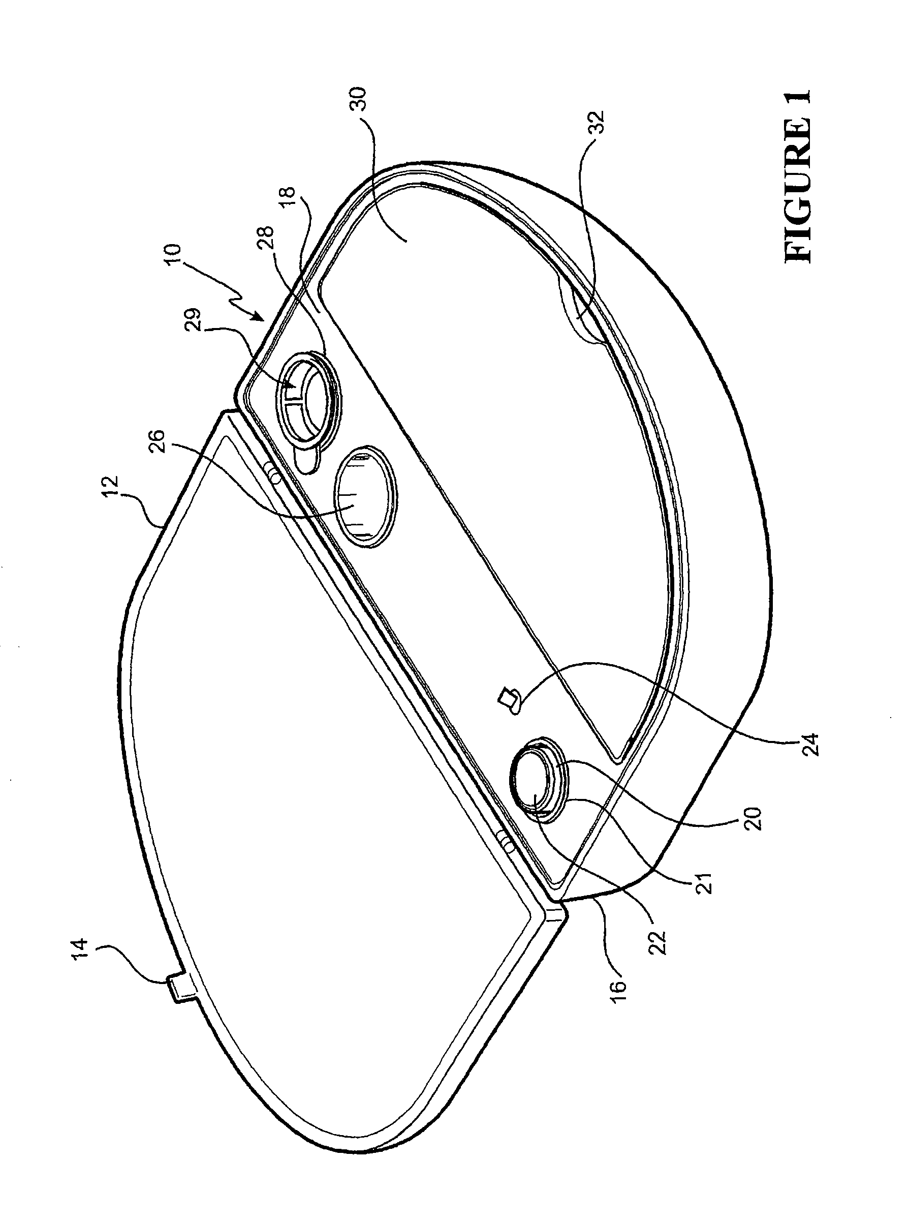
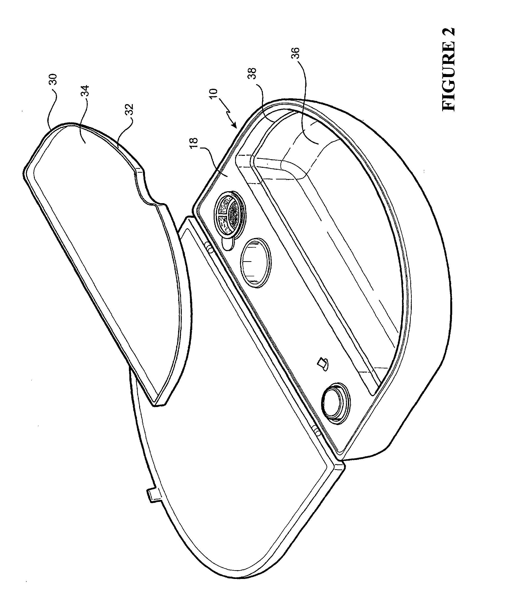
[0128]The embodiment shown in FIG. 1 is directed to a Re-Cell® Rapid Technique cell harvesting apparatus 10 for use in producing a transplantable cellular suspension of living tissue suitable for grafting to a patient.

[0129]As illustrated in FIG. 1 the apparatus includes a closure lid 12 possessing a locking mechanism 14 adapted to releasably engage a base portion 16. The locking mechanism 14 provides a means for closing the apparatus 16 when not in use. Located within the base portion 16 is a first member 18 within which there is provided an aperture 20 in which there is located a vial 22 for the enzyme. Adjacent the aperture there is provided an activation switch 24 capable of activating the heating means (not shown). The first member also provides a fluid containment well 26 and a filter recess 28. As presented in this illustration the filter 29 is shown located in the filter recess. Ordinarily the filter is an optional item included as a separate item in the apparatus.

[0130]The ...


PUM

PropertyMeasurementUnit
suction pressureaaaaaaaaaa
healing timeaaaaaaaaaa
sizeaaaaaaaaaa

Abstract

The present invention provides for methods and devices suitable for producing a transplantable cellular suspension of living tissue suitable for grafting to a patient. In applying the method and / or in using the device, donor tissue is harvested, subjected to a cell dissociation treatment, cells suitable for grafting back to a patient are collected and dispersed in a solution that is suitable for immediate dispersion over the recipient graft site.

Description

RELATED APPLICATIONS[0001]The present invention is a continuation of U.S. patent application Ser. No. 10 / 068,299, filed Feb. 6, 2002, which claims priority to and benefit of Australian Provisional Patent Application PR2989, filed Feb. 7, 2001 and U.S. Provisional Patent Application No. 60 / 281,527, filed Apr. 4, 2001, all of which are incorporated herein by reference in their entirety.FIELD OF THE INVENTION[0002]This invention relates to a simple, rapid and cost effective technique for grafting of cells, and in particular to a device for preparing a suspension of cells from a tissue sample obtained from a donor site and applying that suspension of cells to a recipient site.BACKGROUND[0003]There are many methods of treating wounds known to those skilled in the art. For example, skin grafting techniques exist which aim to reconstruct the skin covering areas of the body where there is either damage or defects to the skin. In general, these types of grafts are classified according to the...

Claims


Application Information

Patent Timeline
Patent Type & Authority Applications(United States)
IPC IPC(8): A61K35/12C12N5/02C12M1/38A61M35/00A61F2/10A61L27/00A61P17/02C12N5/071
CPCA61F2/105C12N5/0629C12N2500/99C12N2509/00C12M47/02C12M45/02C12M45/06C12M45/22A61K35/36C12N2500/98C12M45/09A61P17/02A61P43/00A61M37/00A61M2202/09A61M2205/7545
Inventor WOOD, FIONA M.STONER, MARIE L.
Owner AVITA MEDICAL LTD
Learn More