
System and method for passive catheter tracking with magnetic resonance imaging

Inactive Publication Date: 2010-08-12
NORTHSHORE UNIV HEALTHSYST
View PDF29 Cites 4 Cited by
  • Summary
  • Abstract
  • Description
  • Claims
  • Application Information

AI Technical Summary

Benefits of technology

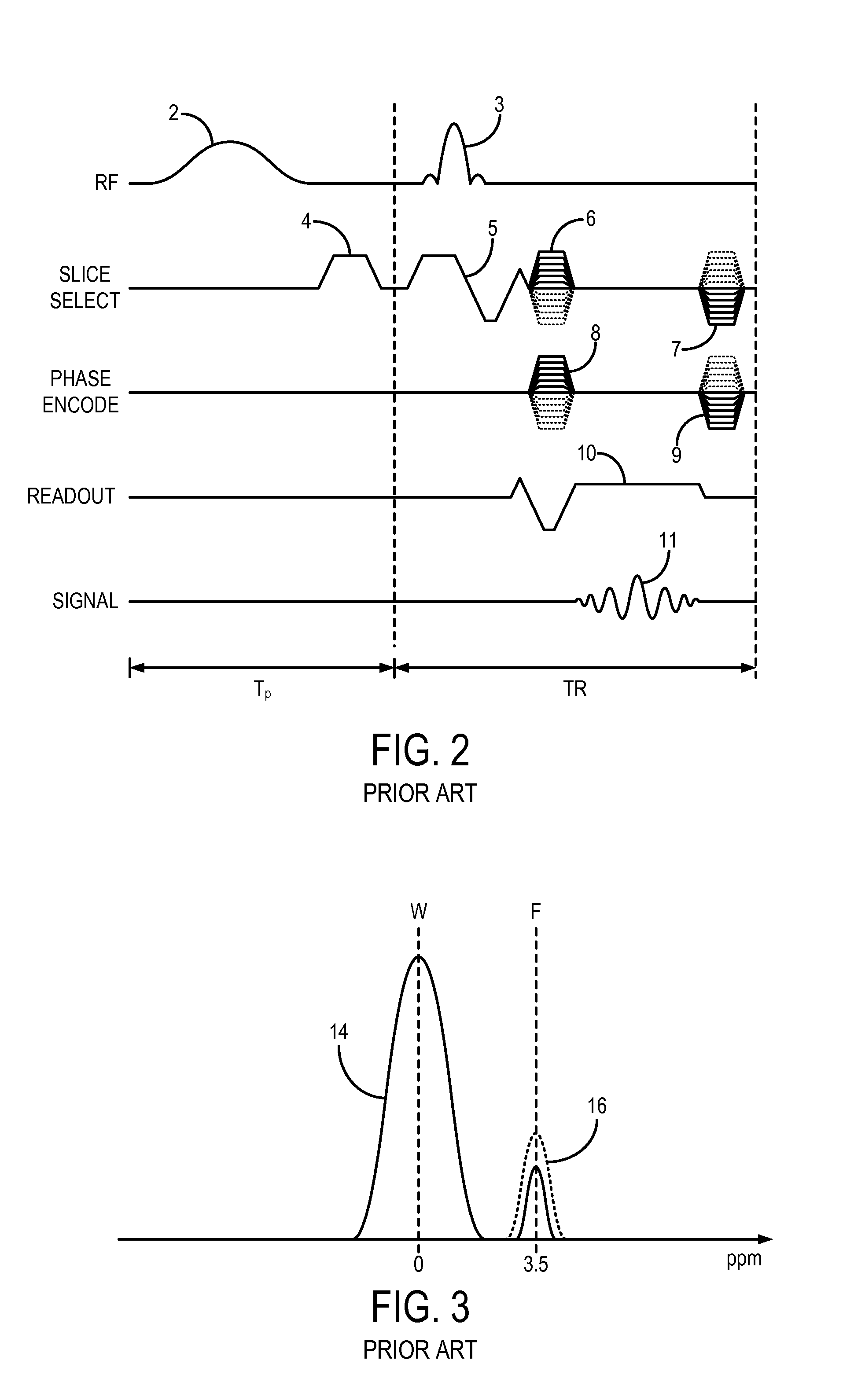
[0034]For example, one such imaging method uses parametric contrast agents, such as those based on gadolinium, which not only shorten the T1 relaxation of adjacent water spins, but they also produce a chemical shift of the Larmor frequency of those water spins. Other imaging agents can also be used to shift the Larmor frequency of adjacent water spins, such as catheters including a frequency shifting agent. For example, a catheter having a coating or filled with material such as gadolinium, can be used. This provides the potential for passive catheter tracking. However, one drawback is the sensitivity of the gadolinium induced frequency shifts to the orientation of the catheter.
[0035]One aspect of the invention is the provision of shaped catheter coatings that shift the resonance frequency and can substantially eliminate the orientation dependence of the frequency shift. For example, the use of a loop in the catheter can overcome these bulk magnetic susceptibility (“BMS”) orientation effects.
[0038]A general focus of the invention is to acquire contrast enhanced MRA images without the need to subtract a mask image. The saturation pulse sequences are performed throughout the acquisition at a rate which keeps the signals from tissues surrounding the subject's vasculature suppressed without significantly affecting the signal from blood that contains the contrast agent. In addition, the contrast agent performs its usual function of shortening the T1 relaxation time of the blood such that many imaging pulse sequences can be performed before the saturated and non-T1 shortened surrounding tissues recover enough to require another saturation pulse sequence. Thus, the scan is not significantly increased in time.

Problems solved by technology

However, one drawback is the sensitivity of the gadolinium induced frequency shifts to the orientation of the catheter.

Method used


Image

  • System and method for passive catheter tracking with magnetic resonance imaging
  • System and method for passive catheter tracking with magnetic resonance imaging
  • System and method for passive catheter tracking with magnetic resonance imaging

Examples


Embodiment Construction

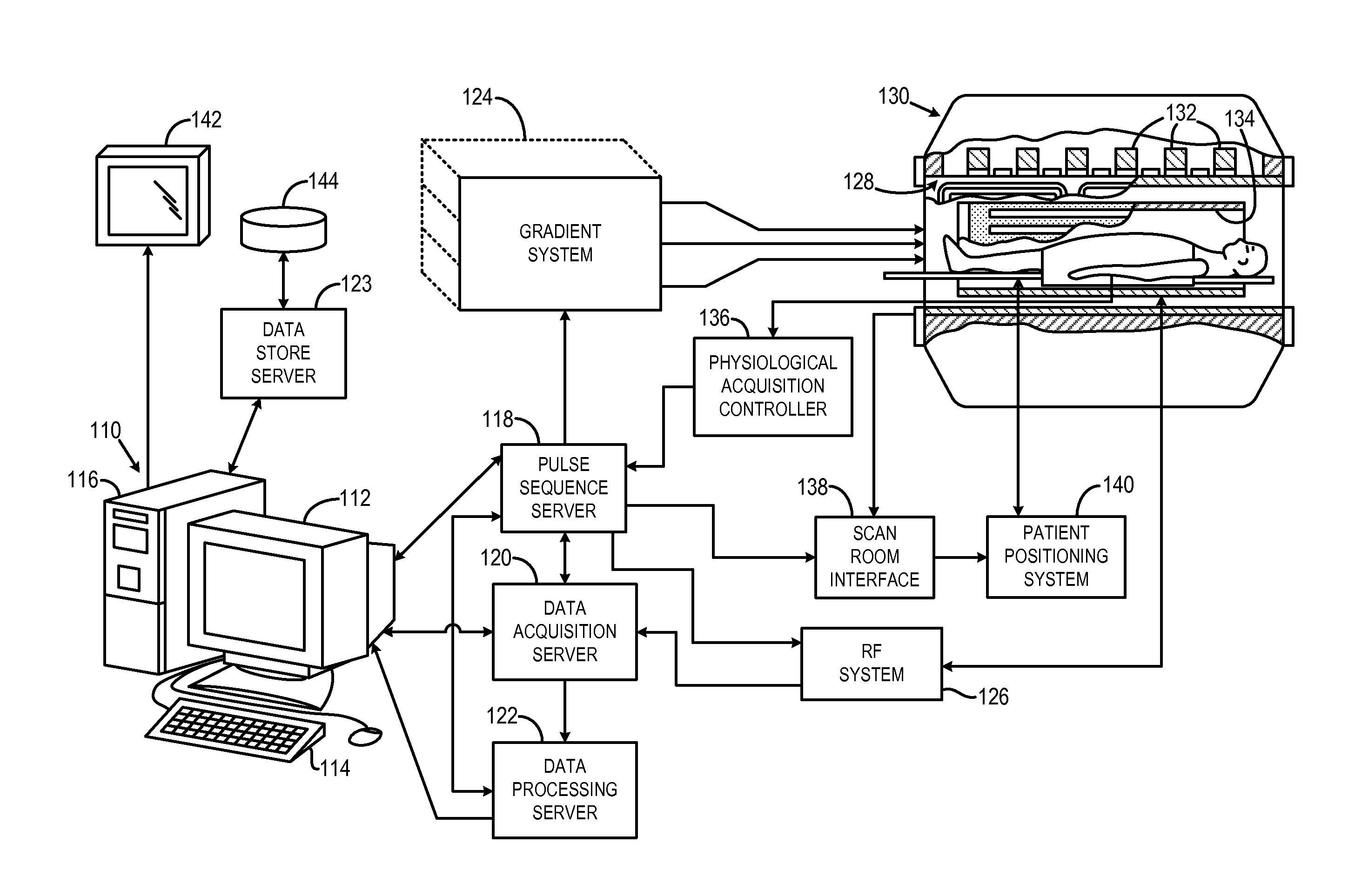
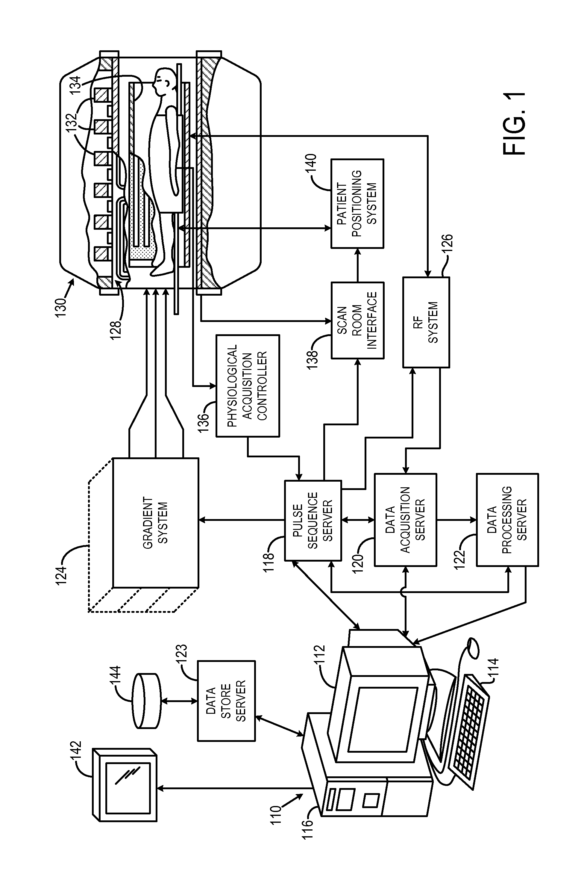
[0052]Referring to FIG. 11, a catheter system 1100 that is amenable to passive tracking with a magnetic resonance imaging (“MRI”) system includes a flexible shaft 1102 having a length suitable for reaching a vasculature of interest from a selected vessel entry point. Exemplary lengths include 80, 110, and 135 centimeters (“cm”). The flexible shaft 1102 extends from a proximal end (shown generally by arrow 1104) towards a distal end (shown generally by arrow 1106), and is composed, for example, of a plastic. In some configurations, the catheter shaft 1102 has a diameter of five French (“5F”); however, it will be appreciated by those skilled in the art that different diameters can be readily employed depending on the desired angiographic procedure. At its proximal end 1104, the shaft 1102 connects to a fixture 1108 that defines an inlet 1109 to the interior of the shaft 1102 and connects that inlet 1109 through a tube 1110 to a valve 1112. The fixture 1108 also provides an opening 111...


PUM


Abstract

Background tissue signals such as water and / or fat are suppressed in an MR image by using an imaging agent that chemically shifts the tissue spins of interest. An imaging pulse sequence is used to acquire the image data using an RF excitation pulse that is tuned to the off-resonance tissue spins of interest with the saturation pulse sequences being interleaved with the imaging pulse sequences to selectively suppress signals from on-resonance background tissues such as water and / or fat.

Description

CROSS-REFERENCE TO RELATED APPLICATIONS[0001]This application is a continuation-in-part of, and incorporates herein by reference, U.S. patent application Ser. No. 12 / 114,990, filed on May 5, 2008, and entitled “Magnetic Resonance Image Acquisition with Suppression of Background Tissues and RF Water Excitation at Offset Frequency,” which is based on, and claims the benefit of, U.S. Provisional Patent Application Ser. No. 60 / 915,781, filed on May 3, 2007, and entitled “Magnetic Resonance Image Acquisition with Suppression of Background Tissues and RF Water Excitation at Offset Frequency.”BACKGROUND OF THE INVENTION[0002]The field of the invention is magnetic resonance imaging (“MRI”) methods and systems and particularly the use of tissue suppression in conjunction with the use of a radio frequency (“RF”) excitation pulse for water at an offset or off-resonance frequency. That is an RF excitation pulse for water at a frequency that is shifted from a nominal Larmor frequency of water.[0...

Claims


Application Information

Patent Timeline
IPC IPC(8): G01R33/48
CPCG01R33/286G01R33/4828G01R33/5607G01R33/5601G01R33/485
Inventor EDELMAN, ROBERT R.
Owner NORTHSHORE UNIV HEALTHSYST
Learn More