

Neural scaffolds
a technology of neural tissue and scaffolds, applied in the field of neural scaffolds, can solve the problem that no biocompatible scaffold has successfully engrafted with neural tissu
- Summary
- Abstract
- Description
- Claims
- Application Information
AI Technical Summary
Benefits of technology
Problems solved by technology
Method used
Image

Examples
example 1
Manufacturing Acellular Spinal Cord and Dura Mater Biological Scaffolds
[0063]In one example, porcine spinal cord was obtained. Using forceps, scissors and a scalpel, dura mater was removed from the spinal cord. The inner dura mater surface was scrapped with scalpel blade to remove any debris. The spinal cord and dura were placed in separate containers and treated in the same manner as listed below. The spinal cord was cut either longitudinally or in cross-section (to increase surface area) and placed in a cassette (e.g., safety container to protect 3-D structure of cord throughout process). Optionally tissue was enzymatically treated using trypsin-EDTA for 30 minutes at 37° C. The tissue was incubated in TRITON X-100™ (4-octylphenol polyethoxylate) solutions at 3% for periods up to 2-3 days at 4° C. This step was repeated with a solution of Triton X-100 at 6% and again with a solution of Triton X-100 at 9%. The spinal cord tissue was incubated in lecithin or lecithin-deoxycholate to...
example 2
Characterization of the Neuronal Scaffolds
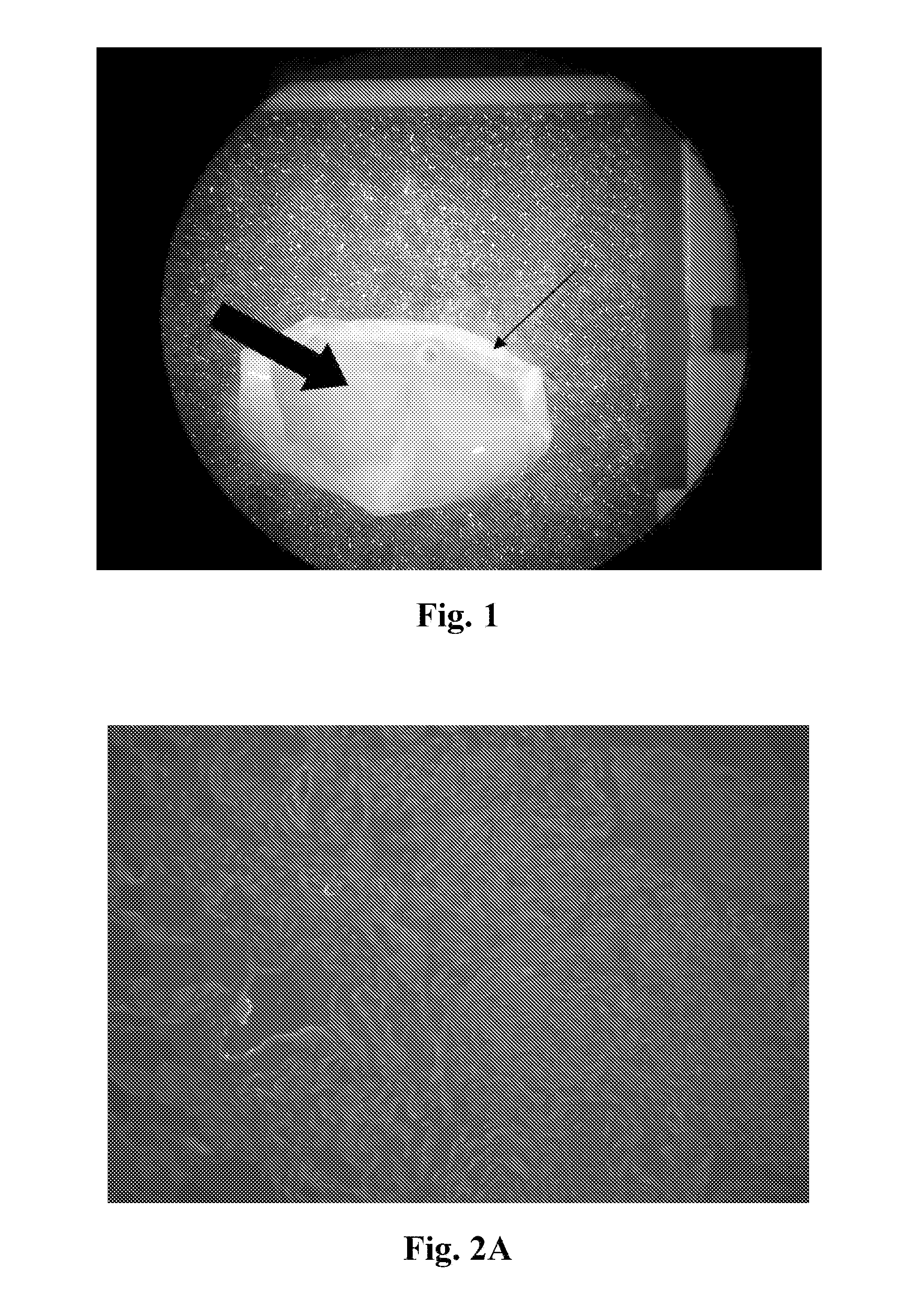
[0064]Described in this example is the characterization of the neuronal scaffolds. After the last step of the process described in the example 1, the acellular spinal cord parenchyma-derived neuronal scaffold is gel-like. FIG. 1 is a digital image showing the gel-like quality of the scaffold. The thick arrow indicates the gel-like substance and the thin arrow indicates the pia matter.
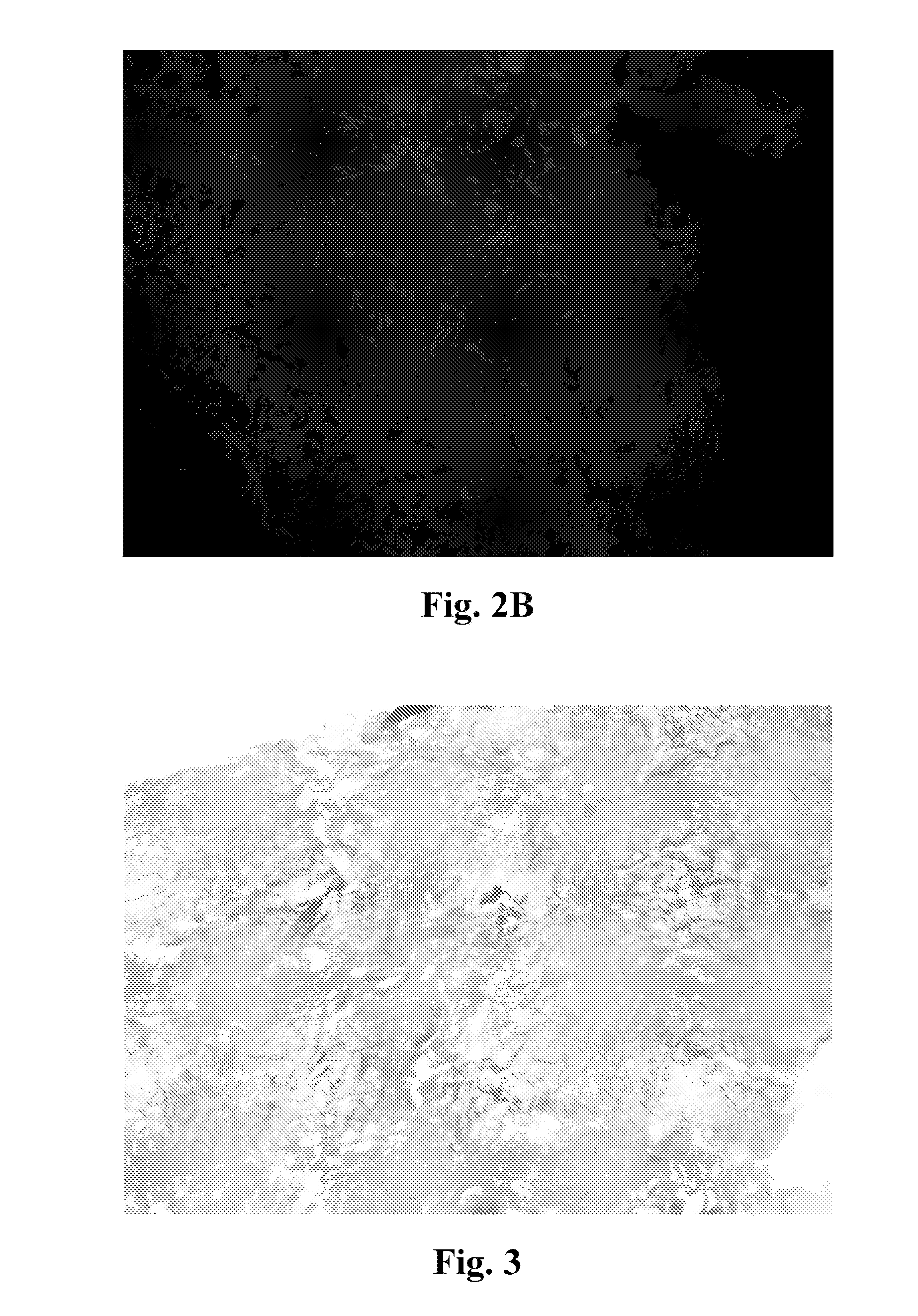
[0065]After forming the neuronal scaffolds, the scaffold is acellular. Various methods were used to determine whether the scaffold was acellular. First, fluorescent stains, such as DAPI (4′,6-diamidino-2-phenylindole), were used to determine whether cells are present. FIGS. 2A-2B and FIG. 8A are fluorescence photomicrographs of the scaffold stained with DAPI, where lack of fluorescence correlated with lack of nuclei and cells within the scaffold. Second, histological stains, such as Masson's trichrome, were also be used. FIG. 3 and FIG. 8B show photomicrographs ...
example 3
Testing for Biocompatibility of the Neuronal Scaffolds
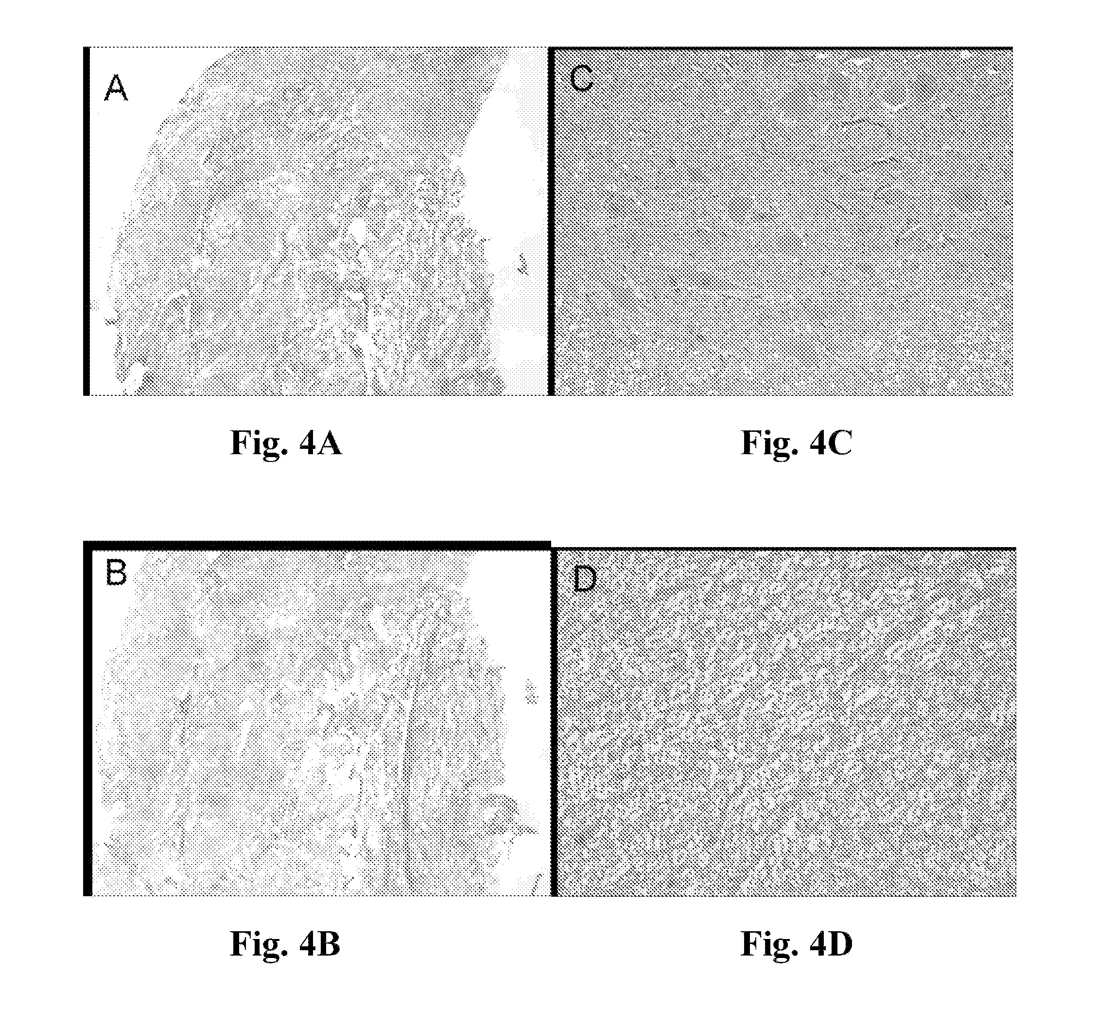
[0067]Biocompatibility of the neuronal scaffold was tested by culturing primary neuronal cells and neuronal cell lines on the scaffold. Rat adrenal pheochromocytoma (PC 12) cells were obtained from ATCC (American Type Culture Collections, Manassas, Va.), ATCC catalog number CRL-1721. The PC12 cells were grown in DMEM with 10% heat-inactivated horse serum, 5% fetal bovine serum, 1% penicillin-streptomycin solution (stock solution: 10,000 units penicillin and 10,000 μg streptomycin / ml) in a humidified incubator at 37° C. supplemented with 5% CO2 in plastic cell culture-treated flasks. Cells were not allowed to become more than 50% confluent in culture flasks before sub-culturing. Prior to seeding on neural scaffolds, adherent PC12 cells were trypsinized (5 ml 0.25% trypsin with EDTA / 75 sq. cm flask) to release and remove cells from culture flasks. Once cells were non-adherent, the trypsin was neutralized with 20 ml culture medium (...
PUM
| Property | Measurement | Unit |
|---|---|---|
| porosity | aaaaa | aaaaa |
| porosity | aaaaa | aaaaa |
| porosity | aaaaa | aaaaa |
Abstract
Description
Claims
Application Information