
Specific amplification of tumor specific DNA sequences
a dna sequence and specific technology, applied in the field of cancer detection and diagnosis, can solve the problems of inability to show a good correlation between tumor size, stage location or type and total cna concentration, and the cost and effectiveness of existing cancer screening methods,
- Summary
- Abstract
- Description
- Claims
- Application Information
AI Technical Summary
Benefits of technology
Problems solved by technology
Method used
Image

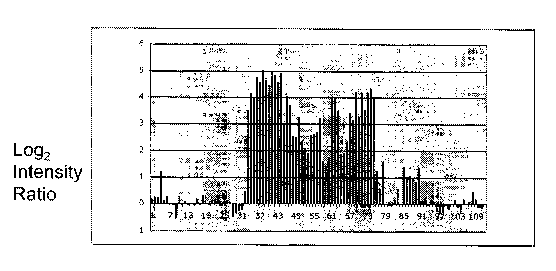
Examples
example 1
Identification of a Tumor Associated Hypomethylated Region
[0055]To test whether methylation profiles of CNA from subjects with ovarian tumors is different from that of normal controls, frozen serum samples were obtained from women who had their blood drawn prior to exploratory surgery for suspected ovarian cancer. Similar samples were obtained from women without cancer. DNA was prepared from 1 ml of cell-free serum by a standard method and the entire resulting sample was subjected to methylation-sensitive amplification as described above. One such pair of samples was submitted to NimbleGen for hybridization to an array that had previously been used for analysis of trophoblast methylation.
[0056]The data indicated that both amplifications (cancer and normal) resulted in measurable signal (defined as >3sd above background) from ˜5% of array addresses. Additionally, in ˜70% of these cases, the log2-ratio of the signals is less than |1.5|, indicating that even though that segment amplifi...
example 2
Development of a Comparison Panel
[0058]In order to facilitate detection and diagnosis using the methods of the present invention, normal and specific cancer patient populations can be compared to develop a methylation profile associated with a particular type of cancer. The methods of the present invention can be used to create such a methylation profile.
[0059]DNA is isolated from the serum or plasma of known cancer patients and normal controls using standard methods (Johnson, K. L., et al., Clin. Chem. 50:516-21 (2004)). Briefly, 10 ml of patient blood is centrifuged two times to remove cells. The resulting plasma is passed over a DNA binding membrane. The DNA is removed from the membrane and the resulting DNA is digested with HpyCh4-IV.
[0060]DNA linkers are annealed and ligated to the digested DNA. The linkers are designed to create a MluI restriction site when ligated to DNA digested with HpyCh4-IV. The linker-mediated PCR is performed as described by Guillaud-Bataille, M., et al...
example 3
Making a Microarray for Detection of Hypomethylated, Tumor-Associated DNA
[0065]The genome is parsed into segments that are bounded by two sites for the methylation sensitive restriction enzyme in question (ACGT for HpyCh4-IV) and less than 500 base pairs long. This provides a list of DNA segments that might be amplified from a serum or plasma DNA sample. An algorithm is used to analyze the sequence of these fragments, with the goal of finding suitable sequence for representation on the microarray. For example, appropriate oligonucleotides will have one or more of the following characteristics: (i) greater than about 40 nucleotides of unique sequence, or greater than about 60 nucleotides of unique sequence; (ii) a GC of about 40% to about 60%, and (iii) should not contain significant repetitive or simple sequences, for example runs of greater than about 15 of a single base. The array contains oligonucleotides chosen in this way with each oligonucleotide on the array representing one ...
PUM
| Property | Measurement | Unit |
|---|---|---|
| Magnetism | aaaaa | aaaaa |
Abstract
Description
Claims
Application Information