Mammary gland content rate estimating apparatus, method and recording medium

a technology of mammary gland and content rate, which is applied in the field of apparatus and a method for estimating can solve the problems of inability to estimate inability to clearly separate the mammary gland region from the adipose region in the two-dimensional image, and changes in area ratio, so as to achieve the effect of accurately and easily estimating the mammary gland content rate for each of the pixels

Active Publication Date: 2010-09-30
FUJIFILM CORP
View PDF8 Cites 27 Cited by
  • Summary
  • Abstract
  • Description
  • Claims
  • Application Information

AI Technical Summary

Benefits of technology

[0013]The presently disclosed subject matter is made in view of the above circumstances, and has an object to provide an apparatus and method for estimating mammary gland content rate, which can estimate a mammary gland content rate for each of pixels easily and precisely without requiring complicated calibration in advance.
[0015]In the first aspect of the invention, the adipose image (image having pixel values in the case where the mammary gland tissue of the breast is all replaced with the adipose tissue) is estimated from the X-ray image of the breast (breast image), and the mammary gland content rate is calculated for each of the pixels based on the relationship between the original breast image and the adipose image. At the time of calculation of the mammary gland content rate, the pixel value of the directly irradiated region (region without X-ray attenuation) in the breast image is used in addition to the breast image and the adipose image. By using the adipose image and the pixel value of the directly irradiated region which are obtained by analyzing the breast image, and the relational expressions between them, the mammary gland content rate can be estimated easily with high precision for each of pixels, without requiring complicated calibration in advance.
[0023]The predetermined value indicating the ratio of the average attenuation coefficients is the value which is hardly influenced by the imaging conditions, but is not completely free from influence. Thus, as the predetermined value indicating the ratio of the average attenuation coefficients, the value (fixed value) indicating the ratio of the average attenuation coefficient of the mammary glands and the average attenuation coefficient of the adipose in the case of imaging the average breast under the average conditions is adopted, and thereby, the error in estimation of the mammary gland content rate can be minimized.
[0034]According to the presently disclosed subject matter, the adipose image (the image having the pixel value in the case where the mammary gland tissue of the breast is all replaced with the adipose tissue) is estimated from the X-ray image (breast image) of the breast, the pixel value of the directly irradiated region without X-ray attenuation is analyzed, and the mammary gland content rate is calculated based on the relationship between the original breast image, the adipose image and the pixel value of the directly irradiated region. Therefore, the mammary gland content rate can be estimated for each of pixels easily and precisely, without requiring complicated calibration in advance.

Problems solved by technology
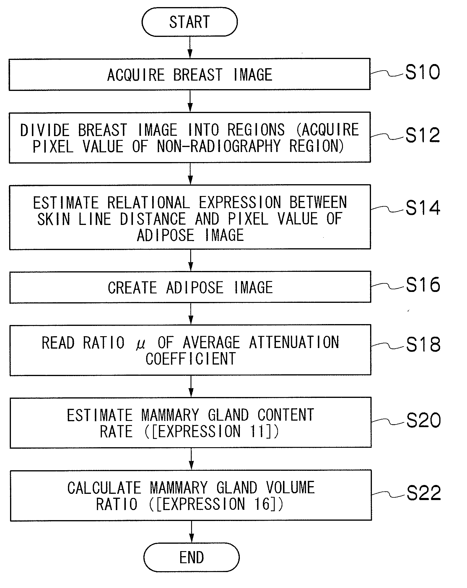
However, since a breast is composed of mammary glands and adipose which are mixed up together, there is the problem that the mammary gland region cannot be clearly separated from the adipose region in the two-dimensional image.
Further, there is also the problem that the area ratio changes depending on the way of compressing a breast and positioning at the time of imaging.
Further, in order to select suitable relational expressions, the information of the breast mass thickness and the like is required, and a mammary gland content rate cannot be estimated with only the image data of the breast image.
Further, the imaging information relating to imaging such as the X-ray tube voltage and the target / filter at the time of radiographing, and breast mass thickness needs to be acquired, and there arises the problem that the volume of mammary glands and the volume ratio cannot be estimated when the imaging information cannot be acquired.

Method used


Image

  • Mammary gland content rate estimating apparatus, method and recording medium
  • Mammary gland content rate estimating apparatus, method and recording medium
  • Mammary gland content rate estimating apparatus, method and recording medium

Examples


application example

(1) Display of Mammary Gland Content Rate or Volume Ratio of Mammary Glands

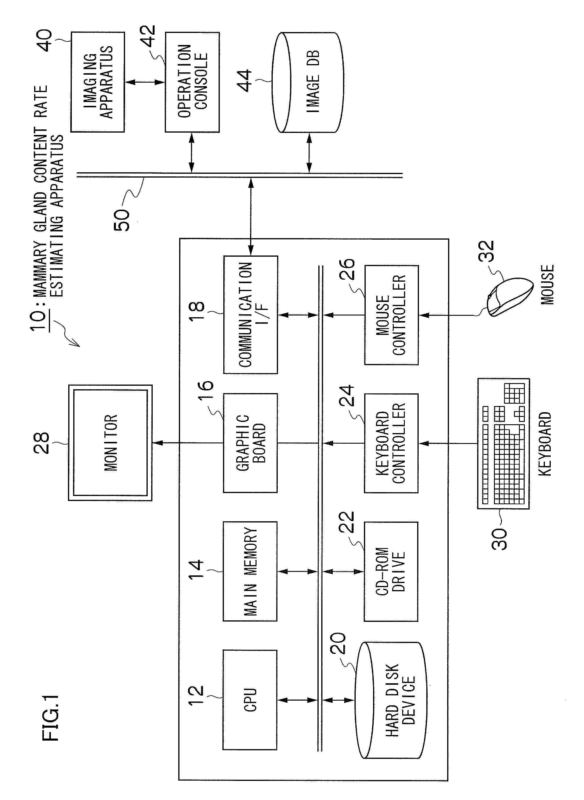
[0134]The mammary gland content rate or the volume ratio of the mammary glands which is calculated as described above may be displayed on the screen of the monitor device. Since the mammary gland content rate can be obtained for the respective pixels, the mammary gland content rate may be displayed in a two-dimensional image form or a graphical form (histogram). Further, one value of the volume ratio of the mammary glands can be obtained for one image, and therefore, the value may be displayed with the image. The information of them is stored in the header of the DICOM file, and can be stored in the image DB 44 together with the taken image. When a doctor interprets the image, the taken image is displayed, and the information of them is displayed at the same time to be the assistance for diagnosis.

(2) Application to Computer-Aided Diagnosis (Computer-Aided Diagnosis: CAD)

[0135]Appearance of a lesion differs b...


PUM


Abstract

A mammary gland content rate estimating apparatus, includes: a breast image acquiring device which acquires a breast image obtained by radiographing a breast by a mammography imaging apparatus; an adipose image estimating device which estimates an adipose image from the acquired breast image based on an assumption that an entire breast is composed of only adipose tissues; a device which acquires a pixel value of a directly irradiated region from the acquired breast image; and a mammary gland content rate calculating device which calculates a mammary gland content rate for each of pixels in the breast image based on the acquired breast image, the estimated adipose image and the acquired pixel value of the directly irradiated region. Accordingly, without requiring complicated calibration in advance, the mammary gland content rate can be estimated for each of pixels easily and precisely.

Description

CROSS-REFERENCE TO RELATED APPLICATIONS[0001]This application claims the priority benefit under 35 U.S.C. §119 of Japanese Patent Application No. 2009-087493 filed on Mar. 31, 2009 and Japanese Patent Application No. 2010-010239 filed on Jan. 20, 2010, which are hereby incorporated in their entirety by reference.BACKGROUND OF THE INVENTION[0002]1. Field of the Invention[0003]The presently disclosed subject matter relates to an apparatus and a method for estimating mammary gland content rate, particularly, to technology for estimating a mammary gland content rate for each pixel in a breast image from only the breast image obtained by radiographing a breast by a mammography imaging apparatus.[0004]2. Description of the Related Art[0005]A breast is mainly constituted of mammary gland tissue and adipose tissue, and the ratio of the mammary glands in the breast is called a mammary gland content rate. The mammary gland content rate is extremely useful in accurately knowing the character o...

Claims


Application Information

Patent Timeline
Patent Type & Authority Applications(United States)
IPC IPC(8): G06T7/00A61B6/00
CPCA61B5/4872A61B6/502G06T7/0014A61B5/4312G06T2207/20021G06T2207/30068G06T2207/10116
Inventor MORITA, JUNYA
Owner FUJIFILM CORP
Why Eureka
Try Eureka