
Devices and methods for delivery of aortic and mitral valve prostheses

a technology for aortic and mitral valves, which is applied in the field of devices and methods for delivery of aortic and mitral valve prostheses, can solve the problems of limited imaging system, limited methods of open heart surgery, and long recovery period, and achieves the limitations of current imaging system, the current imaging system is about 2 mm

Active Publication Date: 2010-09-30
JC MEDICAL INC
View PDF34 Cites 113 Cited by
  • Summary
  • Abstract
  • Description
  • Claims
  • Application Information

AI Technical Summary

Problems solved by technology

Such open heart surgery is particularly invasive and involves a lengthy and difficult recovery period.
A major issue during heart valve replacement is positioning the prosthetic valve within a small, approximately 2-5 mm, range at the target site.
However, there are limitations with these methods and the current imaging systems.
For example, the standard error of the current imaging systems is about 2 mm, and operator handling introduces additional variability.
All these make it very difficult to land a prosthetic valve accurately.
Another critical issue with sutureless valves is valve migration.
The pressure times the valve surface area produces a substantial load force on the prosthetic valve and can cause valve migration towards the aortic arch.
The other cause of valve migration is tilted valve landing.
Other limitations of the related art will become apparent to those of skill in the art upon a reading of the specification and a study of the drawings.

Method used


Image

  • Devices and methods for delivery of aortic and mitral valve prostheses
  • Devices and methods for delivery of aortic and mitral valve prostheses
  • Devices and methods for delivery of aortic and mitral valve prostheses

Examples


Embodiment Construction

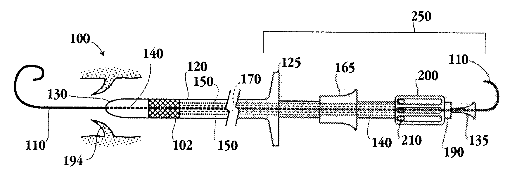
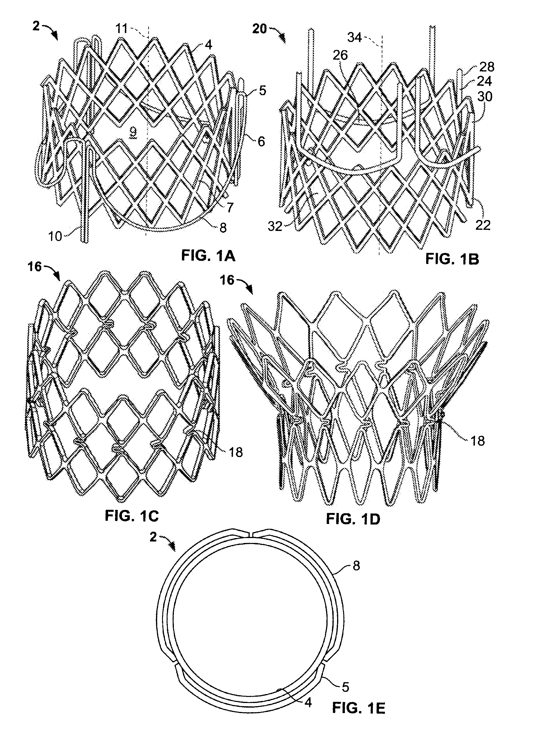
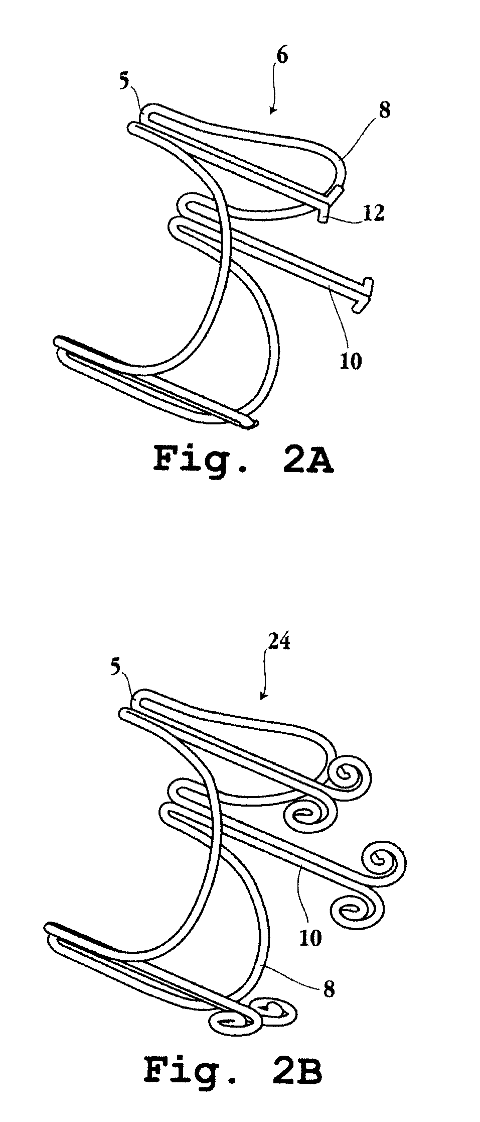
[0159]The present disclosure provides devices, systems and methods for valve replacement, preferably using a minimally invasive surgical technique. While the devices and methods will have application in a number of different vessels in various parts of the body, they are particularly well-suited for replacement of a malfunctioning cardiac valve, and in particular an aortic valve, a pulmonary valve or a mitral valve. The devices and methods are particularly advantageous in their ability to provide a more flexible prosthetic heart valve implantation device, ensure accurate and precise placement of the prosthetic heart valve with reduced reliance on imaging, and provide additional anchoring of the prosthetic valve, reducing the incidence of valve migration. Another advantage is the delivery and implantation of a sutureless valve prosthesis as described herein.

[0160]The present disclosure also provides improved devices and methods for implanting a prosthetic heart valve. In particular, ...


PUM


Abstract

A valve prosthesis and implantation device, and methods for use are provided. The devices described herein may be used for transcatheter delivery of an aortic valve prosthesis or transapical delivery of a mitral valve prosthesis. The implantation device utilizes movable claspers for both positioning and anchoring the valve prosthesis, reducing the extent of imaging needed during the implantation procedure.

Description

PRIORITY CLAIM[0001]This application claims the benefit of U.S. Provisional Application Nos. 61 / 211,430, filed Mar. 30, 2009, 61 / 211,431, filed Mar. 30, 2009; 61 / 211,432, filed Mar. 30, 2009; 61 / 211,433, filed Mar. 30, 2009; and 61 / 228,423, filed Jul. 24, 2009, all of which are incorporated herein by reference in their entirety.TECHNICAL FIELD[0002]The subject matter described herein relates to medical devices and methods for the implantation of a sutureless prosthetic heart valve using minimally invasive procedures.BACKGROUND[0003]Prosthetic heart valves are used to replace damaged or diseased heart valves. In vertebrate animals, the heart is a muscular organ with four pumping chambers: the left and right atria and the left and right ventricles, each provided with its own one-way valve. The natural heart valves are identified as the aortic, mitral (or bicuspid), tricuspid and pulmonary valves. Prosthetic heart valves can be used to replace any of these naturally occurring valves, a...

Claims


Application Information

Patent Timeline
Patent Type & Authority Applications(United States)
IPC IPC(8): A61F2/24
CPCA61F2/2418A61F2/2436A61F2/966A61F2220/0016A61F2220/005A61F2220/0058A61F2220/0066A61F2230/0054A61F2230/0067A61F2250/006A61F2/9517A61F2/9661A61F2/2427
Inventor ZHANG, JI
Owner JC MEDICAL INC
Learn More