Methods for detecting fetal abnormality

a technology of fetal abnormality and methods, applied in biochemistry apparatus and processes, library screening, laboratory glassware, etc., can solve the problems of time-consuming and laborious analysis, potentially harmful to the mother and the fetus, and insufficient accuracy of amniocentesis and chorionic villus sampling (cvs)

Inactive Publication Date: 2010-10-28
THE GENERAL HOSPITAL CORP +1
View PDF3 Cites 9 Cited by
  • Summary
  • Abstract
  • Description
  • Claims
  • Application Information

AI Technical Summary

Problems solved by technology

However, the available methods today, amniocentesis and chorionic villus sampling (CVS) are potentially harmful to the mother and to the fetus.
For example, maternal serum alpha-fetoprotein, and levels of unconjugated estriol and human chorionic gonadotropin can be used to identify a proportion of fetuses with Down's syndrome, however, these tests not one hundred percent accurate.
However, fetal cells represent a small number of cells against the background of a large number of maternal cells in the blood which make the analysis time consuming and prone to error.
These methods suffer from various limitations such as high cost, low yield, need of skilled operators and in some methods lack of specificity.
As a result, no clinically acceptable method for enrichment of rare cell populations, particularly fetal cells, from peripheral blood samples has been devised which yields cell populations sufficient to permit clinical diagnosis.

Method used


Image

  • Methods for detecting fetal abnormality
  • Methods for detecting fetal abnormality
  • Methods for detecting fetal abnormality

Examples


example 1

A Silicon Device Multiplexing 14 Three-Stage Array Duplexes

[0202]FIGS. 11A-11E show an exemplary size-based separation module of the invention, characterized as follows:

[0203]Dimensions: 90 mm×34 mm×1 mm

[0204]Array design: 3 stages, gap size=18, 12 and 8 μm for the first, second and third stage, respectively. Bifurcation ratio=1 / 10. Duplex; single bypass channel

[0205]Device design: multiplexing 14 array duplexes; flow resistors for flow stability

[0206]Device fabrication: The arrays and channels were fabricated in silicon using standard photolithography and deep silicon reactive etching techniques. The etch depth is 150 μm. Through holes for fluid access are made using KOH wet etching. The silicon substrate was sealed on the etched face to form enclosed fluidic channels using a blood compatible pressure sensitive adhesive (9795, 3M, St Paul, Minn.).

[0207]Device packaging: The device was mechanically mated to a plastic manifold with external fluidic reservoirs to deliver blood and buf...

example 2

A Silicon Device Multiplexing 14 Single-Stage Array Duplexes

[0212]FIGS. 13A-13D shows an exemplary device of the invention, characterized as follows.

[0213]Dimensions: 90 mm×34 mm×1 mm

[0214]Array design: 1 stage, gap size=24 p.m. Bifurcation ratio=1 / 60. Duplex; double bypass channel

[0215]Device design: multiplexing 14 array duplexes; flow resistors for flow stability

[0216]Device fabrication: The arrays and channels were fabricated in silicon using standard photolithography and deep silicon reactive etching techniques. The etch depth is 150 Through holes for fluid access are made using KOH wet etching. The silicon substrate was sealed on the etched face to form enclosed fluidic channels using a blood compatible pressure sensitive adhesive (9795, 3M, St Paul, Minn.).

[0217]Device packaging: The device was mechanically mated to a plastic manifold with external fluidic reservoirs to deliver blood and buffer to the device and extract the generated fractions.

[0218]Device operation: An exter...

example 3

Separation of Fetal Cord Blood

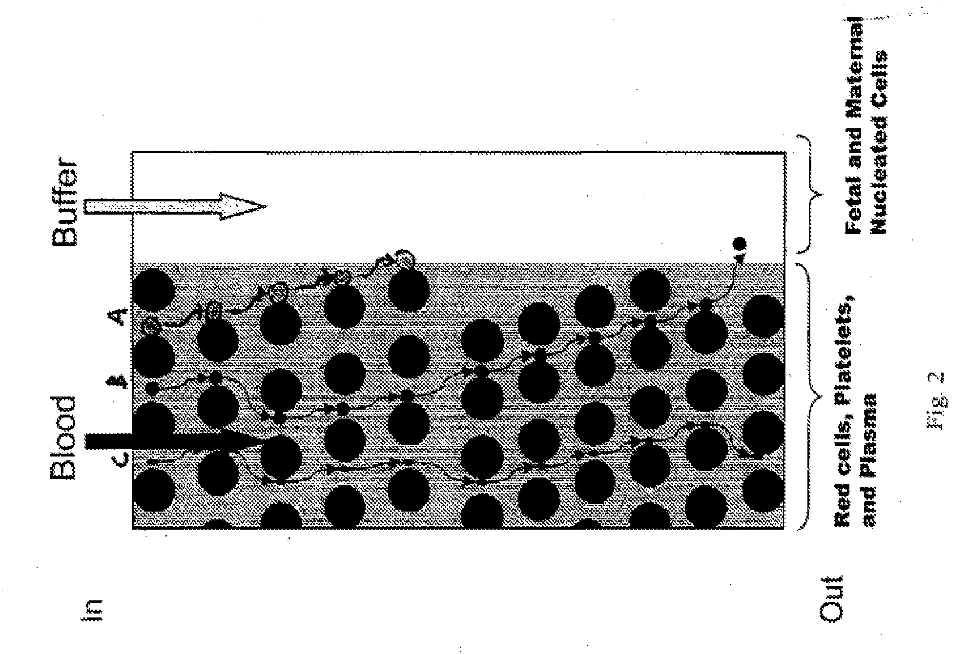
[0222]FIGS. 14A-14D shows a schematic of the device used to separate nucleated cells from fetal cord blood.

[0223]Dimensions: 100 mm×28 mm×1 mm

[0224]Array design: 3 stages, gap size=18, 12 and 8 μm for the first, second and third stage, respectively. Bifurcation ratio= 1 / 10. Duplex; single bypass channel.

[0225]Device design: multiplexing 10 array duplexes; flow resistors for flow stability.

[0226]Device fabrication: The arrays and channels were fabricated in silicon using standard photolithography and deep silicon reactive etching techniques. The etch depth is 140 μm. Through holes for fluid access are made using KOH wet etching. The silicon substrate was sealed on the etched face to form enclosed fluidic channels using a blood compatible pressure sensitive adhesive (9795, 3M, St Paul, Minn.).

[0227]Device packaging: The device was mechanically mated to a plastic manifold with external fluidic reservoirs to deliver blood and buffer to the device and extrac...


PUM

PropertyMeasurementUnit
volumesaaaaaaaaaa
volumesaaaaaaaaaa
volumesaaaaaaaaaa

Abstract

The invention relates to a method of identifying fetal abnormality from a maternal blood sample by capturing an image of a fetal nucleated red blood cell obtained from the maternal blood sample; inputting probe intensities for a plurality of nucleic acid probes that bind fetal nucleic acids of interest; analyzing the probe intensities; and generating a diagnostic output according to results of the analysis. In some embodiments, the probes are specific to a chromosome.

Description

BACKGROUND OF THE INVENTION[0001]Analysis of specific cells can give insight into a variety of diseases. These analyses can provide non-invasive tests for detection, diagnosis and prognosis of diseases, thereby eliminating the risk of invasive diagnosis. For instance, social developments have resulted in an increased number of prenatal tests. However, the available methods today, amniocentesis and chorionic villus sampling (CVS) are potentially harmful to the mother and to the fetus. The rate of miscarriage for pregnant women undergoing amniocentesis is increased by 0.5-1%, and that figure is slightly higher for CVS. Because of the inherent risks posed by amniocentesis and CVS, these procedures are offered primarily to older women, i.e., those over 35 years of age, who have a statistically greater probability of bearing children with congenital defects. As a result, a pregnant woman at the age of 35 has to balance an average risk of 0.5-1% to induce an abortion by amniocentesis agai...

Claims


Application Information

Patent Timeline
Patent Type & Authority Applications(United States)
IPC IPC(8): C40B30/04C12Q1/68
CPCB01L3/502746B01L3/502753B01L3/502761B01L2200/0668B01L2300/0816G01N2015/1486B01L2400/086B82Y15/00B82Y30/00C12Q1/6881B01L2400/0487C12Q2600/156
Inventor BALIS, ULYSSES J.TONER, MEHMETKAPUR, RAVIWALSH, JOHN
Owner THE GENERAL HOSPITAL CORP
Why Eureka
Try Eureka