Rare cell analysis using sample splitting and DNA tags

一种细胞、胎儿细胞的技术,应用在检测或诊断癌症,测定胎儿异常领域,能够解决生存率低、不能及时检测到等问题

Active Publication Date: 2018-05-18
VERINATA HEALTH INC +2
View PDF77 Cites 1 Cited by
  • Summary
  • Abstract
  • Description
  • Claims
  • Application Information

AI Technical Summary

Problems solved by technology

As an example, the earliest stage of lung cancer is asymptomatic and cannot be detected in time to be treated with drugs, resulting in a general 5-year survival rate of less than 15% for lung cancer patients

Method used


Image

  • Rare cell analysis using sample splitting and DNA tags
  • Rare cell analysis using sample splitting and DNA tags
  • Rare cell analysis using sample splitting and DNA tags

Examples


Embodiment 1

[0299] Example 1. Isolation of fetal umbilical cord blood

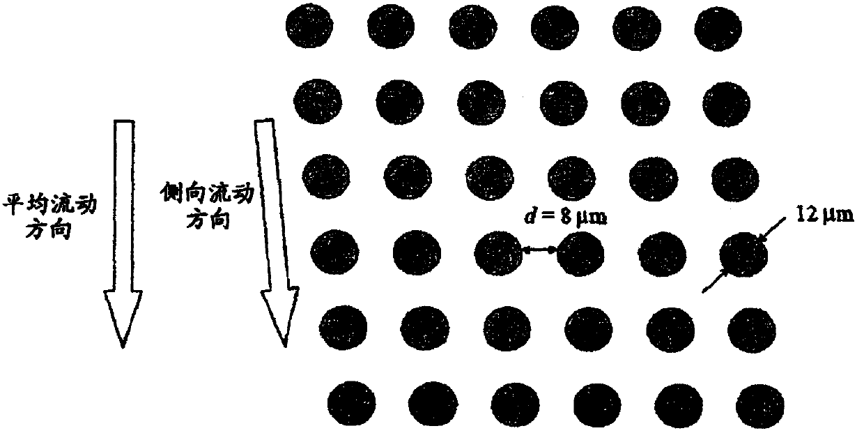
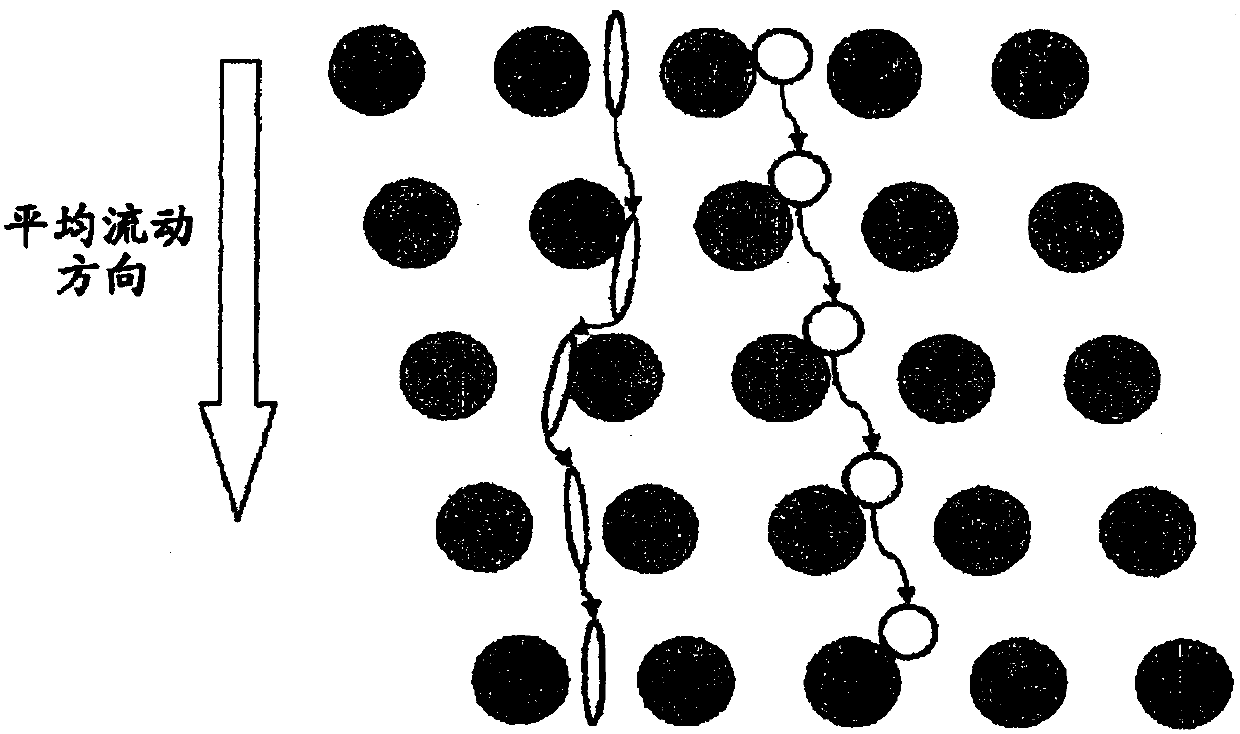
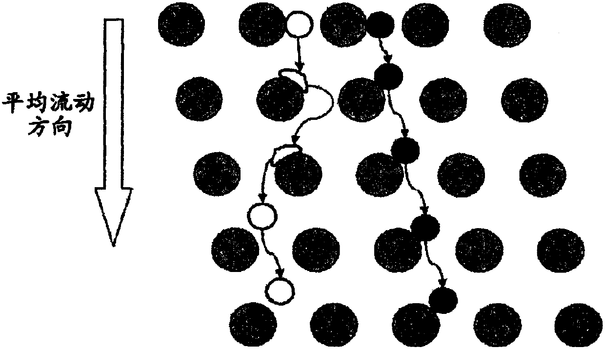
[0300] Figure IE shows a schematic diagram of an apparatus for isolating nucleated cells from fetal cord blood.

[0301] Size: 100mm×28mm×1mm

[0302] Array design: 3 segments, gap size = 18, 12 and 8 μm for the first, second and third segments, respectively.

[0303] Device Fabrication: Arrays and channels are fabricated in silicon using standard photolithography and deep silicon reactive etch techniques. The etching depth is 140 μm. Vias for fluid entry were made using KOH wet etching. The silicon substrate was sealed on the etched surface using a blood-compatible pressure-sensitive adhesive (9795, 3M, St Paul, MN) to create an airtight fluid channel.

[0304] Device Assembly: The device is mechanically coupled to a plastic manifold with external fluid reservoirs to deliver blood and buffer to the device and extract the resulting fractions.

[0305] (00217] Device Operation: An external pressure source was us...

Embodiment 2

[0309] Example 2. Isolation of fetal cells from maternal blood

[0310] The apparatus and methods detailed in Example 1 were used in combination with immunomagnetic affinity enrichment techniques to demonstrate the feasibility of isolating fetal cells from maternal blood.

[0311] Experimental Conditions: Blood was collected from a consenting maternal donor carrying a male fetus into K 2 EDTA vacutainer tubes (366643, Becton Dickinson, Franklin Lakes, NJ) followed by elective termination of pregnancy immediately thereafter. Undiluted blood was processed using the apparatus described in Example 1 at room temperature and within 9 hours of drawing. The nucleated and non-nucleated cells (erythrocytes and platelets) of the blood were separated, and the plasma was passed into a calcium-free and Magnesium Dulbecco's phosphate-buffered saline (14190-144, Invitrogen, Carlsbad, CA) was in the buffer stream. Subsequently, the nucleated cell fraction was labeled with anti-CD71 microbea...

Embodiment 3

[0314] Example 3. Confirmation of the presence of male fetal cells in enriched samples

[0315] Confirmation of the presence of male fetal cells in the enriched samples was performed using qPCR with primers specific for DYZ, a marker repeated in high copy number on the Y chromosome. Following enrichment of fnRBCs by any of the methods described herein, the resulting enriched fnRBCs were partitioned by splitting the sample into 100 PCR wells. Enriched samples can be screened by FISH for the presence of any fnRBCs containing aneuploidy of interest prior to partitioning. Because of the low number of fnRBCs in maternal blood, only a fraction of wells contained a single fnRBC (other wells were expected to be negative for fnRBC). Cells were fixed in 2% paraformaldehyde and stored at 4°C. Cells in each partition were pelleted and resuspended in 5 μl PBS plus 1 μl 20 mg / ml proteinase K (Sigma #P-2308). Cells were lysed by incubation at 65°C for 60 minutes, followed by inactivatio...


PUM


Abstract

The present invention provides systems, apparatuses, and methods to detect the presence of fetal cells when mixed with a population of maternal cells in a sample and to test fetal abnormalities, e.g.aneuploidy. The present invention involves labeling regions of genomic DNA in each cell in said mixed sample with different labels wherein each label is specific to each cell and quantifying the labeled regions of genomic DNA from each cell in the mixed sample. More particularly the invention involves quantifying labeled DNA polymorphisms from each cell in the mixed sample.

Description

[0001] cross reference [0002] This application is based on the divisional application of Chinese Patent Application No. 200780030309.3. This application claims the benefit of U.S. Provisional Application No. 60 / 804,819, filed June 14, 2006, and U.S. Provisional Application No. 60 / 820,778, filed July 28, 2006, which applications are incorporated herein by reference. Background of the invention [0003] Analysis of specific cells can provide insight into a variety of diseases. These assays provide non-invasive tests for the detection, diagnosis and prognosis of diseases such as cancer or fetal abnormalities, thereby eliminating the risk of invasive diagnostics. With regard to fetal abnormalities, available prenatal diagnostics, such as amniocentesis and chorionic villus sampling (CVS), can be harmful to both mother and fetus. Pregnant women undergoing amniocentesis have an increased miscarriage rate of 0.5-1%, and with CVS this figure is slightly higher. Because of the uniq...

Claims


Application Information

Patent Timeline
Patent Type & Authority Applications(China)
IPC IPC(8): C12Q1/6883C12Q1/6844C12Q1/6809
CPCC12Q1/6883C12Q2600/156C12Q2600/158C12Q2600/16B01L3/502761C12Q1/6809G01N2015/1006Y10T436/143333G01N2015/1029C12Q1/6869C12Q1/6881
Inventor D·肖马克M·富克斯N·X·克鲁格M·托纳D·格雷R·卡普尔Z·王
Owner VERINATA HEALTH INC
Why Eureka
Try Eureka