B7-H1 (CD274) Antagonists Induce Apoptosis of Tumor Cells

a tumor cell and apoptosis technology, applied in the field of b7h1, can solve the problems of adverse side effects, and achieve the effects of restoring the killing of cancer cells, increasing the resistance of cancer cells, and inhibiting, reducing, or blocking b7-h1 mediated signal transduction

Inactive Publication Date: 2010-11-11
THE JOHN HOPKINS UNIV SCHOOL OF MEDICINE
View PDF5 Cites 32 Cited by
  • Summary
  • Abstract
  • Description
  • Claims
  • Application Information

AI Technical Summary

Benefits of technology

[0009]Compositions and methods for restoring killing of cancer cells are provided. Preferably, the compositions are administered to a subject in an effective amount to antagonize, inhibit, reduce, or block B7-H1 mediated signal transduction in cancer cells expressing B7-H1. It has been discovered that B7-H1 transmits an anti-apoptotic signal in cancer cells that increases the resistance of the cancer cells to CTL mediated cytolysis and to Fas induced cell death. It is believed that blocking the transmission of the anti-apoptotic signal by B7-H1 increases the susceptibility of the cancer cells to apoptosis and CTL cytolysis thereby enhancing the death of cancer cells. Preferred compounds or B7-H1 antagonists include antibodies that bind to B7-H1, B7-H1 receptors, or ligands of B7-H1. Representative ligands or counter-receptors include B7-1 and PD-1. Additional B7-H1 antagonists include small molecules, for example small molecules that bind the cytoplasmic portion of B7-H1 or the extracellular portion of B7-H1 and inhibit, reduce, block or interfere with B7-H1 signal transduction.

Problems solved by technology

Although many cancer therapies are available, they are usually associated with adverse side-effects.

Method used


Image

  • B7-H1 (CD274) Antagonists Induce Apoptosis of Tumor Cells
  • B7-H1 (CD274) Antagonists Induce Apoptosis of Tumor Cells
  • B7-H1 (CD274) Antagonists Induce Apoptosis of Tumor Cells

Examples


example 1

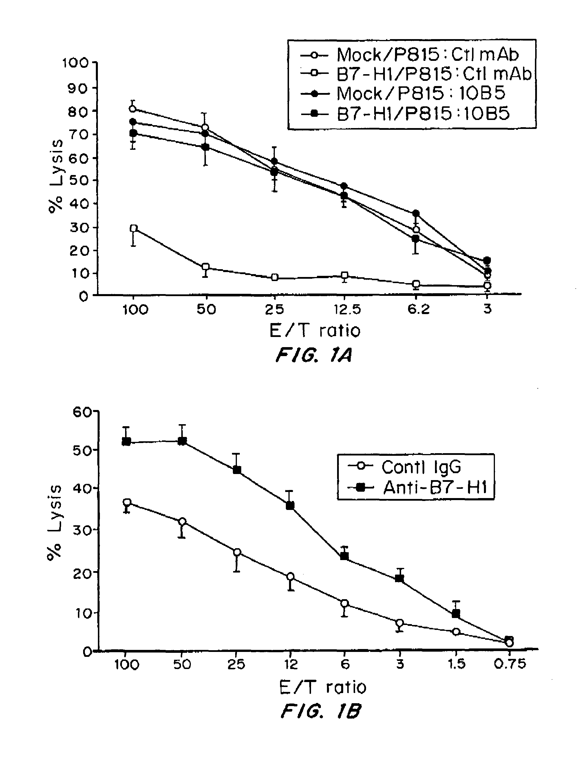
Expression of B7-H1 Confers Resistance of Tumor Cells to Specific CTL-Mediated Lysis

Mice and Tumor Lines

[0129]Female DBA / 2, C57BL / 6 (B6) mice were purchased from the National Cancer Institute (Frederick, Md.). Age-matched mice, 6 to 10 weeks old, were used for all experiments. 2C transgenic mice (a gift from Dr. Larry Pease, Mayo Clinic, Rochester, Minn.) and PD-1 deficient mice in B6 background (a gift from Dr. Tasuko Honjo, Kyoto University, Kyoto, Japan) were described previously16. The 2C×PD-1KO mice were obtained by backcrossing between 2C transgenic mice and PD-1KO mice. All mice were maintained in the Animal Facility at Johns Hopkins Hospital under approved protocol by the Institutional Animal Care and Use Committee. P815 mastocytoma cells were purchased from the American Type Culture Collection (Rockville, Md.). A subclone, which does not express B7-H1, even in the presence of IFN-gamma or activated T cells, was selected before transfection. Stable transfectants of P815 line...

example 2

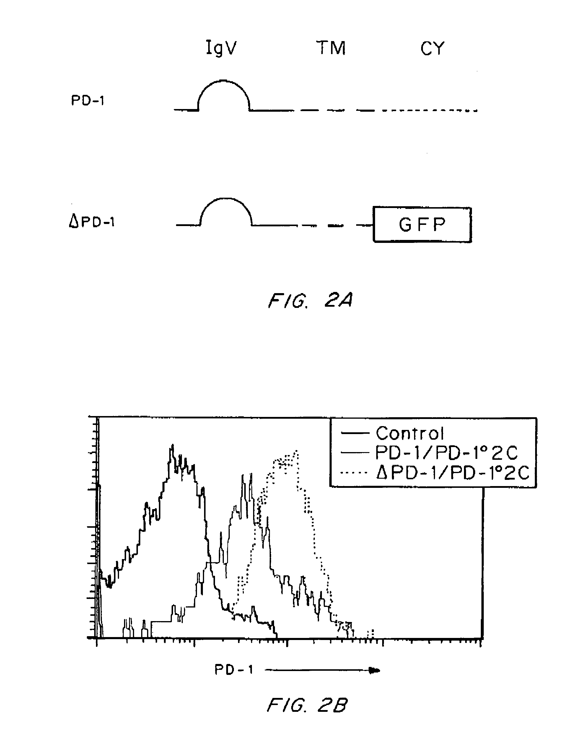
PD-1 Signaling is not Required for Molecular Shielding of Tumor Cells from T Cell Killing

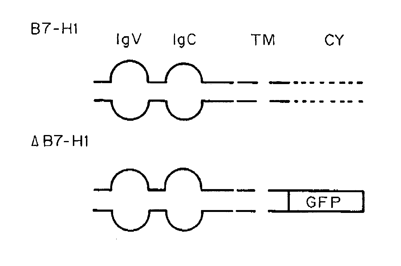
[0135]A previous study showed that interaction between B7-H1 and PD-1 is required for the formation of a molecular shield (Hirano, F., et al. Cancer Res., 65:1089-1096 (2005)). It is widely accepted that signaling from B7-H1 to PD-1 on T cells delivers a negative signal to suppress T cell responses (Freeman, G. J., et al. J Exp Med., 192:1027-1034 (2000)). Therefore, one possible explanation for molecular shield is tumor-associated B7-H1 signaling through PD-1 into T cells, leading to transient loss of T cell cytolytic activity. To test this possibility, a truncated PD-1 was prepared as described in Example 1, in which the intracellular domain of PD-1 was replaced by green fluorescence protein (GFP) gene (FIG. 2A) to eliminate its intracellular signaling. This truncated PD-1 (ΔPD-1) as well as wild type PD-1 (PD-1) was inserted into lentiviral vectors for efficient T cell transduction. To elimin...

example 3

Intracellular Domain of B7-H1 is Required for Molecular Shielding

Monoclonal Antibodies, Fusion Proteins and Flow Cytometry Analysis

[0136]Purified mAb against mouse H-2Dd and CD95 (clone Jo2) was purchased from PharMingen (San Diego, Calif.) and H-2Ld from BioLegend (San Diego, Calif.). Rat mAb (clone TY25) against B7-DC, FITC- or phycoerythrin-conjugated goat anti-mouse antibodies and FITC-conjugated goat anti-hamster antibodies were purchased from eBioseience (San Diego, Calif.). An Armenian hamster mAb (clone 10135) against mouse B7-H1 (Hirano, F. et al., Cancer Res., 65:1089-1096 (2005)), a hamster mAb (clone G4) against mouse PD-1 (Hirano, F. et al., Cancer Res., 65:1089-1096 (2005)), a rat mAb (clone 2A) against mouse CD137 (Wilcox, R. A., et al., J Clin Invest., 109:651-659 (2002)), mouse PD-1Ig fusion protein (Hirano, F. et al., Cancer Res., 65:1089-1096 (2005)) and mouse B7-H1Ig fusion protein (Hirano, F. et al., Cancer Res., 65:1089-1096 (2005)) were all described previousl...


PUM

PropertyMeasurementUnit
molecular weightaaaaaaaaaa
solubleaaaaaaaaaa
surface molecular signaturesaaaaaaaaaa

Abstract

Compositions and methods for restoring killing of cancer cells are provided. Preferably, the compositions are administered to a subject in an effective amount to antagonize, inhibit, reduce, or block B7-H1 mediated signal transduction in cancer cells expressing B7-H1. It has been discovered that B7-H1 transmits an anti-apoptotic signal in cancer cells that increases the resistance of the cancer cells to CTL mediated cytolysis and to Fas induced cell death. It is believed that blocking the transmission of the anti-apoptotic signal by B7-H1 increases the susceptibility of the cancer cells to apoptosis and CTL cytolysis thereby enhancing the death of cancer cells. Preferred compounds or B7-H1 antagonists include antibodies that bind to B7-H1, B7-H1 receptors, or ligands of B7-H1 such as PD-I or B7-1. Additional B7-H1 antagonists include small molecules, for example small molecules that bind the cytoplasmic portion of B7-H1 or the extracellular portion of B7-H1 and inhibit, reduce, block or interfere with B7-H1 signal transduction. Methods for treating one or more symptoms associated with cancer or hyperproliferation are also provided.

Description

GOVERNMENT SUPPORT[0001]This invention was made with government support under grant numbers CA85721, CA97085 and CA113341 awarded by National Institutes of Health. The government has certain rights in the invention.FIELD OF THE INVENTION[0002]The invention is generally related to methods and compositions for treating or preventing aberrant cellular proliferation.BACKGROUND OF THE INVENTION[0003]Cancer has an enormous physiological and economic impact. For example a total of 1,437,180 new cancer cases and 565,650 deaths from cancer are projected to occur in the United States in 2008 (Jemal, A., Cancer J Clin, 58:71-96 (2008)). The National Institutes of Health estimate overall costs of cancer in 2007 at $219.2 billion: $89.0 billion for direct medical costs (total of all health expenditures); $18.2 billion for indirect morbidity costs (cost of lost productivity due to illness); and $112.0 billion for indirect mortality costs (cost of lost productivity due to premature death). Althoug...

Claims


Application Information

Patent Timeline
Patent Type & Authority Applications(United States)
IPC IPC(8): A61K39/395A61K38/02A61P35/00
CPCC07K16/2827A61K2039/505A61P35/00A61P37/04
Inventor CHEN, LIEPING
Owner THE JOHN HOPKINS UNIV SCHOOL OF MEDICINE
Why Eureka
Try Eureka