Method of maxillary sinus bone grafting for placement of implant

a maxillary sinus bone and implant technology, applied in the field of maxillary sinus bone grafting for implant placement, can solve the problems of long implant that cannot be placed into lack of bone in which the implant can be placed, and difficulty in carrying out the implantation of the upper posterior teeth, etc., to achieve fast, safe, and alleviate the pain of the patien

Active Publication Date: 2010-11-18
LEE DAL HO
View PDF17 Cites 19 Cited by
  • Summary
  • Abstract
  • Description
  • Claims
  • Application Information

AI Technical Summary

Benefits of technology

[0033]According to the present invention, upon placement of the implant, the vertical hole can be formed and enlarged with ease in the maxillary sinus floor, the maxillary membrane can be easily lifted in an always-stable state using the piezotome and gel type bone graft material while preventing the membrane from becoming damaged, and even the bone graft material of coarse bone meal can be uniformly diffused and infused, thereby ensuring that the implant is placed in a fast, safe manner while allowing a patient to keep relaxed and alleviating the pain the patient feels, resulting in shortening a curing time and maximizing effects of the placement of the implant.

Problems solved by technology

However, since many of the patients have an oral structure in which is difficult to place an implant, some of dentists tend to avoid treating such patients.
This is because the upper posterior teeth are weak and the maxillary sinus exists near the upper posterior teeth, so that a long implant cannot be placed into the upper posterior teeth.
That is, since the maxillary sinus existing in the maxillary is a space surrounded by a mucous membrane, when a tooth is lost, the maxillary sinus physiologically falls down and widens greatly and the downward extension of the maxillary sinus and the osteolysis owing to the loss of teeth occur, resulting in a lack in the amount of bone into which the implant can be placed, making it difficult to carry out the implantation for the upper posterior teeth.
However, such a lateral window technique is very difficult to do and is a careful, time-intensive process because especially during the windowing the maxillary sinus is often punctured in the process of using the round bur, the piezo sawtooth, or the diamond tip, resulting in the symptoms of swelling and pain.
However, despite that such a crestal approaching technique has the advantage of few occurrences of edema occurring in patients after operation because of the narrow target section of the operation, a long time is spent performing the operation since a dentist cannot directly observe the maxillary membrane so that he must operate very carefully while checking the target section using X-rays, a patient feels very unpleasant owing to the striking which is done during the operation, and particularly when the drill comes into point contact with the maxillary membrane as the tip thereof rotates is a vertical load concentrated upon the contacted maxillary membrane thereby easily fracturing the same.
That is, while the special drill or reamer does not fracture the sinus floor of the maxillary membrane since it is not hammered (so that it causes only a little pain to the patient), it has a very small operating force so as to be insufficient to puncture the maxillary membrane, so that it takes too long of a time to remove the same.
Further, if the maxillary membrane is of an irregular shape or is severely inclined, the drill or reamer can puncture the maxillary membrane while removing the sinus floor, making it difficult to maintain in place the bone graft material for forming the bone material.
This allows the implant to be exposed to the outside in the maxillary membrane without being placed in the bone, thereby increasing the possibility of infection and degrading the capability of supporting the implant because of an insufficient amount of support bone.
However, carrying out the placement of an implant using such a conventional piezoelectric device has problems in that even experienced dentists in performing the placement find it difficult to lift the maxillary membrane in an always-safe state without damaging it, and in that in case of puncturing the vertical hole into which the implant is placed, a narrow drill should be used, and in that while in the process of placing the bone graft material into the section of the root of the artificial tooth, the bone graft material is placed into the section while being loaded in a bone carrier, in case of using the bone carrier, a tip size of a protrusion in front of a syringe barrel thereof is too large or small for the punctured hole, so that during injection the bone graft material escapes outside or is not uniformly dispensed into the hole to a sufficient depth.
Particularly, in case of the placement of the bone graft material into an upper molar using a common syringe, problems arise because inconvenience is caused by the placement, a lot of time is required for the placement, the placement is difficult to carry out and thus efficient placement cannot be implemented in an easy state, and because of the unnatural forcible placement, whereby if the bone graft material is of coarse particles, the graft and lifting become difficult owing to the roughness of the particles, which may cause the maxillary membrane and also the gums to be damaged, and further because although coarse bone meal is effectively used in the placement of the bone graft material rather than fine bone meal, such coarse bone meal cannot be used in common syringes.

Method used


Image

  • Method of maxillary sinus bone grafting for placement of implant
  • Method of maxillary sinus bone grafting for placement of implant
  • Method of maxillary sinus bone grafting for placement of implant

Examples


Embodiment Construction

[0046]Description will now be made of exemplary embodiments of the present invention with reference to the accompanying drawings.

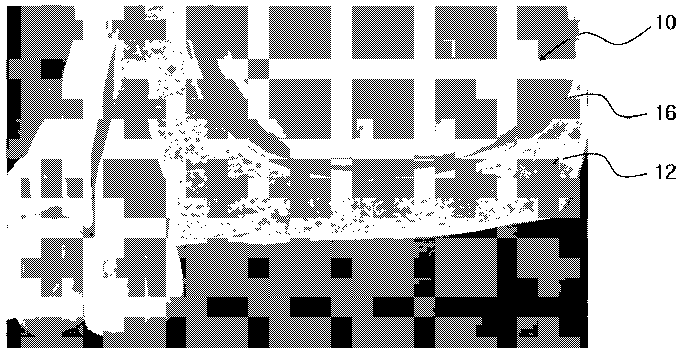
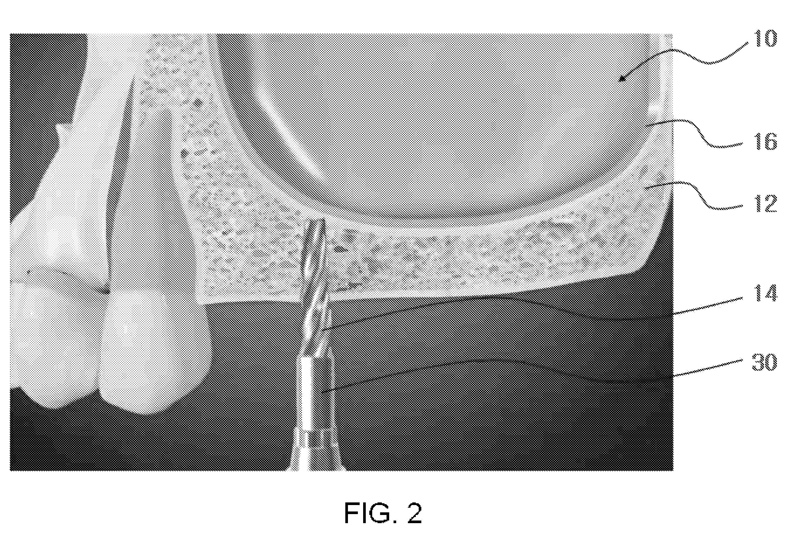
[0047]As illustrated in FIGS. 1 to 14, a method of maxillary sinus bone grafting for placement of an implant according to the present invention includes a first step of forming a vertical hole in a maxillary sinus floor of a maxillary sinus for placement of the implant, a second step of lifting a maxillary membrane via the vertical hole of the maxillary sinus floor, a third step of enlarging a diameter of the vertical hole with the maxillary membrane lifted, a fourth step of inserting the bone graft material via the enlarged vertical hole, a fifth step of pushing the inserted bone graft material in a space between maxillary sinus floor and the maxillary membrane, and a sixth step of after the bone graft material has been cured, placing the implant into the vertical hole.

[0048]First, in FIG. 1, upon placement of an implant in an upper posterior region, if a...


PUM


Abstract

A method of maxillary sinus bone grafting for placement of an implant in which upon placement of the implant, a vertical hole is formed and enlarged with ease in a maxillary sinus floor, a maxillary membrane is easily lifted in an always-stable state using a piezotome and gel type bone graft material while preventing the membrane from becoming damaged, and even the bone graft material of coarse bone meal is uniformly diffused and infused, thereby ensuring that the implant is placed in a fast, safe manner while allowing a patient to keep relaxed and alleviating the pain the patient feels, resulting in shortening a curing time and maximizing effects of the placement of the implant.

Description

BACKGROUND OF THE INVENTION[0001]1. Field of the Invention[0002]The present invention relates to a method of maxillary sinus bone grafting for implant placement by which an artificial tooth is effectively placed into the mouth of a patient, and more particularly, to a method of maxillary sinus bone grafting for implant placement by which an implant can be implanted in a fast, safe manner while allowing a patient to stay relaxed, resulting in maximizing effects of the placement.[0003]2. Description of the Related Art[0004]While an implant generally refers to replacement of a specific section of a human body, it means grafting of an artificial tooth in dentistry. The implant is a hi-tech operation method by which upon the occurrence of the loss of the root of a tooth, an artificial dental root made of titanium, which is not rejected by the human body, is implanted in the alveolus from which the tooth was removed, and an artificial tooth then is fixed thereto to recover an original fun...

Claims


Application Information

Patent Timeline
Patent Type & Authority Applications(United States)
IPC IPC(8): A61C13/00
CPCA61C8/0092A61C8/0089
Inventor LEE, DAL HO
Owner LEE DAL HO
Why Eureka
Try Eureka