

Method for transducing cells with primary cilia
a technology of primary cilia and aav, which is applied in the direction of biocide, animal repellents, drug compositions, etc., can solve the problems of cyst formation, cell dysfunction, disorganization and death of these cells,
- Summary
- Abstract
- Description
- Claims
- Application Information
AI Technical Summary
Problems solved by technology
Method used
Image

Examples
example 1
Effects of Viral Infection and Mutant EFEMP1 Expression On hfRPE Cells
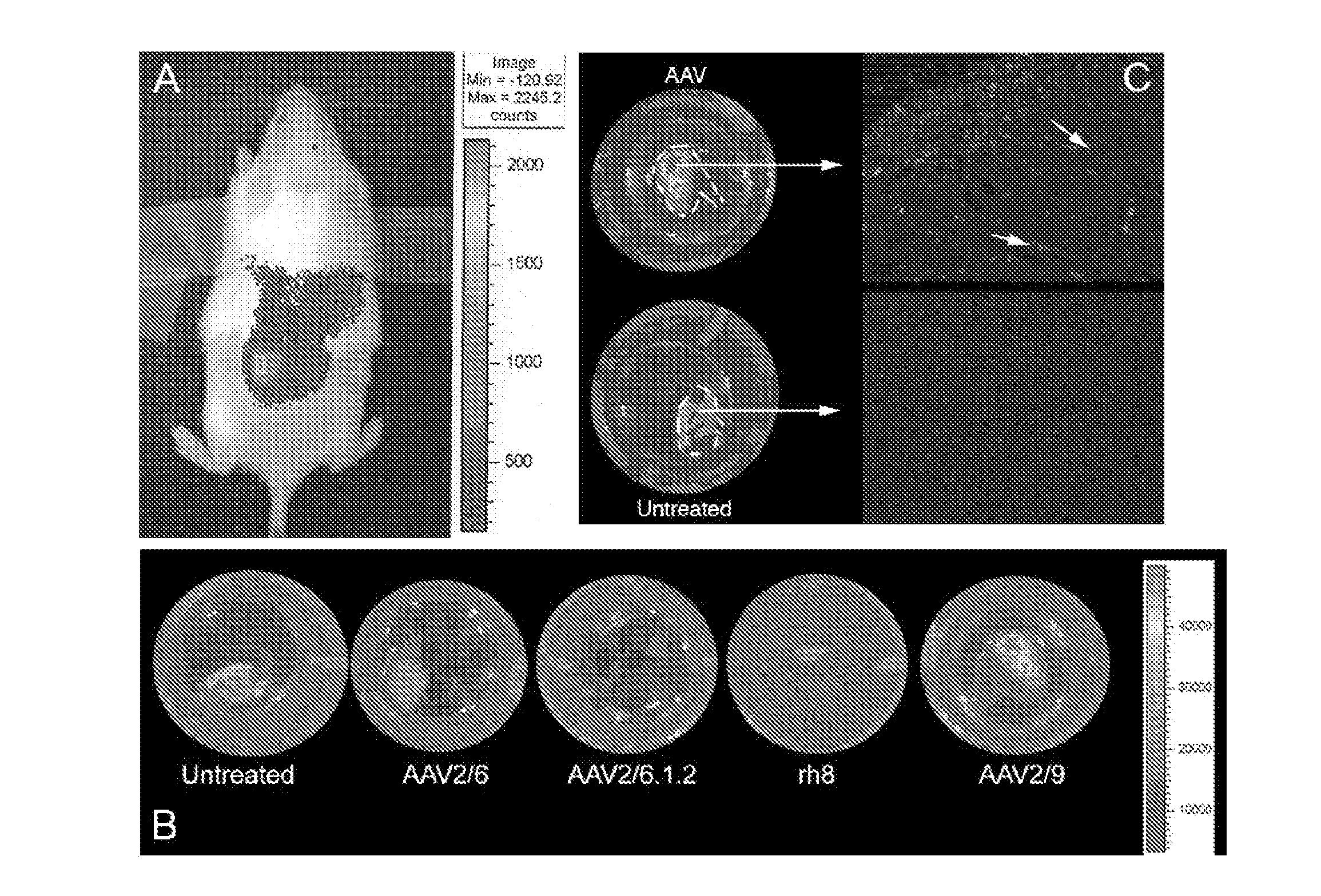
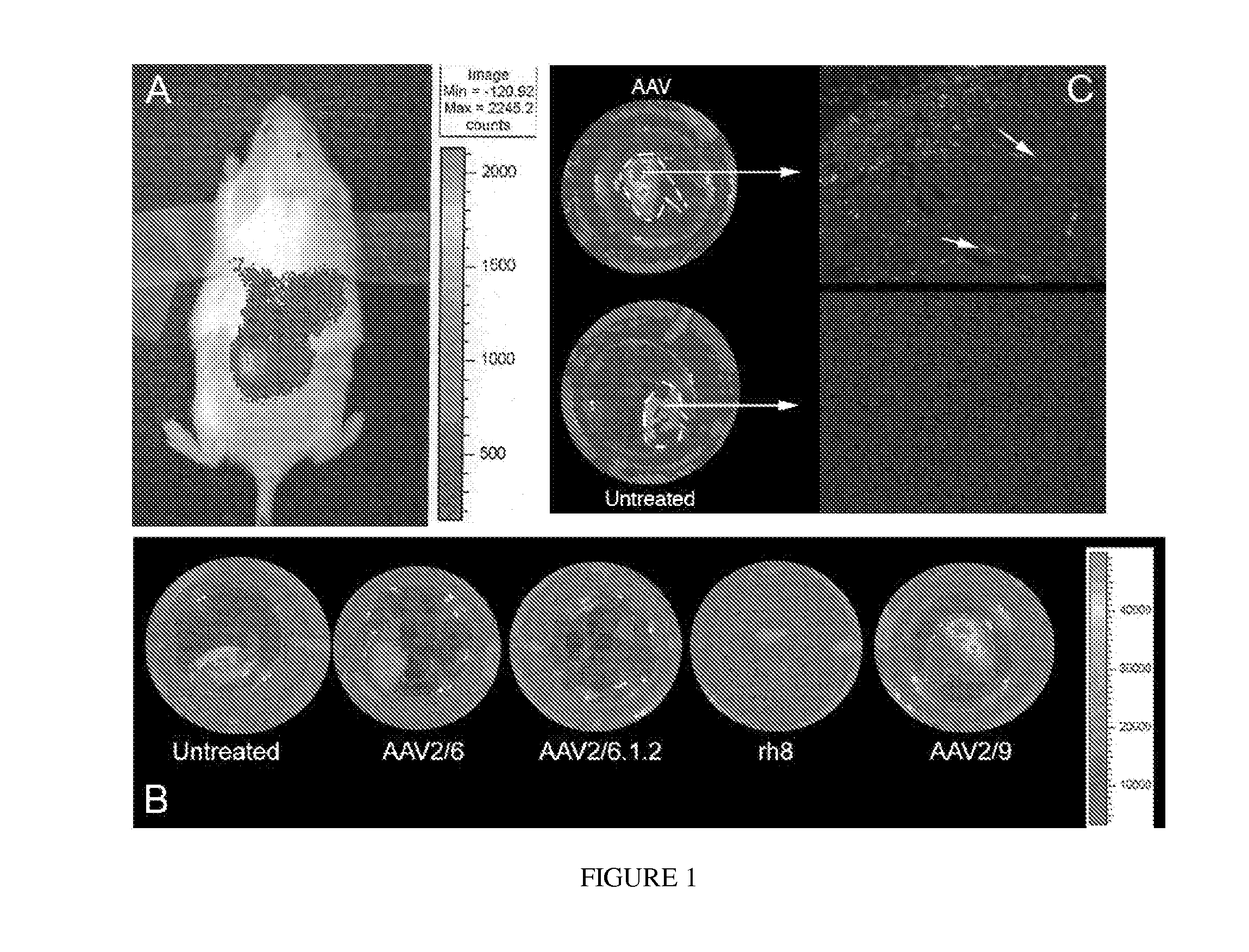
[0140]In order to express EFEMP1wt-FLAG and EFEMP1mut-FLAG in hfRPE cells, genes encoding these fusion proteins were delivered by infecting the cells with recombinant AAV serotype 2 / 1 vectors (rAAV2 / 1). This serotype of rAAV was shown to transduce RPE cells at a high level in vivo. Using the AAV2 / 1.VMD2.EGFP vector, it was found that an MOI of 106 vg / cell was sufficient to transduce 80-90% of RPE cells in culture. Using this MOI, we infected differentiated hfRPE cell monolayers with AAV2 / 1 vectors expressing EGFP, hEFEMP1wt-FLAG, or hEFEMP1mut-FLAG under the control an RPE-specific promoter (VMD2). Cells were maintained for 3-4 weeks prior to analysis to allow for maximal activation of the VMD2 promoter.
[0141]Then transduced hfRPE cells were analyzed to determine if any adverse effects of viral infection or overexpression of EFEMP1mut could be detected. Light microscopy after eight weeks post-infection showed that...
example 2
Establishment of hfRPE Monolayers
[0143]RPE cells were removed from human fetal eyes at 15-17 weeks of gestation and cultured in hfRPE growth media for at least four weeks. The cells were then removed, seeded on to Transwell-Clear membranes, and grown for another four weeks until they gained a cobblestoned appearance consistent with a differentiated epithelial cell monolayer. Pigmentation, which is markedly diminished immediately following seeding on Transwell filters, increased in these cells with time. Transepithelial resistance (TER) readings were found to increase during this time, suggesting that the monolayers had differentiated and that tight junctions had formed (FIG. 3). Monolayers with TER>200 Qcm2 are well-differentiated, as evidenced by their morphological characteristics and ATP-induced fluid transport properties, and were used for further analyses.
example 3
Polarized Secretion of EFEMP1 by hfRPE Cells
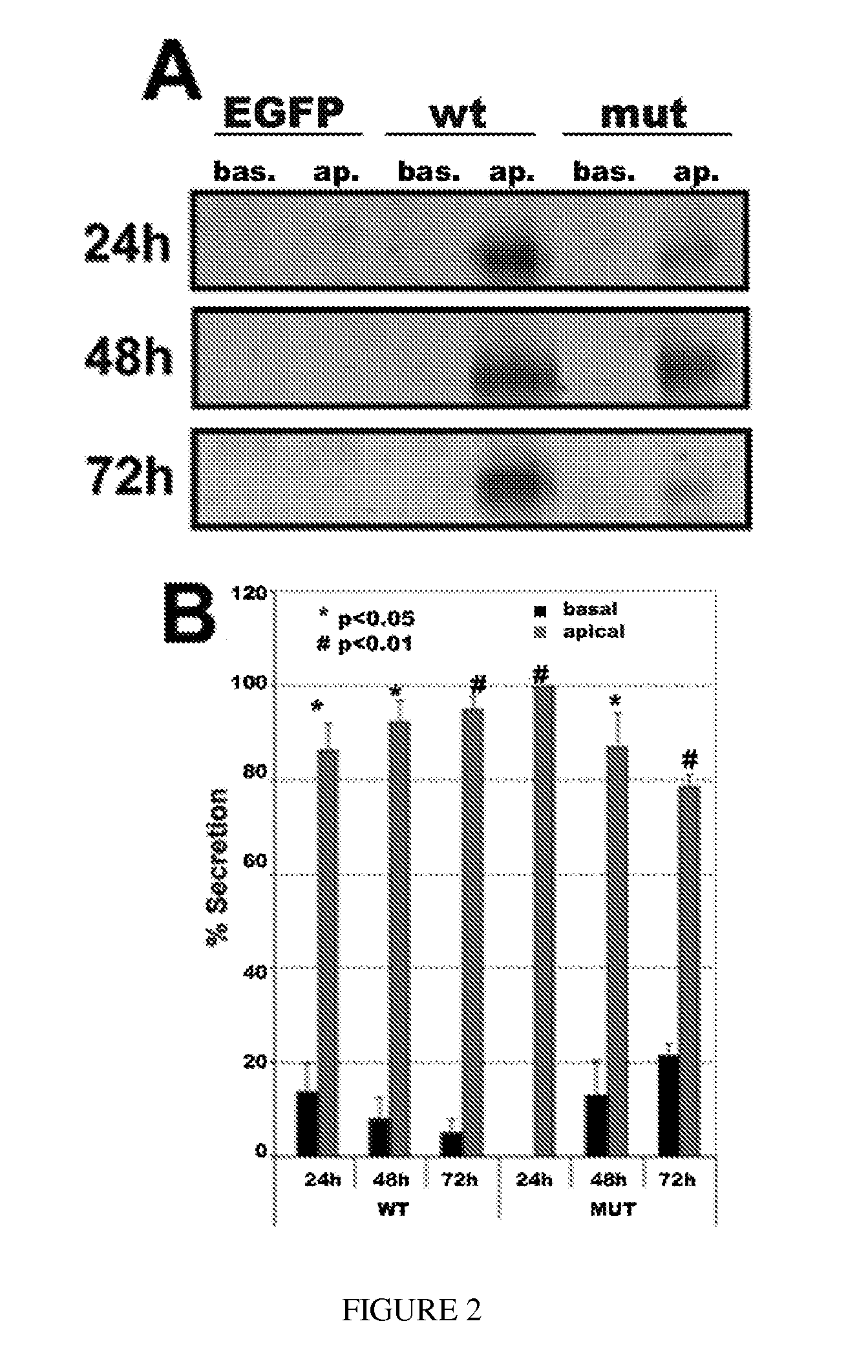
[0144]In individuals with ML, localization of the protein is altered, and EFEMP1 resides outside the basolateral membrane of the RPE, adjacent to drusen. To test whether RPE cells secrete EFEMP1 apically, and that the R345W mutation in the protein that leads to ML misdirects this secretion basolaterally, hfRPE cell monolayers were transduced with rAAV2 / 1 vectors expressing EFEMP1wt-FLAG or EFEMP1mut-FLAG, and secretion of the proteins into the apical and basolateral media at various timepoints was analyzed. As a control epithelial cell line, MDCK cell monolayers transduced with AAV2 / 5 vectors expressing EGFP, EFEMP1wt-FLAG, or EFEMP1mut-FLAG were used. Apical and basolateral media was removed from the cells, replaced with serum-free medium, and cultured for 24-72 hours. EFEMP1-FLAG was immunoprecipitated from the media and detected by immunoblotting. As shown in FIG. 6A (left panels), contrary to our hypothesis, EFEMP1wt-FLAG was secreted ...
PUM
| Property | Measurement | Unit |
|---|---|---|
| pH | aaaaa | aaaaa |
| pH | aaaaa | aaaaa |
| total volume | aaaaa | aaaaa |
Abstract
Description
Claims
Application Information