Medical diagnosis support device, image processing method, image processing program, and virtual microscope system

a medical diagnosis and support device technology, applied in the field of medical diagnosis support devices, image processing methods, image processing programs, virtual microscope systems, can solve the problems of difficult to make judgment on translocation, difficult to carry out multiple staining of the same specimen, and difficult to perform multiple staining with respect to the same specimen, etc., to achieve easy and precise determination of chromosome abnormality and/or gene amplification, easy and precise determination of markers

Inactive Publication Date: 2010-12-23
OLYMPUS CORP +1
View PDF18 Cites 17 Cited by
  • Summary
  • Abstract
  • Description
  • Claims
  • Application Information

AI Technical Summary

Benefits of technology

[0056]According to the present invention, in a case where respective cells are in different conditions, a user such as a doctor can easily confirm a marker by his / her eyes because only the marker is identified and displayed. Further, a user such as a doctor can easily confirm whether a marker state is positive / negative by his / her eyes because a positive marker state and a negative marker state can be easily determined, respectively. As a result, it is possible to easily and precisely determine chromosome abnormality and / or gene amplification related to cancer or genetic disease.
[0057]Further, according to the present invention, a user such as a doctor can easily confirm a cell by his / her eyes because a marker and a cell are each extracted in an intensified state, respectively, and a cell state of the extracted cell is identified and displayed, based on the extracted marker. Yet further, a user such as a doctor can easily confirm whether a cell state is positive / negative by his / her eyes because a positive cell state and a negative cell state are identified and displayed such that these two states can be easily determined, respectively. As a result, it is possible to easily and precisely determine chromosome abnormality and / or gene amplification related to cancer or genetic disease.

Problems solved by technology

It has been conventionally difficult to carry out multiple staining with respect to the same one specimen by CISH.
Therefore, making judgment on a marker in CISH is more difficult in FISH.
In the case of Dual CISH, in particular, since the two colors of multiple staining are mixed, markers for each staining cannot be easily identified and thus judgment on translocation cannot be made easily.

Method used


Image

  • Medical diagnosis support device, image processing method, image processing program, and virtual microscope system
  • Medical diagnosis support device, image processing method, image processing program, and virtual microscope system
  • Medical diagnosis support device, image processing method, image processing program, and virtual microscope system

Examples


first embodiment

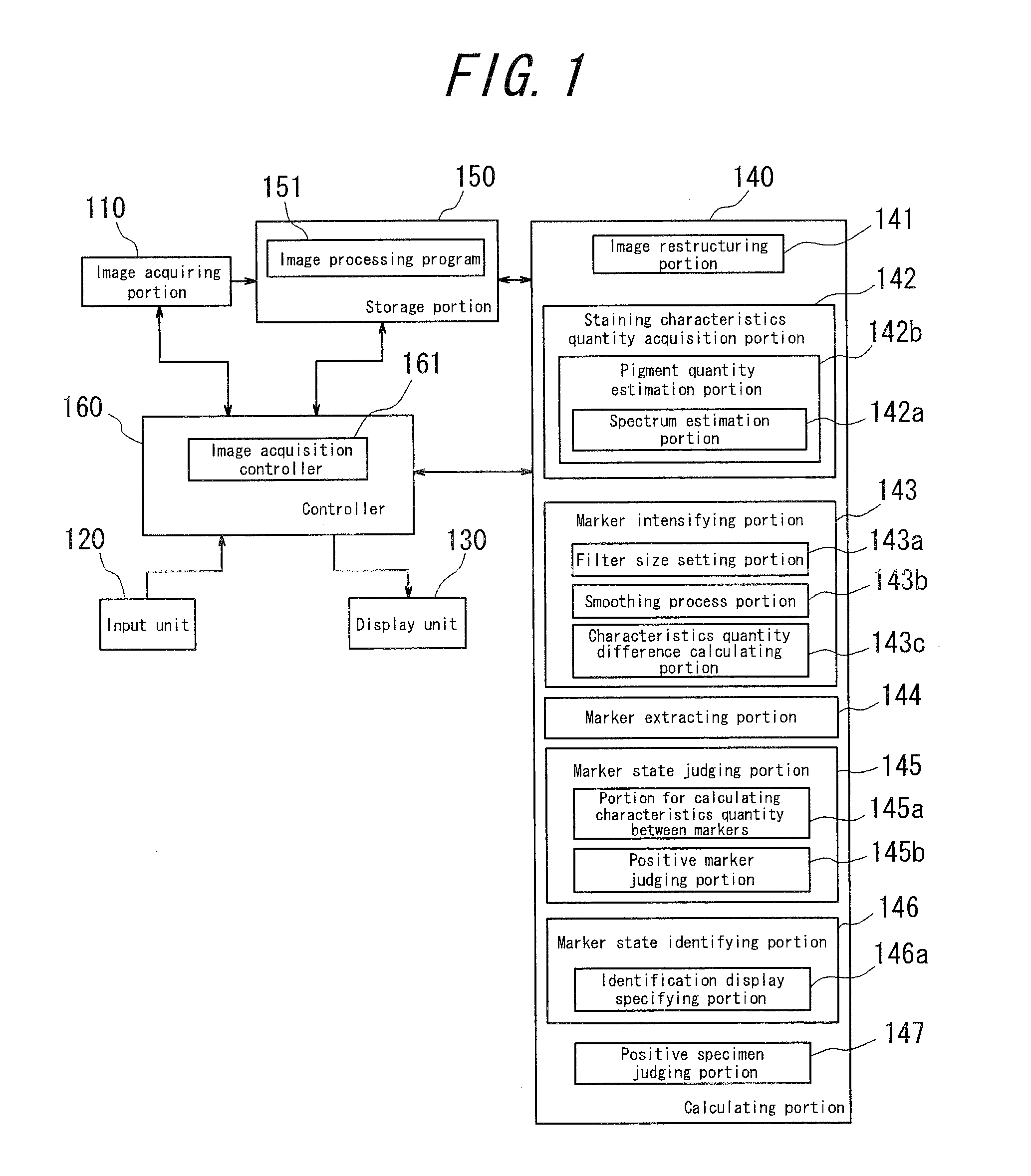
[0085]FIG. 1 is a block diagram showing a functional constitution of main parts of a medical diagnostics support device according to a first embodiment of the present invention. The medical diagnosis support device is structured to include a computer such as a personal computer and provided with an image acquiring portion 110 including a microscope, an input portion 120, a display portion 130, a calculation portion 140, a storage portion 150, and a controller 160 for controlling the respective portions.

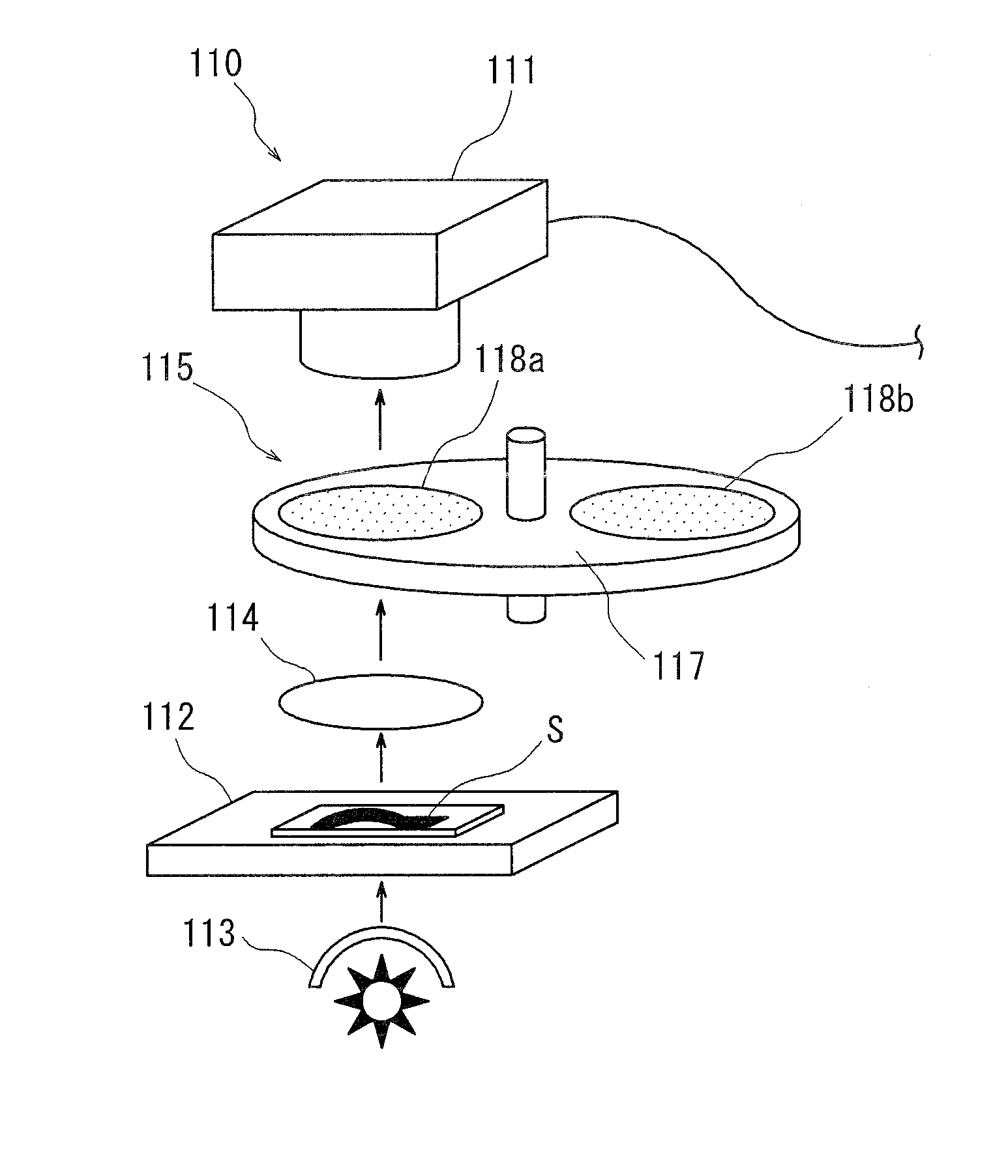
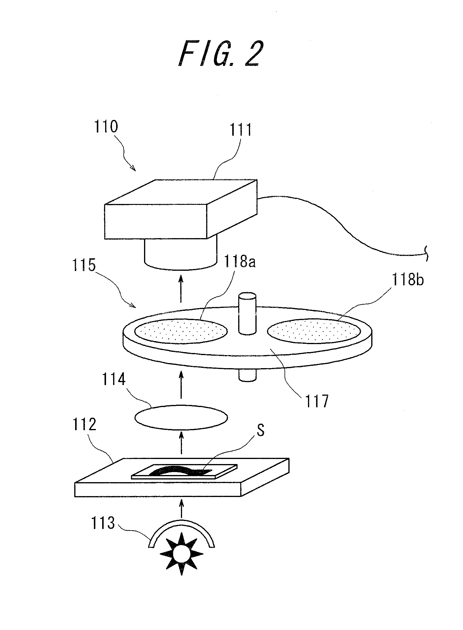
[0086]The image acquisition portion 110 acquires a multiband image (6 band image in the present embodiment) of a target stained specimen (which will be referred to as a “target specimen” hereinafter) by a microscope. FIG. 2 schematically shows a structure of main parts of the image acquisition portion 110. As shown in FIG. 2, the image acquisition portion 110 includes: a RGB camera 111 equipped with an image pickup element such as CCD (charge coupled devices) or CMOS (complementary me...

second embodiment

[0137]The medical diagnosis support device according to a second embodiment of the present invention differs from the aforementioned first embodiment in that the former employs as characteristics quantity between markers a ratio of an area where markers of respective stainings are superposed on each other, with respect to an area of a marker where no such superposition is observed, in judging a maker state at step S111 in FIG. 7. Specifically, there is employed a ratio of an area where marker I and marker J are superposed on each other as shown in FIG. 18(b) with respect to an area of marker I or marker J as shown in FIG. 18(a). A normal or translocation state is then determined based on comparison of the area ratio with a predetermined threshold value, as described above.

[0138]The aforementioned area ratio can be obtained by following formula (23). In formula (23), overlay_ratei represents a superposed area ratio of marker I and overlay(x, y) represents whether the pixels are super...

third embodiment

[0140]The medical diagnosis support device according to a third embodiment of the present invention differs from the aforementioned first embodiment in that the former carries out the steps S103 to S109 in FIG. 7, based on multiband images of a target specimen S at respective depths acquired by the image acquisition portion 110, to extract a marker of each staining at the respective depths.

[0141]Thereafter, in step S111 of FIG. 7, the distance between the centers of gravity of two different markers at different depths is calculated as characteristics quantity between the markers by following formula (24), so that a normal or translocation marker state can be determined based on comparison of the distance between the centers of gravity thus calculated and a predetermined threshold value, as described above. FIG. 19 shows an example of “distanceij” as the distance between the centers of gravity of markers I, J at different depths (in z direction) calculated by formula (24). Since othe...


PUM


Abstract

The present invention provides a medical diagnosis support device, which is capable of acquiring information to support medical diagnosis for easily and precisely determining chromosome abnormality and / or gene amplification related to cancer or genetic disorder.The medical diagnosis support device for acquiring information to support medical diagnosis from an image of a specimen stained by multiple staining, the image is obtained by photographing the stained specimen with transmitted light, the device comprises: staining characteristics quantity acquisition means for acquiring characteristics quantity of each staining, based on a pixel value of the image of the stained specimen; marker intensifying means for intensifying a marker, based on the characteristics quantity of each staining thus acquired; marker extracting means for extracting the marker of each staining, based on the characteristics quantity in which the marker has been thus intensified; marker state judging means for judging a state of the marker, based on the marker of each staining thus extracted; and marker state identifying and displaying means for identifying and displaying the marker state, based on the judgment result.

Description

CROSS-REFERENCE TO RELATED APPLICATION[0001]The present application claims priority from Japanese Application No. 2009-145754, filed on Jun. 18, 2009, the content of which is incorporated herein by reference.BACKGROUND OF THE INVENTION[0002]1. Field of the Invention[0003]The present invention relates to a medical diagnosis support device, an image processing method, a image processing program and a virtual microscope system, for acquiring information to support medical diagnosis from a stained specimen image obtained by photographing a stained specimen.[0004]2. Description of Related Art[0005]Spectral transmittance spectrum is a physical quantity which represents a physical property inherent to a subject to be photographed. Spectral transmittance is a physical quantity which represents a ratio of transmitted light to incident light at a wavelength. Unlike color information such as RGB value which varies depending on changes in illumination light, the spectral transmittance is inform...

Claims


Application Information

Patent Timeline
Patent Type & Authority Applications(United States)
IPC IPC(8): G06K9/00
CPCG06K9/0014G06T7/0012G06T2207/30024G06T2207/10056G06T2207/10024G06V20/695
Inventor OTSUKA, TAKESHIYAMADA, TATSUKIISHIKAWA, YUICHITAKEUCHI, KENGO
Owner OLYMPUS CORP
Why Eureka
Try Eureka