

Detection of worsening renal disease in subjects with systemic lupus erythematosus
a systemic lupus and renal disease technology, applied in the field of treatment of systemic lupus erythematosus, can solve the problems of insufficient high-quality biomarkers, insensitive renal biomarkers, and inability to detect active sle nephritis early detection and diagnosis, so as to improve the diagnostic capability, improve the activity of renal disease activity, and improve the effect of diagnosis and determination
- Summary
- Abstract
- Description
- Claims
- Application Information
AI Technical Summary
Benefits of technology
Problems solved by technology
Method used
Image

Examples
example 1
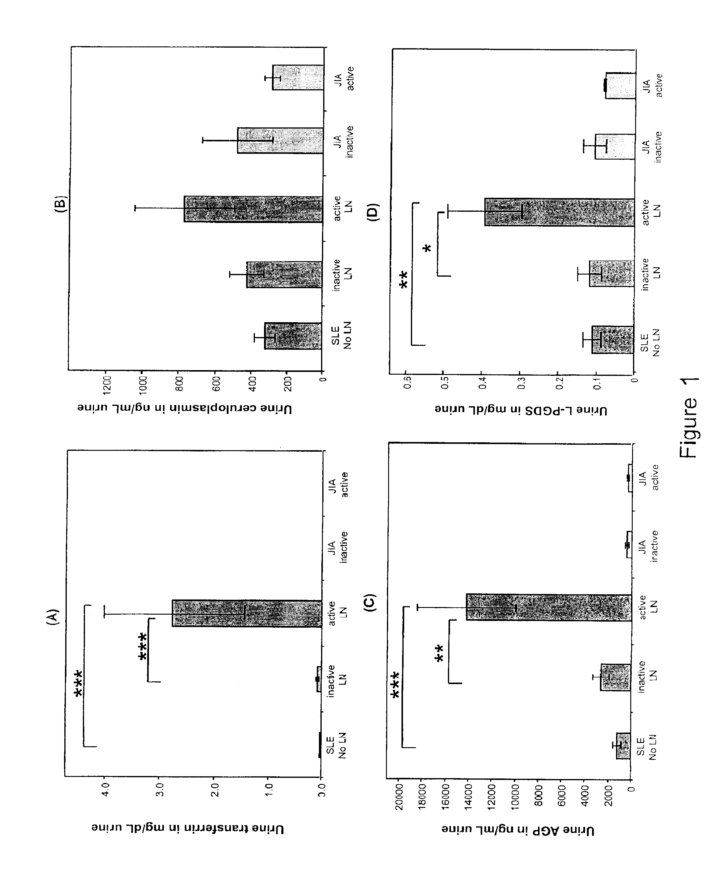
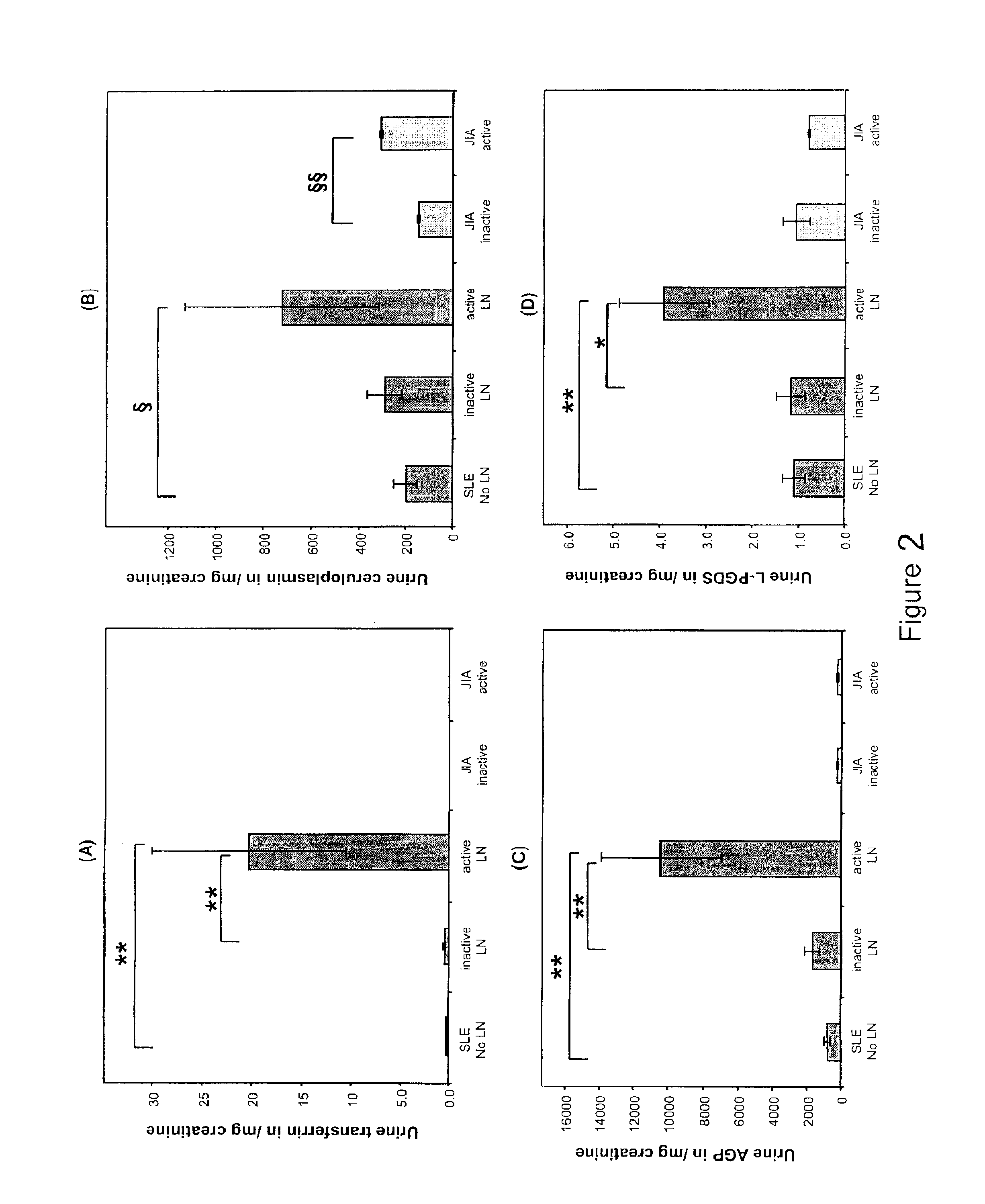
LN Panel Proteins
[0078]Children diagnosed with SLE (4) prior to the age of 16 years (n=98) were studied every 3 months for up to 18 months. At each study visit, blood and random spot urine samples for research were obtained, and information on the following laboratory measures was collected: BUN (urea), serum creatinine, serum complement levels C3 and C4, presence of anti-dsDNA antibodies, urine protein:creatinine ratio (normal <0.2), and creatinine clearance approximated according to the Schwartz formula. At the participating centers, kidney biopsies are obtained in SLE patients when abnormal urinalyses cannot be explained by mechanisms other than SLE. Thus all children without kidney biopsies were considered to have SLE without LN. The study was approved by the Institutional Review Board (IRB) of the Cincinnati Children's Hospital Medical Center, and the IRBs of all other participating centers, with informed consent obtained prior to any study-related procedures.
[0079]At each stud...
example 2
Urinary NGAL
[0098]Study—With approval of the institutional review boards of the participating institutions, children fulfilling American College of Rheumatology Classification Criteria for SLE prior to the age of 16 years (cSLE) were studied during routine visits to the pediatric rheumatology clinics. A convenience sample of 30 children diagnosed with juvenile idiopathic arthritis (JIA) was recruited as disease controls. Samples of healthy controls (n=50) were obtained from The Cincinnati Genomic Control Cohort assembled by the Cincinnati Children's Hospital Medical Center. Children diagnosed with cSLE were studied longitudinally every 3 months, while for JIA and healthy controls cross-sectional samples were available only. Random urine samples were collected on all subjects.
[0099]NGAL levels in urine were quantified by ELISA using an NGAL ELISA KIT (KIT 036; AntibodyShop, Grusbakken, Denmark) that specifically detects human NGAL. The assay was performed as per manufacturer's protoc...
example 3
Urinary NGAL in Worsening LN
[0121]147 children with cSLE were enrolled at nine centers in North America, and assessed for urine and plasma NGAL by ELISA every 3 months (a total of 510 visits). The global and LN disease course was determined in 3 ways: MD assessment of global / renal cSLE, and the extrarenal and renal domain scores by the SLEDAI-2K and BILAG indices. Mixed effect models were used to test for significant differences between groups adjusted for age, race, and sex. The level of NGAL in the urine and plasma / serum was detected at levels by ELISA kits (NGAL ELISA KIT 036; AntibodyShop, Grusbakken, Denmark) and methods well known and disclosed herein. The change in disease activity compared between the last visit (0 months) and the visit prior (−3 months) was categorized based on: the status of the LN diagnosis as active LN, improved or better LN, inactive LN, and worsening active LN; and urine level NGAL, standardized for urinary creatinine (U-NGAL crea) and plasma level NGA...
PUM
| Property | Measurement | Unit |
|---|---|---|
| time | aaaaa | aaaaa |
| time | aaaaa | aaaaa |
| time | aaaaa | aaaaa |
Abstract
Description
Claims
Application Information