Medical image processing apparatus, method, and program

a technology of image processing and image enhancement, applied in image analysis, image enhancement, instruments, etc., can solve the problems of not directly detecting an abnormal pattern itself, not being able to detect a pattern present in an area not contrast enhanced, and ignoring abnormal patterns

Inactive Publication Date: 2011-01-27
FUJIFILM CORP
View PDF6 Cites 39 Cited by
  • Summary
  • Abstract
  • Description
  • Claims
  • Application Information

AI Technical Summary

Benefits of technology

[0042]According to the medical image processing apparatus and method, and a computer readable recording medium on which is recorded a program for causing a computer to realize a function corresponding to the invention, a medical image representing a subject is obtained, a lung field area is extracted from the obtained medical image, a contour of the extracted lung field is compared with a comparison contour obtained by causing an active curve, having an initial shape in the lung field area and repeats deformation according to a certain deformation trend, to converge after repeating the deformation and a depressed portion of the lung field is detected, and the detected depressed portion is determined as a first pleural plaque candidate. This allows a depressed portion to be detected from a contour of a lung field, and the depressed portion is determined as a pleural plaque candidate. Thus, pleural plaque candidate detection may be performed on the entire contour of a lung field without administering a contrast agent to a patient, whereby the burden on the patient may be reduced and the diagnosis may be performed accurately.
[0043]If the medical image processing apparatus of the present invention includes a learned image storage unit that records a learned image obtained by learning image data that include lung field related images representing normal lung field areas, and an anatomical difference detection unit that compares the detected lung field area with the learned image and detects an anatomical information difference between them, and the pleural plaque candidate detection unit is a unit that further determines the detected anatomical information difference as a second pleural plaque candidate, a pleural plaque candidate may be detected based not only on a depressed portion but also on a comparison with a normal learned image so that the diagnosis may be performed more accurately.
[0044]Further, if the obtained medical image includes a lung field peripheral area between at least either one of a rib and sternum and a lung field, and the anatomical difference detection unit is a unit that compares, instead of the lung field area, at least either one of a signal value of the lung field peripheral area and a thickness of the lung field peripheral area of the medical image with the learned image, pleural plaque candidate detection is performed based not only on a depressed portion but also on a comparison of the lung field peripheral area with that of a normal learned image so that a pleural plaque candidate may be detected accurately.
[0045]Still further, if the medical image processing unit of the present invention further includes an uneven portion detection unit that detects, by calculating roughness of a peripheral wall of the lung field area of the detected lung field area and comparing a calculated roughness value with a predetermined value, an uneven portion of the peripheral wall of the lung field area, and the pleural plaque candidate detection unit is a unit that further determines the detected uneven portion as a third pleural plaque candidate, pleural plaque candidate detection is performed based not only on a depressed portion but also on uneven information, so that a pleural plaque candidate may be detected accurately.
[0046]Further, if the medical image includes a plurality of two-dimensional image or a three-dimensional image, and the depressed portion detection unit is a unit that detects the depressed portion from the plurality of two-dimensional image or the three-dimensional image, pleural plaque candidate detection is performed based on a plurality of information sources, so that a pleural plaque candidate may be detected accurately.
[0047]Still further, if the medical image includes a plurality of two-dimensional image and a three-dimensional image, and the depressed portion detection unit is a unit that detects the depressed portion from a first two-dimensional image and obtains, according to the detection of the depressed portion, another comparison contour calculated using the three-dimensional image or a second two-dimensional image, which is different from the first two-dimensional image, that includes an adjacent area of the depressed portion from the anatomical information extraction unit, and further detects another depressed portion in the adjacent area of the depressed portion using the another comparison contour, and the pleural plaque candidate detection unit is a unit that determines the depressed portion and the another depressed portion as the first pleural plaque candidate, then, when a depressed portion is detected from a first two-dimensional image, the depressed portion information may be detected, according to the detection, from the three-dimensional image or a second two-dimensional image, which may eliminate the need to perform the detection of a depressed portion each time for each of the two-dimensional images or the three-dimensional image and a depressed portion may be detected accurately without excessively increasing the load in terms of calculation and time.

Problems solved by technology

There have been possibilities that such abnormal patterns are overlooked due to the difference in image interpretation ability among radiological readers who observe / interpret radiation images or misapprehended due to subjective judgment of the radiological readers.
The method disclosed in Japanese Unexamined Patent Publication No. 2009-089847, however, can not directly detect an abnormal pattern itself because an abnormal pattern is found by radiological reading first and then an abnormal region is confirmed.
The method disclosed in U.S. Pat. No. 7,583,829 has a problem that an abnormal pattern can be detected by image diagnosis only in a limited area adjacent to a contrast enhanced blood vessel and an abnormal pattern present in an area not contrast enhanced can not be detected.
Further, the method requires CT imaging by administering a contrast agent, causing a problem of a burden on a patient.

Method used


Image

  • Medical image processing apparatus, method, and program
  • Medical image processing apparatus, method, and program
  • Medical image processing apparatus, method, and program

Examples


first embodiment

[0062]Hereinafter, the present invention will be described with reference to the accompanying drawings.

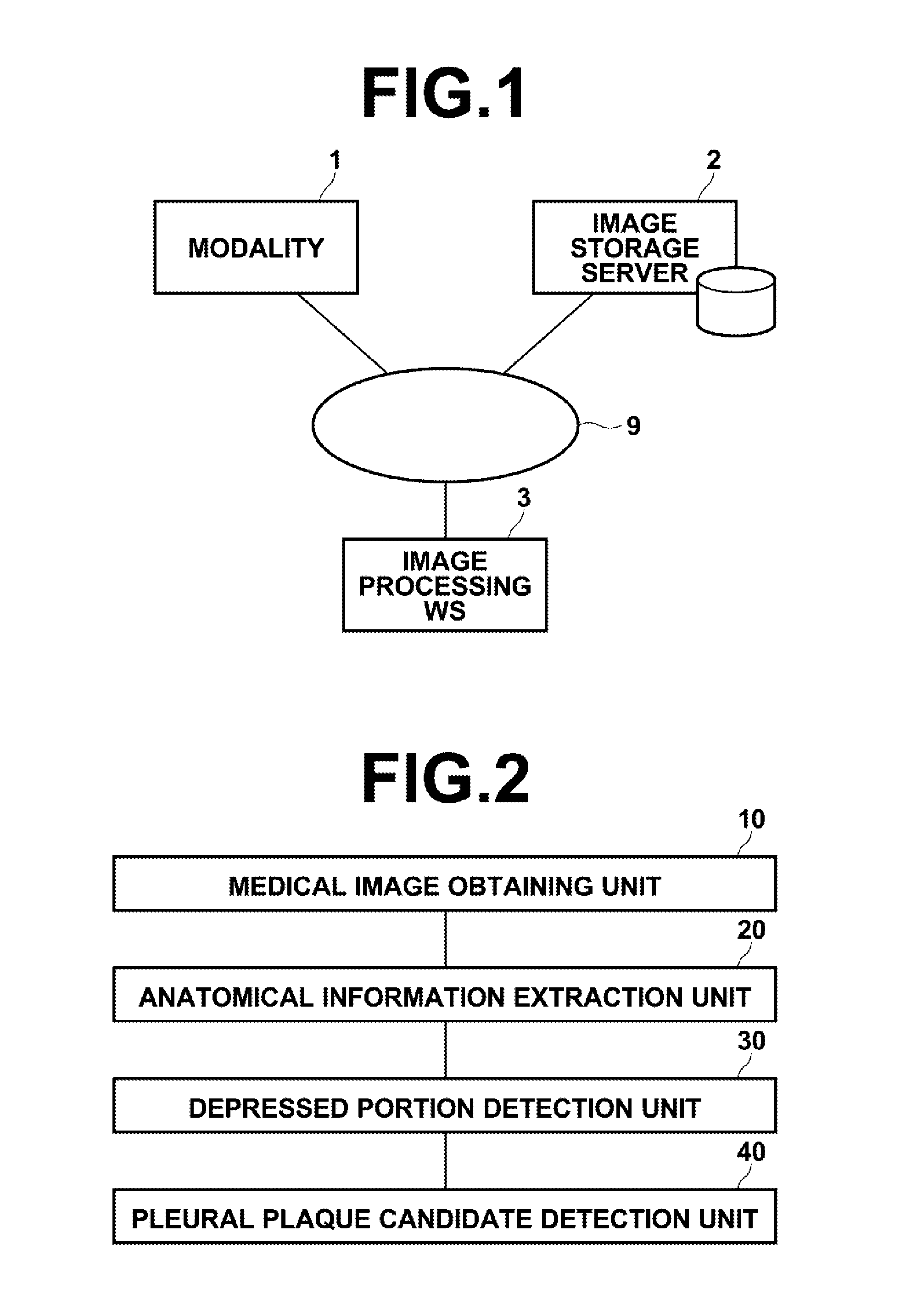
[0063]FIG. 1 is a hardware configuration diagram of a medical image processing apparatus, illustrating an overview thereof. As shown in FIG. 1, the system includes modality 1, image storage server 2, and image processing workstation 3 communicatably linked via network 9.

[0064]Modality 1 is equipment for obtaining a medical image V representing a test body, and more specifically it is CT equipment, MRI equipment, PET, ultrasonic diagnostic equipment, or the like.

[0065]Image storage server 2 is a computer for storing / managing a medical image V obtained by modality 1 and a medical image V generated in workstation 3 by image processing, and includes an external large capacity recording medium and database management software (for example, ORDB (object relational database) management software).

[0066]In the present embodiment, a software program that provides a function to detect a pleur...

second embodiment

[0110]Hereinafter, the present invention will be described.

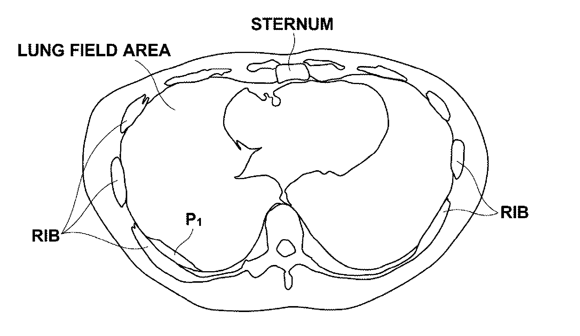
[0111]As described above, the pleural plaque is irregular white plate like thickening developed in the parietal pleura, which characteristically appears in morphology, size, and peripheral or inner density in a diagnostic image. The second embodiment focuses attention on the morphology and density of a pleural plaque and performs determination of a pleural plaque candidate using a normal medical image, in addition to the detection of a first pleural plaque candidate based on the depressed portion described in the first embodiment. Note that the hardware configuration is identical to that of the first embodiment.

[0112]A configuration related to a medical image processing function according to the second embodiment of the present invention will now be described.

[0113]FIG. 5 is a block diagram illustrating a portion of workstation 3 related to the medical image processing function according to the second embodiment of the prese...

third embodiment

[0132]Hereinafter, a third embodiment will be described.

[0133]As described above, the pleural plaque, in general, is irregular white plate like thickening developed in the parietal pleura. There may be a case in which such thickening outside of a lung is developed in patches and in such a case a pleural plaque may be detected as an uneven portion of the parietal pleura.

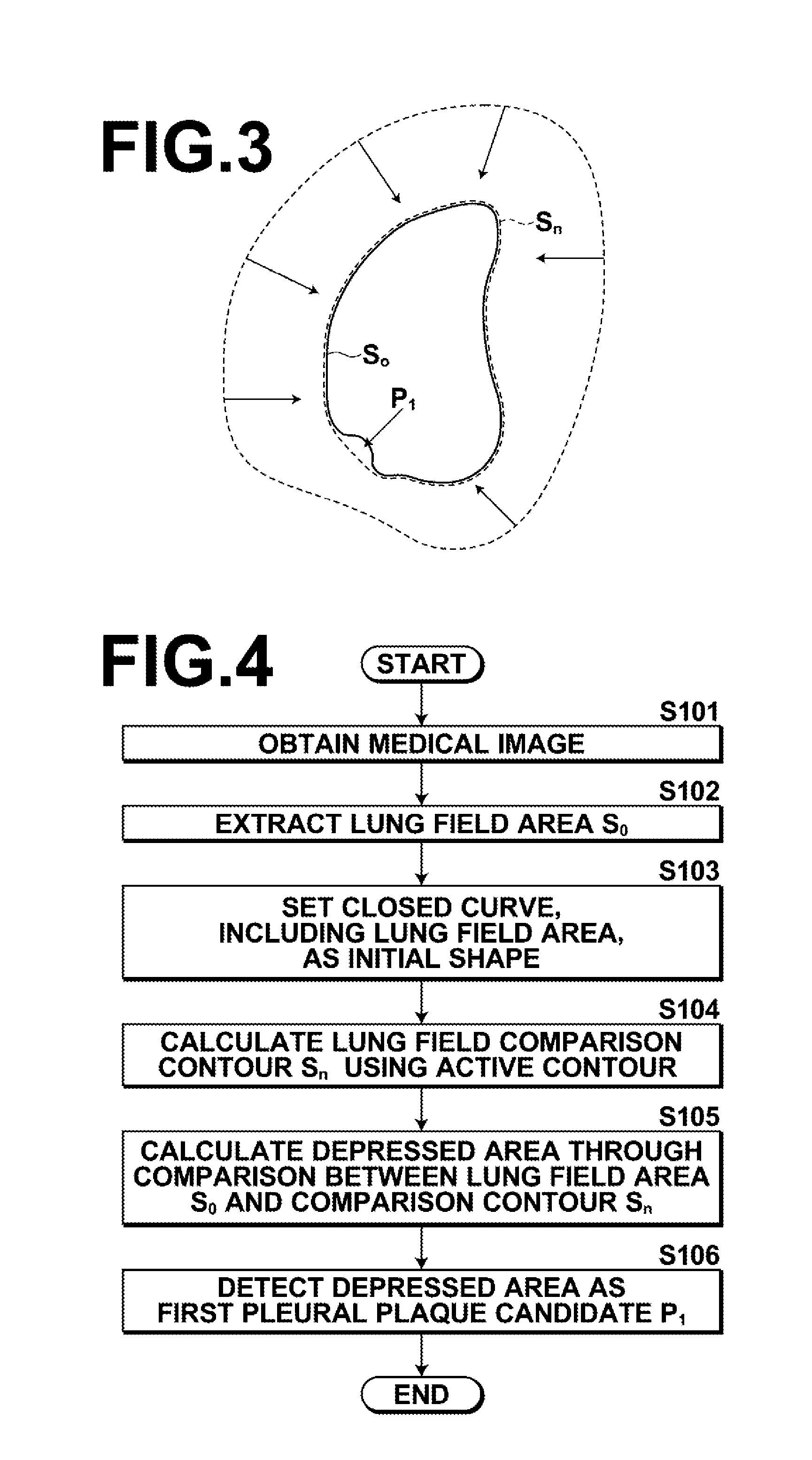
[0134]Consequently, in the third embodiment, a portion of a detected lung field contour S0 having an unevenness value greater than a threshold value is detected as a third pleural plaque candidate, thereby providing diagnostic assistance.

[0135]In the first embodiment, a first pleural plaque candidate P1 is detected based on a depressed portion, while in the third embodiment, a pleural plaque candidate is detected by taking into account an uneven portion of a lung field contour in addition to a depressed portion. Note that the hardware configuration is identical to that of the first embodiment. Further, it is possible ...


PUM


Abstract

A method of detecting a pleural plaque candidate from a medical image, which includes the steps of obtaining a medical image representing a subject, extracting a lung field area from the obtained medical image, comparing a contour of the extracted lung field with a comparison contour obtained by causing an active curve, having an initial shape in the lung field area and repeats deformation according to a certain deformation trend, to converge after repeating the deformation and detecting a depressed portion of the lung field, and determining the detected depressed portion as a first pleural plaque candidate.

Description

BACKGROUND OF THE INVENTION[0001]1. Field of the Invention[0002]The present invention generally relates to a medical image processing apparatus and method, and more particularly to a medical image processing apparatus and method for detecting a pleural plaque (pleural thickening plaque). The invention also relates to a computer readable recording medium on which is recorded a program for causing a computer to realize a function corresponding to the invention.[0003]2. Description of the Related Art[0004]Recently, there has been a demand, in the medical field, for measures to detect a pleural plaque (pleural thickening plaque) that causes calcified thickening of parietal pleura by image diagnosis.[0005]In order to find a pleural plaque, as is common to a general diagnosis of a lesion site, a radiological reader interprets a medical image of a subject to find a lesion site or observes the state of the lesion site to find an abnormal tumor pattern. There have been possibilities that suc...

Claims


Application Information

Patent Timeline
Patent Type & Authority Applications(United States)
IPC IPC(8): G06K9/00
CPCG06T7/0014G06T2207/30061G06T2207/20116G06T2207/10116
Inventor MIZUNO, OSAMU
Owner FUJIFILM CORP
Why Eureka
Try Eureka