Method and apparatus for visually supporting an electrophysiological catheter application

a technology of electrophysiological catheter and catheter application, which is applied in the field of method and an apparatus for visually supporting an electrophysiological catheter application, can solve problems such as desired successful therapy, and achieve the effect of better catheter application and better catheter guidance planning

Inactive Publication Date: 2011-01-27
SIEMENS AG
View PDF16 Cites 25 Cited by
  • Summary
  • Abstract
  • Description
  • Claims
  • Application Information

AI Technical Summary

Benefits of technology

[0017]The object of the present invention is to specify a method and an apparatus for controlling or monitor...

Problems solved by technology

The result is ablation lesions of varying effectiveness (e.g. incomplete rather than—as desired—complete ablation lines...

Method used


Image

  • Method and apparatus for visually supporting an electrophysiological catheter application
  • Method and apparatus for visually supporting an electrophysiological catheter application
  • Method and apparatus for visually supporting an electrophysiological catheter application

Examples


Embodiment Construction

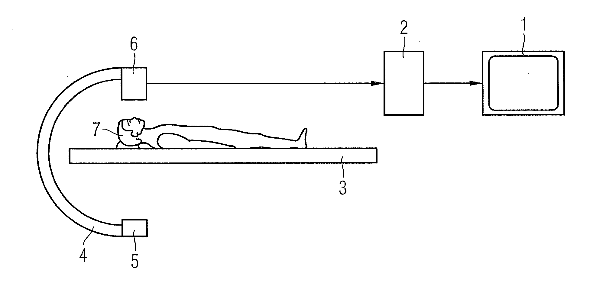
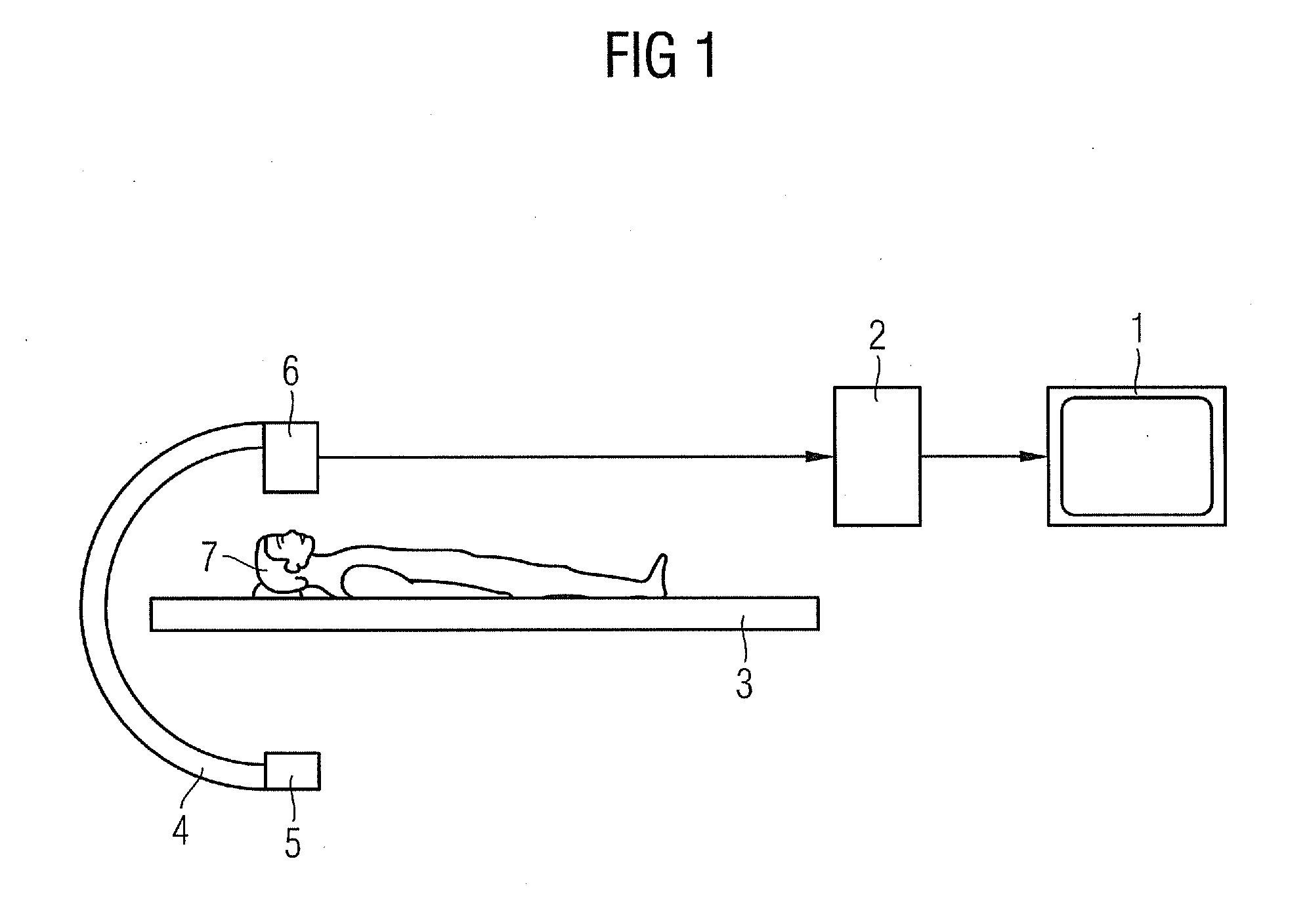
[0032]By way of example FIG. 1 shows an x-ray diagnosis facility having a C-arm 4 that is supported in a rotatable manner on a stand (not shown), at the ends of which C-arm 4 are disposed an x-ray radiation source 6, for example an x-ray emitter, and an x-ray image detector 5.

[0033]The x-ray image detector 5 can be a rectangular or square, flat semiconductor detector, which is preferably made of amorphous silicon (aSi).

[0034]In the beam path of the x-ray radiation source 6 is a patient support couch 3 for holding a region of a patient 7 to be examined. An image system 2 is connected to the x-ray diagnosis facility to receive and process the image signals of the x-ray image detector 5. The processed image signals can then be displayed on a display apparatus 1 connected to the image system 2.

[0035]The x-ray radiation source 6 emits a beam bundle from a beam focal point of the x-ray radiation source 6, said beam striking the x-ray image detector 5.

[0036]The x-ray radiation source 6 and...


PUM


Abstract

A method for visually supporting an electrophysiological catheter application is provided. An electroanatomical 3D mapping data of a region of interest in the heart is visualized. A 3D image data of the region of interest is captured before the catheter application. A 3D surface profile of objects in the region of interest is extracted from the 3D image data by segmentation. The electroanatomical 3D mapping data and 3D image data forming at least the 3D surface profile is assigned by registration and visualized superimposed on one another. Characteristic parameters are measured for catheter guidance during the catheter application. The characteristic parameters are compared with at least one predefined threshold value and regulation data for catheter guidance is generated as a function of the comparison result. The regulation data is integrally displayed and represented in the superimposed visualization.

Description

CROSS REFERENCE TO RELATED APPLICATIONS[0001]This application claims priority of German application No. 10 2009 034 245.1 filed Jul. 22, 2009, which is incorporated by reference herein in its entirety.FIELD OF THE INVENTION[0002]The invention relates to a method and an apparatus for visually supporting an electrophysiological catheter application as claimed in the respective preambles of the independent claims.BACKGROUND OF THE INVENTIION[0003]The treatment of cardiac arrhythmia has changed significantly since the introduction of the technique of catheter ablation by means of high-frequency current. In this technique an ablation catheter is introduced under x-ray monitoring into one of the heart chambers, via veins or arteries, and obliterates the tissue causing the cardiac arrhythmia by means of high-frequency current. Ablation procedures, for example in the left atrium, to treat atrial fibrillation, are carried out on the basis of electrophysiological and anatomical criteria. This...

Claims


Application Information

Patent Timeline
IPC IPC(8): G06K9/00
CPCA61B6/12A61B6/4441A61B6/466A61B6/503G06T2210/41A61B8/0891A61B18/1492A61B2019/465G06T19/00A61B6/504A61B2090/065
Inventor RAHN, NORBERTTILL, DIETRICH
Owner SIEMENS AG
Why Eureka
Try Eureka