Systems and methods for efficient imaging

a technology of efficient imaging and imaging data, applied in the field of analysis, processing, viewing, and transport of medical and surgical imaging information, can solve the problems of inefficient page method, radiologist time, and “homegrown” tools used for analyzing and processing imaging data

Inactive Publication Date: 2011-02-03
DATAPHYSICS RES
View PDF28 Cites 93 Cited by
  • Summary
  • Abstract
  • Description
  • Claims
  • Application Information

AI Technical Summary

Benefits of technology

[0027]Where appropriate, the system can utilize context-based (e.g., image-based icons instead of text) presentation of information to speed communication with the user. These abstracting icons can provide graphical summary to radiologist so that in most cases they don't have to take time to open the whole folder to access and assess information.

Problems solved by technology

Furthermore, existing tools used for analyzing and processing imaging data are “home grown” and not optimized to present the relevant clinical information to streamline the cognitive process of diagnosis and thus minimize radiologist time.
The page method is an inefficient artifact left over from the days of reading a few x-rays at a time.
Using the page method this review is tedious and error-prone, and does not scale well to the large, and ever-increasing, number of images for each examination.
The scrolling method still lacks a three-dimensional image, can be time consuming, can be difficult even for trained radiologists to comprehend—and is especially difficult for a non-radiologist to understand—and does not include substantial longitudinal and volumetric quantitative analytical tools.
Because of the proliferation of medical imaging and the increased number of two-dimensional images for each examination, using existing methods radiologist are expected to shortly reach a point where a radiologist's daily work load becomes unsustainable.
Theses tasks still consume valuable time from the main task of diagnosis.
These interface devices have served well for general computing tasks but they are not optimal for the specialized task of patient study interpretation.
The currently available systems follow predefined hanging protocols, but their static and rigid format for presentation and orientation of an imaging corpus can take a large amount of time.
Mostly these protocols have been designed in support of patient studies with a few two-dimensional images (e.g., x-ray film) and are inadequate and not easily scalable for the current and upcoming needs.

Method used


Image

  • Systems and methods for efficient imaging
  • Systems and methods for efficient imaging
  • Systems and methods for efficient imaging

Examples


Embodiment Construction

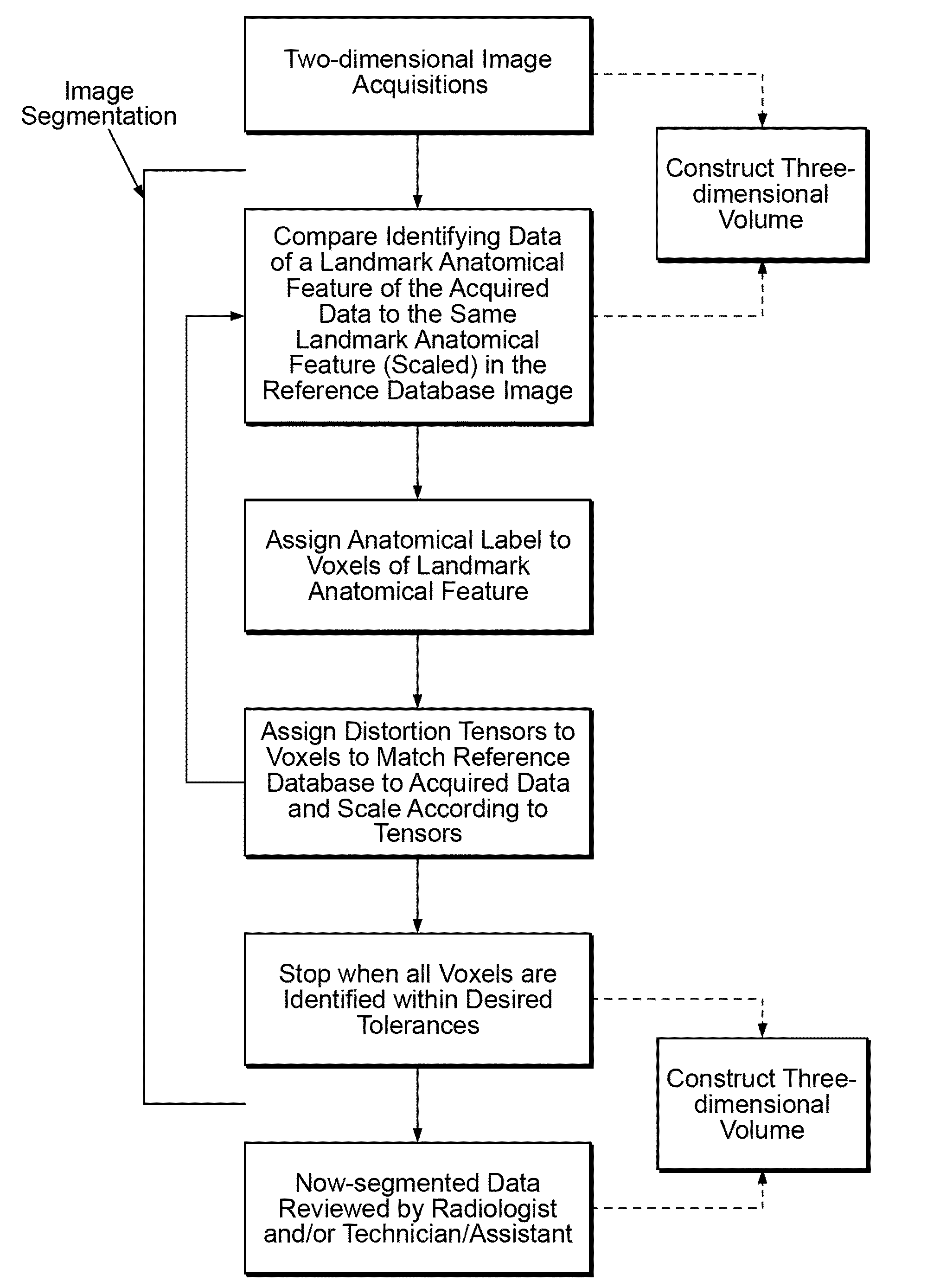
[0053]The systems and methods disclosed herein can be used to process information (e.g., examination data files containing radiological data sets) for medical and / or surgical imagining techniques used for diagnosis and / or therapy. The systems can be computer hardware having one or more processors (e.g., microprocessors) and software executed on computer hardware having one or more processors and the methods can be performed thereon. The systems can have multiple computers in communication (e.g., networked), for example including a client computer and a server computer.

[0054]The examination data files can contain radiological data, modality information, ordering physicians notes, reason / s for the examination, and combinations thereof. The radiological data can include a corpus of data from the radiological examination and processing of the data from the radiological examination. The corpus can include data that represents images (e.g., PACS images, multiplanar images), objects, datas...


PUM


Abstract

A system and methods for more efficient review, processing, analysis and diagnoses of medical imaging data is disclosed. The system and methods include automatically segmenting and labeling imaging data by anatomical feature or structure. Additional tools that can improve the efficiency of health care providers are also disclosed.

Description

CROSS-REFERENCE TO RELATED APPLICATIONS[0001]This application is a continuation of PCT Application No. PCT / US2008 / 013318, filed Dec. 2, 2008, which claims the benefit of U.S. Provisional App. No. 60 / 992,084, filed Dec. 3, 2007, both of which are incorporated herein in their entireties.BACKGROUND OF THE INVENTION[0002]1. Field of the Invention[0003]The invention relates to the analysis, processing, viewing, and transport of medical and surgical imaging information.[0004]2. Description of Related Art[0005]Proliferation of noninvasive medical examination imaging (e.g., computed tomography (CT), magnetic resonance imaging (MRI), positron emission tomography (PET)) coupled with a shortage of accredited radiologists, especially in the U.S., has increased the value of radiologists' time. Increased resolution of imaging technology is also driving the quantity of information needing review by radiologists. Hence, the demand for radiologists' time is expected to increase further for the fores...

Claims


Application Information

Patent Timeline
Patent Type & Authority Applications(United States)
IPC IPC(8): A61B5/05G06K9/00G16H30/20G16H30/40G16H50/20
CPCG06F19/321G06T7/0012G06T19/00G06T2219/028G06T2207/30004G06T2210/41G06T2219/004G06T2207/10081G16H30/40G16H30/20G16H50/20G06V10/20
Inventor DOUGLAS, STEVEN K.RODER, HEINRICHTSYPIN, MAXIM M.ABHYANKAR, VISHWAS G.RIEGEL, STEPHENSCHUSTER, JAMES A.WOLFE, GENE J.
Owner DATAPHYSICS RES
Why Eureka
Try Eureka