Automated monitoring of myocardial function by ultrasonic transducers positioned on the heart

an ultrasonic transducer and myocardial function technology, applied in the field of automatic monitoring of myocardial function and myocardial function monitoring system, can solve the problems of limited sensitivity and specificity of conventional monitoring techniques such as ecg and blood pressure monitoring, and the regional myocardial ischaemia induced by this occlusion is difficult to detect, so as to induce ischaemia and increase the contraction of a hypokinetic heart.

Inactive Publication Date: 2011-02-24
UNIV OSLO HF
View PDF14 Cites 3 Cited by
  • Summary
  • Abstract
  • Description
  • Claims
  • Application Information

AI Technical Summary

Benefits of technology

[0044]an alarm state initiated if the generated parameter or graphical representation passes the threshold value.
[0045]For the care unit, incorporation of an alarm in the monitoring may be implemented by further comprising input means allowing an operator to set a threshold value for the generated parameter or graphical representation and means for generating an alarm, wherein electronic processing unit further comprises software means for monitoring the generated parameter or graphical representation and activating the means for generating an alarm if the generated parameter or graphical representation passes the threshold value.
[0046]The method and care unit may not only be used for monitoring a state of a patient in order to detect ischaemia or dysfunction due to graft occlusion. In another embodiment, the method and care unit is used to monitor the effect or lack of effect from a medical intervention. Here, the parameter or graphical representation is generated over a period of time including or following intravenous administration of a fluid or a medicament affecting the global hypokinetic heart function. A change in the global hypokinetic heart function during this period of time may be followed and optionally quantified using the parameter or graphical representation. Such a medicament could be infusion of adrenaline to increase the contraction of a hypokinetic heart. This drug may induce ischaemia if given in a too large dose. The ultrasonic measurements of cardiac function would precisely show when the function is normalized and / or if ischaemia occurs and a correct lowest medicament dose can be determined.
[0050]using the read value, or a derived value or function, to indicate regional cardiac ischaemia or estimate global hypokinetic heart function.
[0051]In yet another embodiment, the invention provides the use of a TDI signal for indicating regional cardiac ischaemia or estimating global hypokinetic heart function in accordance with the above method.
[0052]The basic idea of the invention is to use TDI signals from one or more miniaturized ultrasonic transducers fastened to the myocardium of a patient to automatically generate parameters or graphical representations that indicate or correlate with myocardial ischaemia or dysfunction. This has the advantage over standard methods using a manually operated ultrasound probe that it can be used continuously over longer periods of time, and thereby be used in post-operation monitoring of patients. In relation to previous disclosures of miniaturized ultrasonic transducers fastened to the heart, the invention provides the advantage of generating parameters or graphical representations that indicate or correlate with myocardial ischaemia or dysfunction.

Problems solved by technology

The regional myocardial ischaemia induced by this occlusion is often difficult to detect with present bedside monitoring techniques.
Conventional monitoring techniques as ECG and blood pressure monitoring are of limited sensitivity and specificity.
Established modalities as CT and MR are not suitable for continuous post-operative monitoring.
However, such techniques require a skilled operator and do therefore not allow continuous monitoring of myocardial function.
It is a disadvantage of the above prior art techniques that no automated quantification of the signals is provided, which is needed if it shall be used in a postoperative care unit, especially where a multiple of patient data are continuously monitored.

Method used


Image

  • Automated monitoring of myocardial function by ultrasonic transducers positioned on the heart
  • Automated monitoring of myocardial function by ultrasonic transducers positioned on the heart
  • Automated monitoring of myocardial function by ultrasonic transducers positioned on the heart

Examples


Embodiment Construction

[0060]The invention relates to the analysis and treatment of data from a miniaturized ultrasonic transducer fastened to the myocardium of a patient. Hence, the operative procedure of fastening the transducer is a separate and preceding step which is not part of the invention or covered by the present application / patent.

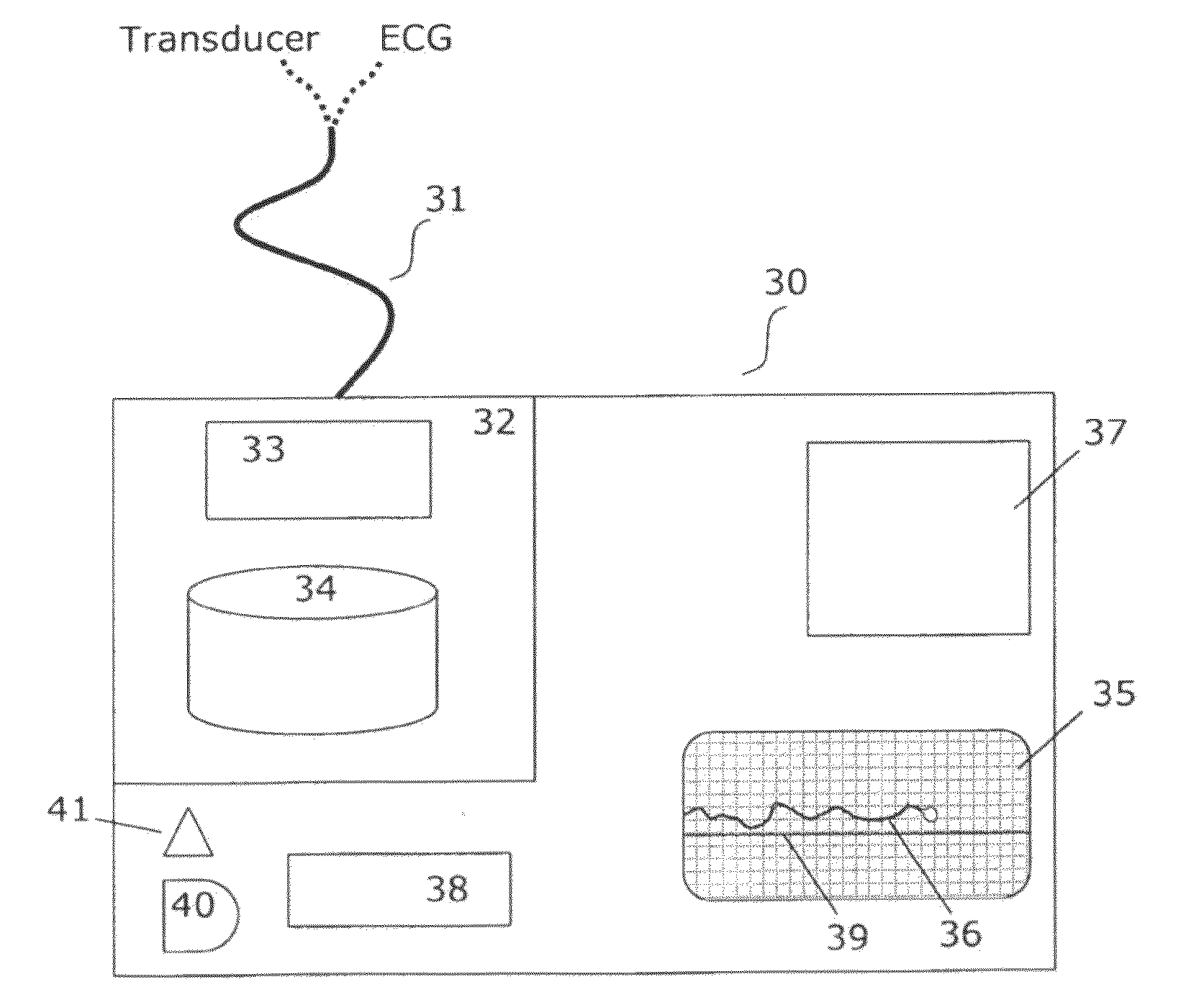
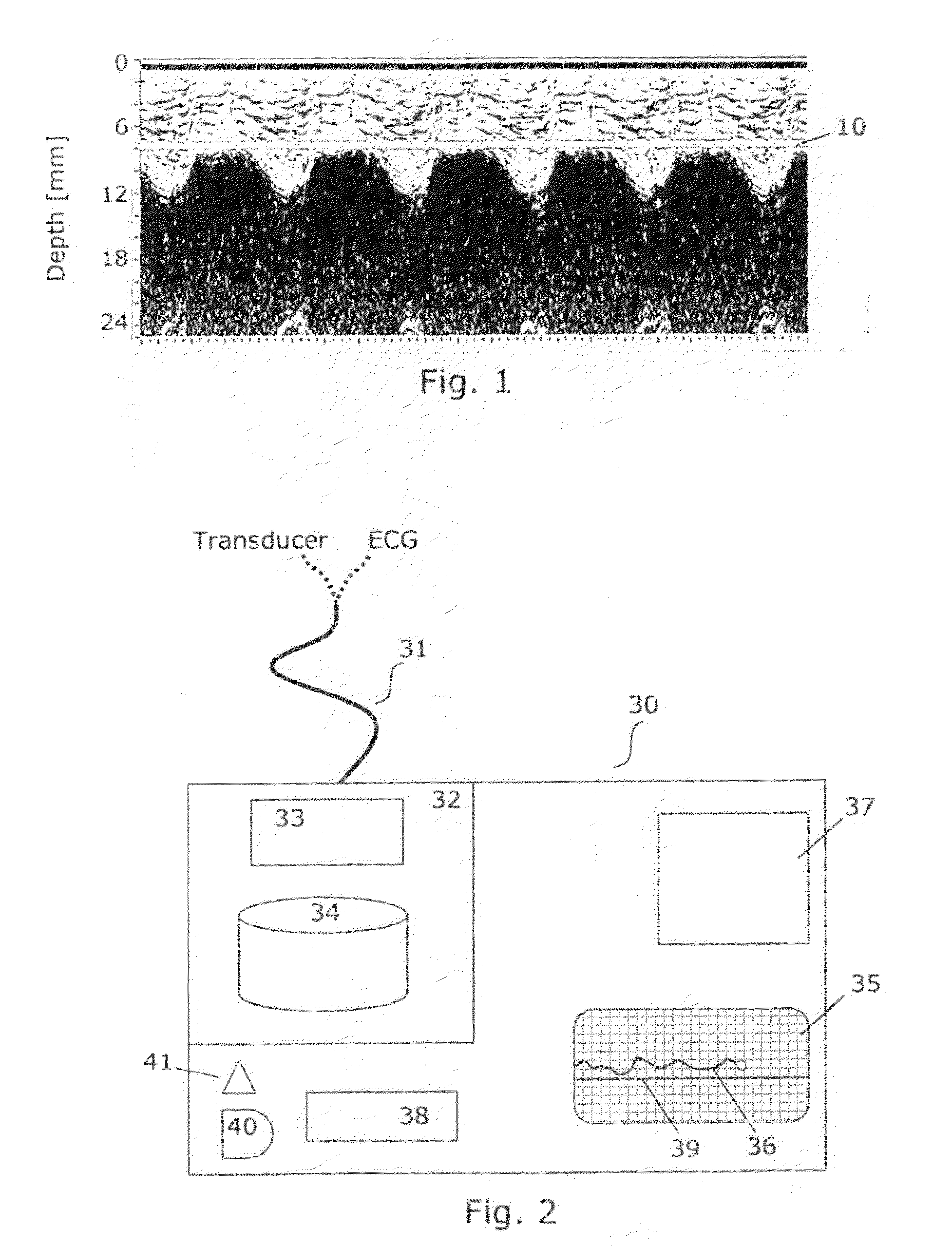
[0061]Miniaturized ultrasonic transducers are known from a number of applications, both medical and non-medical, see e.g. US 2006 / 0116584. When the ultrasonic transducer is sutured to the heart during the operation an optimal position on the wall and depth of measurement is secured by using M-mode echo signals as guidance shown in FIG. 1. An optimal position has been obtained when relatively distinct outer and inner surface lines of the wall are seen. Since the miniature ultrasound transducer is a regional monitor of myocardial function, it may be preferable to have two or three miniature ultrasound transducers in order to monitor global function or to confirm the rea...


PUM


Abstract

The invention relates to a method and a post-operative care unit for analysing and quantifying an ultrasound tissue Doppler imaging (TDI) signal from a transducer fastened on the myocardium to obtain a parameter indicating regional cardiac ischaemia or correlates with global hypokinetic heart function. This has the advantage over manually operated probes that it can be automated and used continuously over long time. According to the method, a TDI signal trace corresponding to at least one of tissue velocity, strain or strain rate is extracted and correlated with an electrocardiogram to define subsections within a cardiac cycle in the extracted trace corresponding to the early systolic phase and the post-systolic phase. Then, a velocity, strain or strain rate is read in at least the post-systolic phase of the extracted trace, and a parameter which is a function of one of these readings and which indicates ischaemia or global hypokinetic function is generated.

Description

FIELD OF THE INVENTION[0001]The present invention relates to estimating cardiac pumping capacity, in particular a myocardial function monitoring system applying data recorded by ultrasonic transducers positioned on the heart.BACKGROUND OF THE INVENTION[0002]During and after cardiac surgery, it is of interest to monitor the performance of the heart and its responses to various forms of treatment. For example, an important mechanism for haemodynamic instability in the post operative period after aorta-coronary bypass surgery is graft occlusion. The regional myocardial ischaemia induced by this occlusion is often difficult to detect with present bedside monitoring techniques. Also, a sensitive technique for detecting regional ischaemia is strongly warranted during the surgical procedure in off-pump by-pass procedures.[0003]There is a need for new monitoring techniques of regional myocardial function in the postoperative period after cardiac surgery. Conventional monitoring techniques a...

Claims


Application Information

Patent Timeline
Patent Type & Authority Applications(United States)
IPC IPC(8): A61B8/00
CPCA61B8/0883A61B8/12A61B8/488A61B8/4488A61B8/485A61B8/42
Inventor ELLE, OLE JAKOBFOSSE, ERIKIHLEN, HALFDANESPINOZA, ANDREASHOFF, LARS
Owner UNIV OSLO HF
Why Eureka
Try Eureka