

Novel biomarker for the prognosis of breast cancer
a breast cancer and prognosis technology, applied in the field of breast cancer prognosis, can solve the problems of increased survival and decreased mortality, and achieve the effect of improving the prognosis of breast cancer patients
- Summary
- Abstract
- Description
- Claims
- Application Information
AI Technical Summary
Benefits of technology
Problems solved by technology
Method used
Image

Examples
example 1
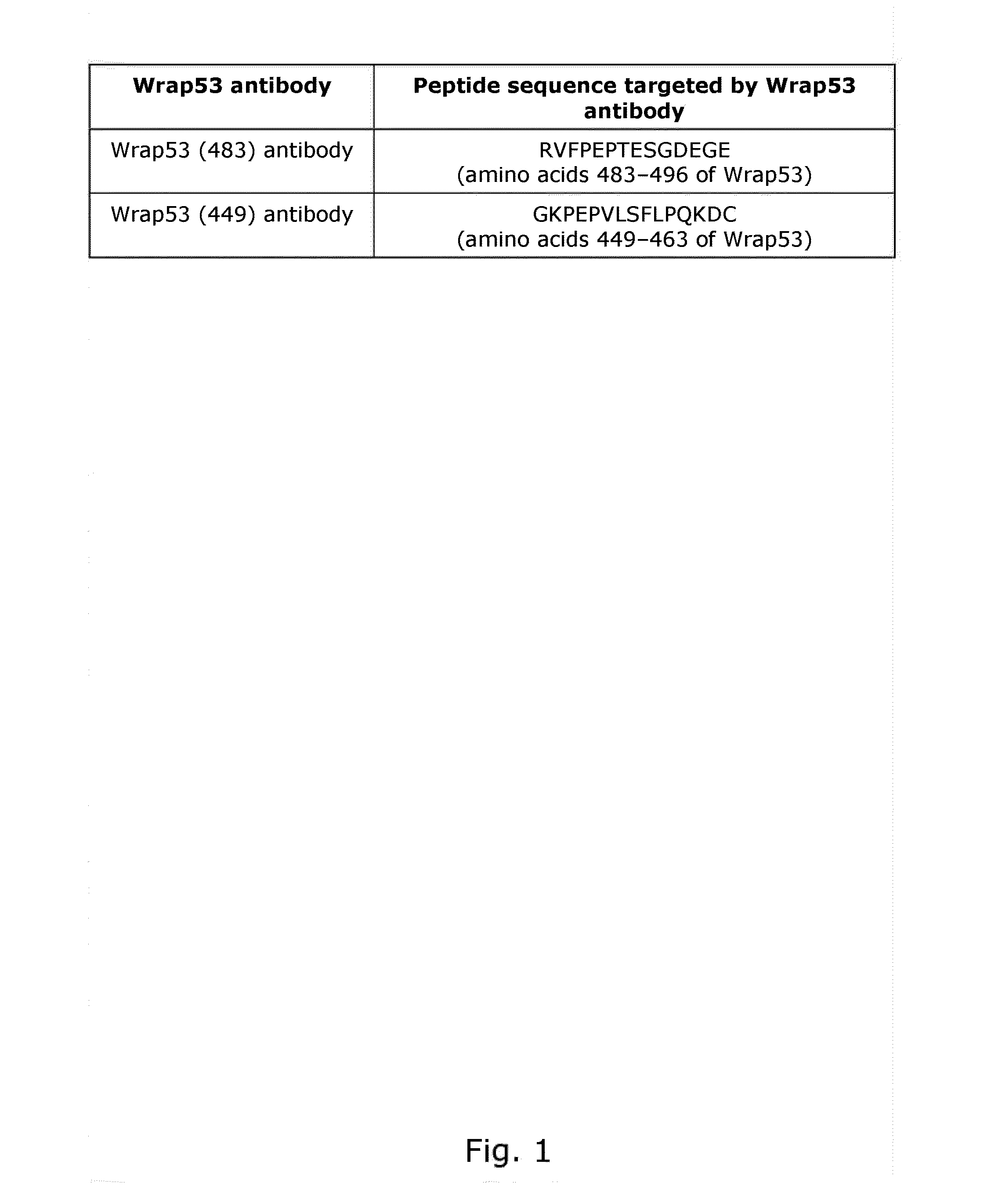
[0222]The Wrap53 protein is detected using Wrap53 (483) and Wrap53 (4491 antibodies
Material and Methods:
[0223]To generate the Wrap53 (483) antibody rabbits were immunized with a KLH-conjugated Wrap53 peptide (NH2-) (C) RVFPEPTESGDEGE (SEQ ID NO. 6) (—CONH2), corresponding to amino acids 483-496 of full-length Wrap53 protein, followed by affinity IgG purification (Innovagen AB, Sweden). A Cysteine (C) residue was added in the N terminus to facilitate coupling of the peptide to the carrier molecule.
[0224]To generate the Wrap53 (449) antibody rabbits were immunized with a KLH-conjugated Wrap53 peptide (Ac-) GKPEPVLSFLPQKDC (SEQ ID NO. 7) (—COOH), corresponding to amino acids 449-463 of full-length Wrap53 protein, followed by affinity IgG purification (Innovagen AB, Sweden).
Conclusion:
[0225]The above peptide sequences (FIG. 1) are unique for the Wrap53 protein and therefore the Wrap53 (483) and (449) antibodies should specifically recognize the Wrap53 protein.
example 2
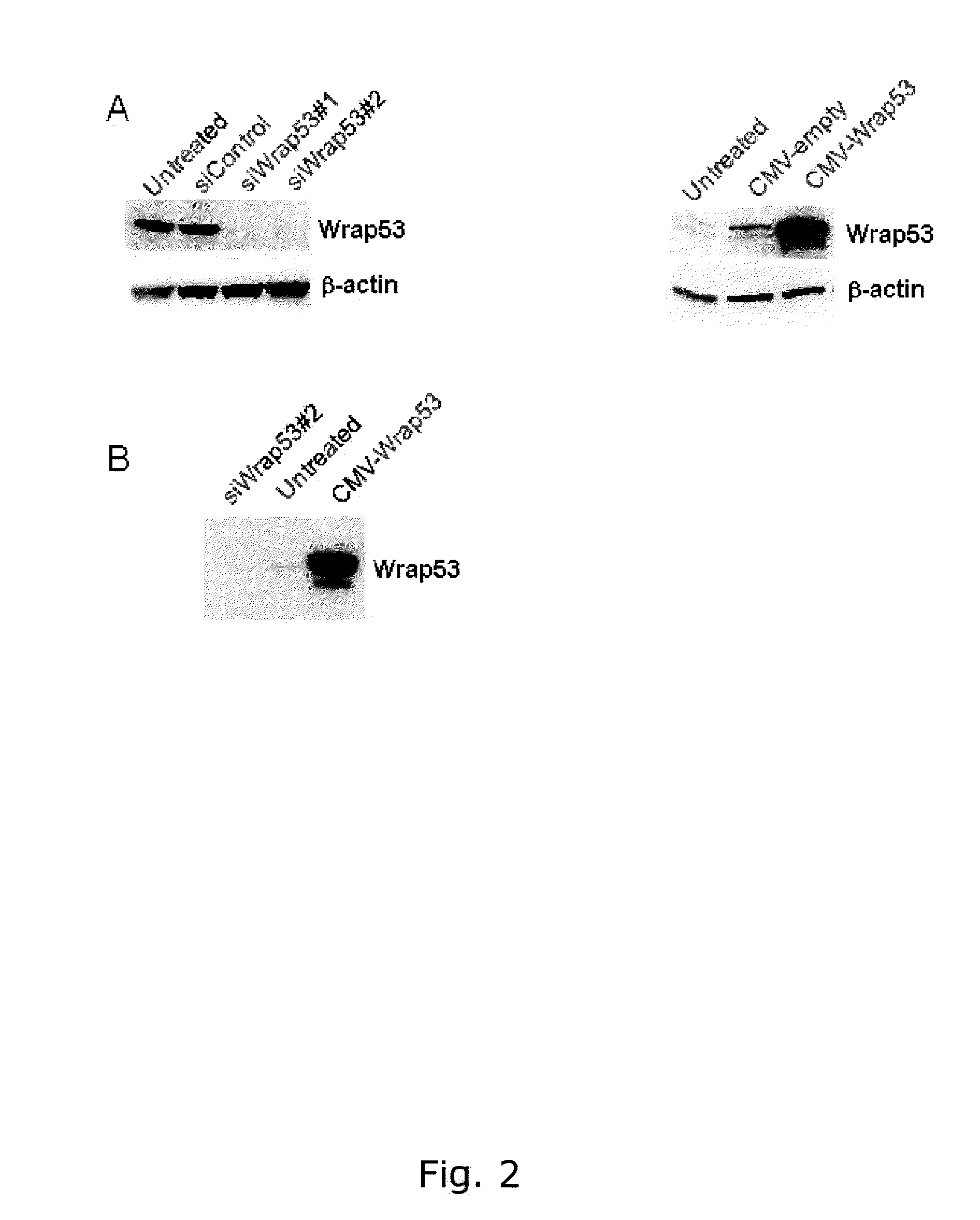
[0226]The Wrap53 (483) and Wrap53 (449) antibodies specifically detects the Wrap53 protein.
Material and Methods:
[0227]Cell extracts for Western blot analysis were prepared by harvesting cells, wash once in PBS and lyse the cells in ice cold WB lysis buffer (100mM Tris-HCL pH 8, 150mM NaC1, 1% NP-40, 1% PMSF, 1% protease inhibitor cocktail) for 30 minutes on ice. Lysates were centrifuged at 14000rpm for 15 minutes at 4° C. and protein concentrations were determined using Bradford assay (Biorad). Western blot analysis was performed according to standard procedures. Wrap53 knockdown was performed by transfected cells with 10-20nM of Wrap53-specific siRNA oligos using Oligofectamine (Invitrogen®) transfection reagent in accordance with the supplier's recommendations. The two siRNA oligonucleotides siWrap53#1 (targets Wrap53 sequence 5′- CCGGGAGAACCCGATTCATAT -3′, (SEQ ID NO. 8) cat# SI00388941) and siWrap53#2 (targets Wrap53 sequence 5′-AACGGGAGCCTTTCTGAAGAA -3′, (SEQ ID NO. 9) cat# SI0...
example 3
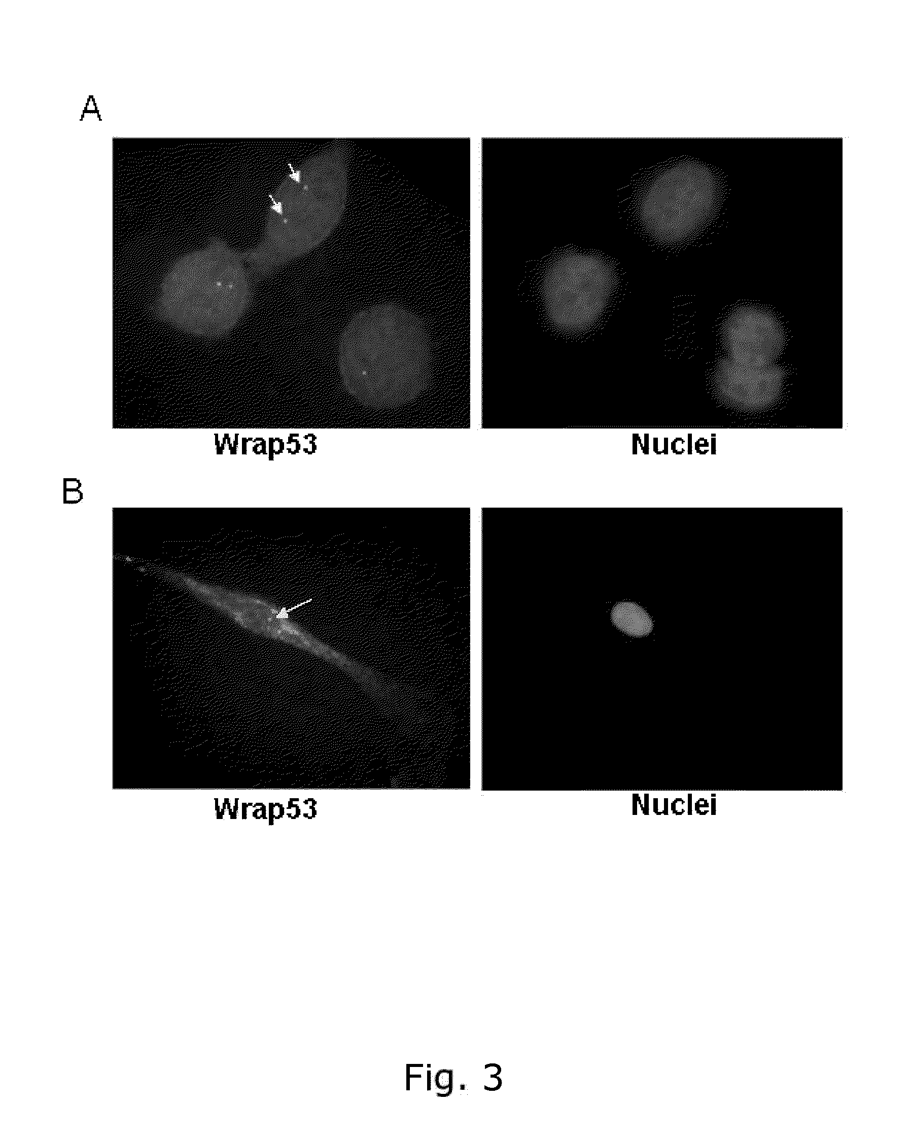
[0229]Wrap53 protein is expressed in the nucleus and in the cytoplasm in breast cancer cells (MCF-7) in normal fibroblasts (HDF)
Material and Methods:
[0230]For immunofluorescence (IF) experiments, cells were grown on sterilized cover slips and fixed with 100% MeOH for 20 minutes at −20° C. The cells were then permeabilized with 0.1% Triton X-100 for 5 minutes at RT, followed by 30 minutes of blocking in blocking buffer (2% BSA, 5% glycerol, 0,2% Tween20, 0,1% NaN3). Cover slips were subsequently incubated for 1 hour in primary antibody and 40 minutes in secondary antibody diluted in blocking buffer. The cover slips were mounted with Vectorshield mounting medium with DAPI (Vector laboratories). Images were acquired with a Zeiss Axioplan 2 microscope, equipped with an AxioCam HRm Camera using 43 or 60 oil immersion lenses, and processed using Axiovision Release 4.7. A rabbit polyclonal Wrap53 (1-50) antibody from Bethyl Laboratories (#A301-442A) was used for the staining.
Conclusions:
[0...
PUM
| Property | Measurement | Unit |
|---|---|---|
| Ratio | aaaaa | aaaaa |
| Level | aaaaa | aaaaa |
Abstract
Description
Claims
Application Information