Method and apparatus for diagnosing and treating vascular disease

a vascular disease and vascular disease technology, applied in the field of vascular disease treatment, can solve the problems of no device that enables a physician to simultaneously diagnose and treat vascular disease, increased risk of future thrombosis, and injury to the femoral vein

Inactive Publication Date: 2011-04-28
LARSON +1
View PDF10 Cites 31 Cited by
  • Summary
  • Abstract
  • Description
  • Claims
  • Application Information

AI Technical Summary

Problems solved by technology

Duplex ultrasound is also used to position the tip of the ablation catheter, for example below the level of the terminal valve of the saphenofemoral junction prior to starting an ablation procedure, in that if the catheter extends into the femoral vein it may cause injury to the femoral vein.
Blood thinners are often utilized to reduce the risk of clots forming pulmonary emboli, but do not typically remove an existing clot.
After effective thrombolysis, post therapy imaging modalities facilitate evaluation of the vein wall and its structure (if the vein is narrowed or damaged, there is an increased risk of future thrombosis).
Currently, there is no device that enables a physician to simultaneously image, evaluate and diagnose vascular disease and then perform treatment and outcome assessment with the same instrument, nor is there an instrument that integrates the user interface of the imaging and therapeutic modalities, either for usefulness in the operation of the system or the interactive integration to improve on optimizing a therapeutic procedure.

Method used


Image

  • Method and apparatus for diagnosing and treating vascular disease
  • Method and apparatus for diagnosing and treating vascular disease
  • Method and apparatus for diagnosing and treating vascular disease

Examples


Embodiment Construction

[0043]The present invention will be discussed with reference to the accompanying Figures which represent the invention by way of example only.

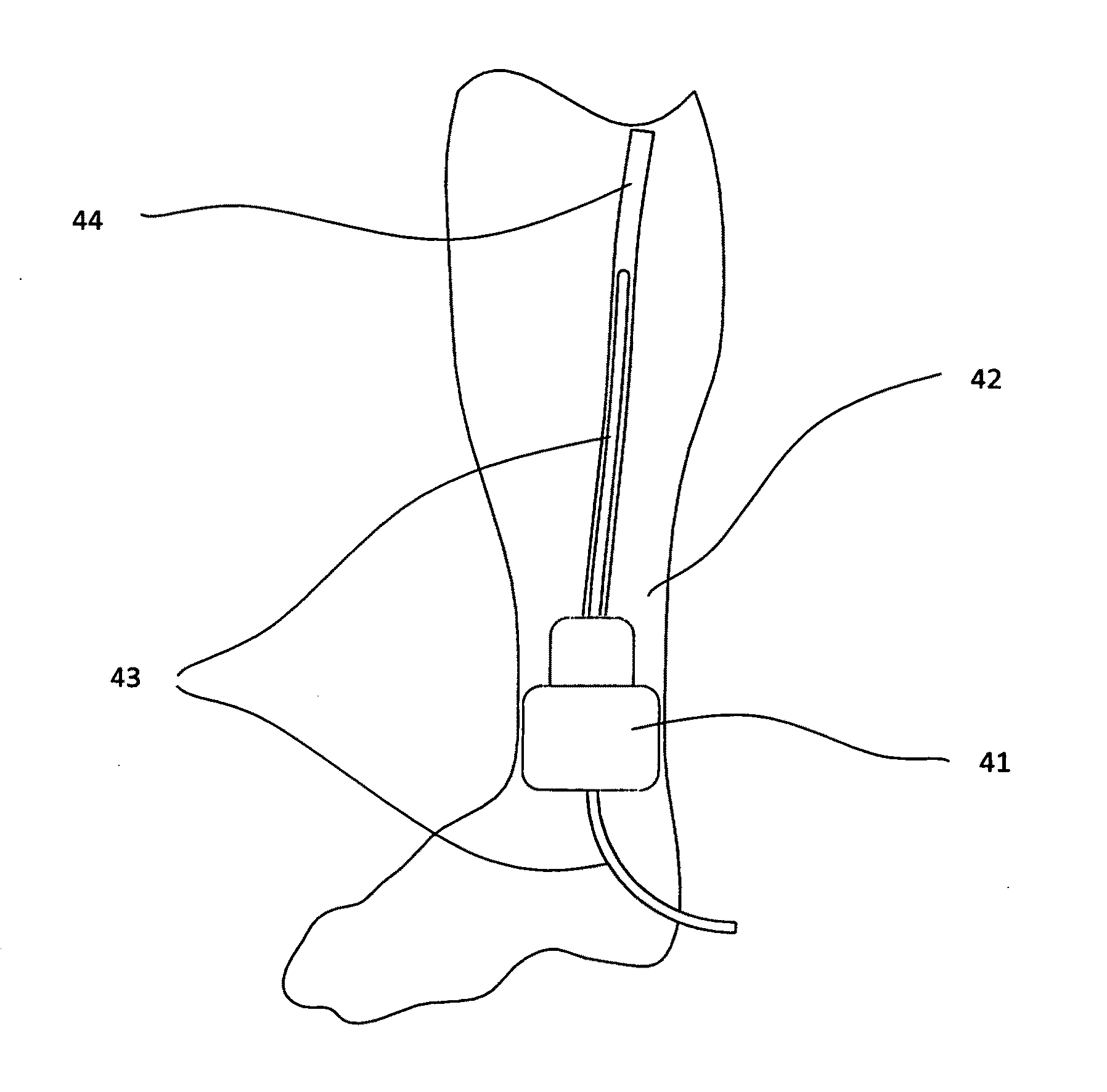
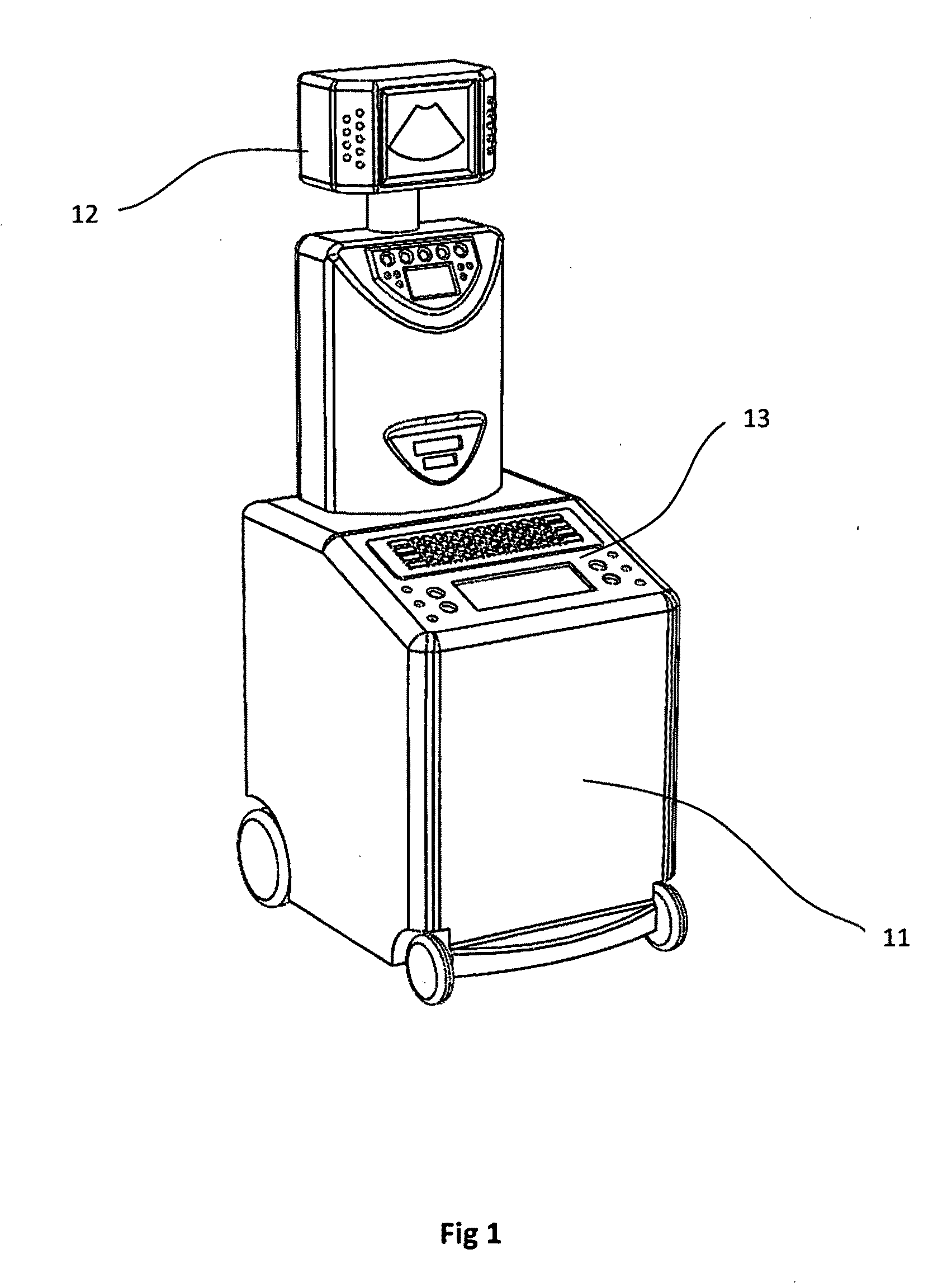
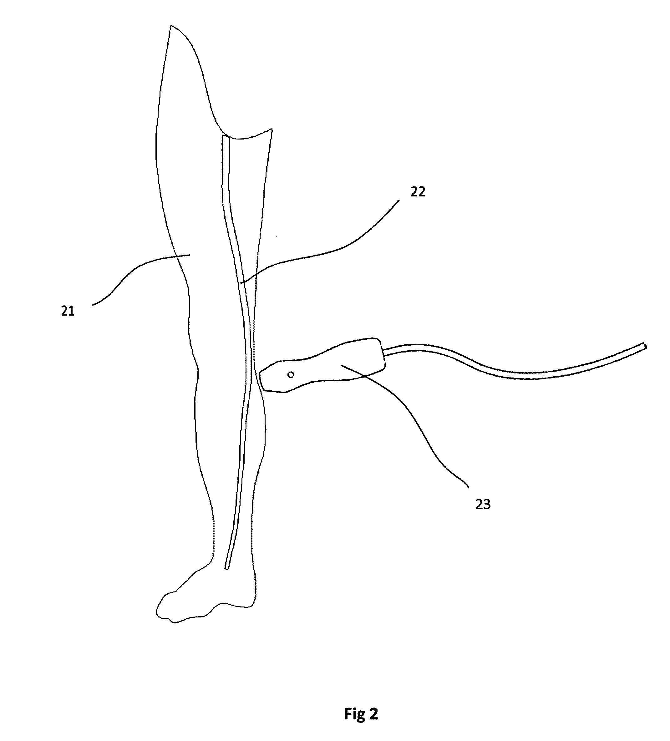
[0044]FIGS. 1 and 2 show an ultrasound imaging system with a patient contact ultrasound transducer 23 (such as a linear array) adjacent a leg for imaging a portion of a vein, such as the saphenous vein 22 of a right leg 21 and providing Doppler flow and vein diameter information to an integrated system controller located in a console unit 11. The console unit 11 includes a display 12, which can show both imaging and therapy data, and a common user interface 13. The positional and morphological information may be combined with thermal dose data and fed to a therapeutic vein ablation device, e.g. for treatment of varicose veins, or the thrombolytic device for treating blood clots.

[0045]Information, such as the position and thermal dose of a catheter, may be sent to the common display 12 and user interface 13 of the system. This data is then util...


PUM


Abstract

Imaging and therapy for the treatment of venous disease are integrated into the same device, such that there is a common user interface and / or display, which may be passive or interactive between image and therapy parameters.

Description

[0001]This application claims the benefit of U.S. Provisional Patent Application No. 61 / 279,832 filed Oct. 26, 2009, the entire disclosure of which is hereby incorporated by reference.FIELD OF THE INVENTION[0002]The present invention is directed to the treatment of vascular disease and in particular discloses a method and apparatus wherein imaging and therapy are integrated into a single device.BACKGROUND OF THE INVENTION[0003]The application of ultrasound imaging is expanding rapidly in today's society as a diagnostic test used to evaluate the health of blood vessels, in particular peripheral vascular disease. Imaging, evaluation, treatment and subsequent monitoring of diseases such as varicose veins and deep vein thrombosis (DVT) can often be performed as an outpatient procedure. What once was performed only in hospitals is now being utilized in physician offices, vascular suites, and even vein clinics. Vascular ultrasound is used for both diagnoses of vascular disease and to eval...

Claims


Application Information

Patent Timeline
Patent Type & Authority Applications(United States)
IPC IPC(8): A61B8/14
CPCA61B5/02007A61N7/022A61B8/0833A61B8/0841A61B8/0858A61B8/13A61B8/4405A61B8/463A61B17/22004A61B18/1492A61B18/24A61B2018/00404A61B2019/5276A61B2019/528A61B8/06A61B2090/3784A61B2090/378
Inventor LARSON, MARIAN L.LARSON, EUGENE A.
Owner LARSON
Why Eureka
Try Eureka