Gold nanoparticle imaging agents and uses thereof

a nanoparticle and imaging agent technology, applied in the field of nanoparticles and nanoparticlebased molecular imaging, can solve the problems of ineffective detection of imaging modalities, inability to distinguish between benign and cancerous tumors, and inability to specific target patients

Inactive Publication Date: 2011-05-12
UNIV OF MARYLAND BALTIMORE COUNTY
View PDF4 Cites 10 Cited by
  • Summary
  • Abstract
  • Description
  • Claims
  • Application Information

AI Technical Summary

Benefits of technology

[0010]In one embodiment of the invention, there is provided an imaging agent of the invention is a renin-angiotensin system (RAS) targeted molecule. In specific embodiments, the gold nanoparticle is coated with the RAS targeted molecule). In further specific embodiments, a RAS targeted molecule is a metal-coated angiotensin converting enzyme (ACE) inhibitor. In other certain embodiments, a RAS targeted molecule of the invention is useful for generating imaging agents for diagnosing or monitoring disease. In other certain embodiments of the invention, a kit comprising a RAS targeted molecule of the invention useful for generating imaging agents for diagnosing or monitoring disease is provided.

Problems solved by technology

However, these imaging modalities are not efficient in detecting tumors and metastases that are smaller than 0.5 cm, and they can barely distinguish between benign and cancerous tumors.
Present CT imaging agents are predominantly based on iodine containing molecules, which are effective in absorbing X-rays but are nonspecifically targeted because they cannot be conjugated to or otherwise associated with most biological components or cancer markers, and they allow only very short imaging times due to rapid clearance by the kidneys.
Therefore, the prior art is deficient in specifically targeted molecular imaging agents.

Method used


Image

  • Gold nanoparticle imaging agents and uses thereof
  • Gold nanoparticle imaging agents and uses thereof
  • Gold nanoparticle imaging agents and uses thereof

Examples


example 1

Materials

[0039]All glassware used was cleaned with freshly prepared aqua regia solution (HCl / HNO3, 3:1), then rinsed thoroughly with ultrapure water before use. The water was distilled and subsequently purified to 18 mΩ ultrapure water quality using the Milli-Q academic system. Gold chloride (HAuCl4.H2O) and trisodium citrate (Na3C6H5O7.2H2O), obtained from Electron Microscopy Sciences, and sodium hydroxide (NaOH), from Fisher Scientific, were of analytical grade and were used without further purification. Tween 20 was purchased from Aldrich. Lisinopril dehydrate was obtained from Waterstone Technology LCC. The Millex-LCR 0.45 μm syringe filters were obtained from Millipore. The TEM carbon coated 200 mesh copper grids were purchased from Ted Pella, Inc.

example 2

Preparation of Gold Nanoparticles

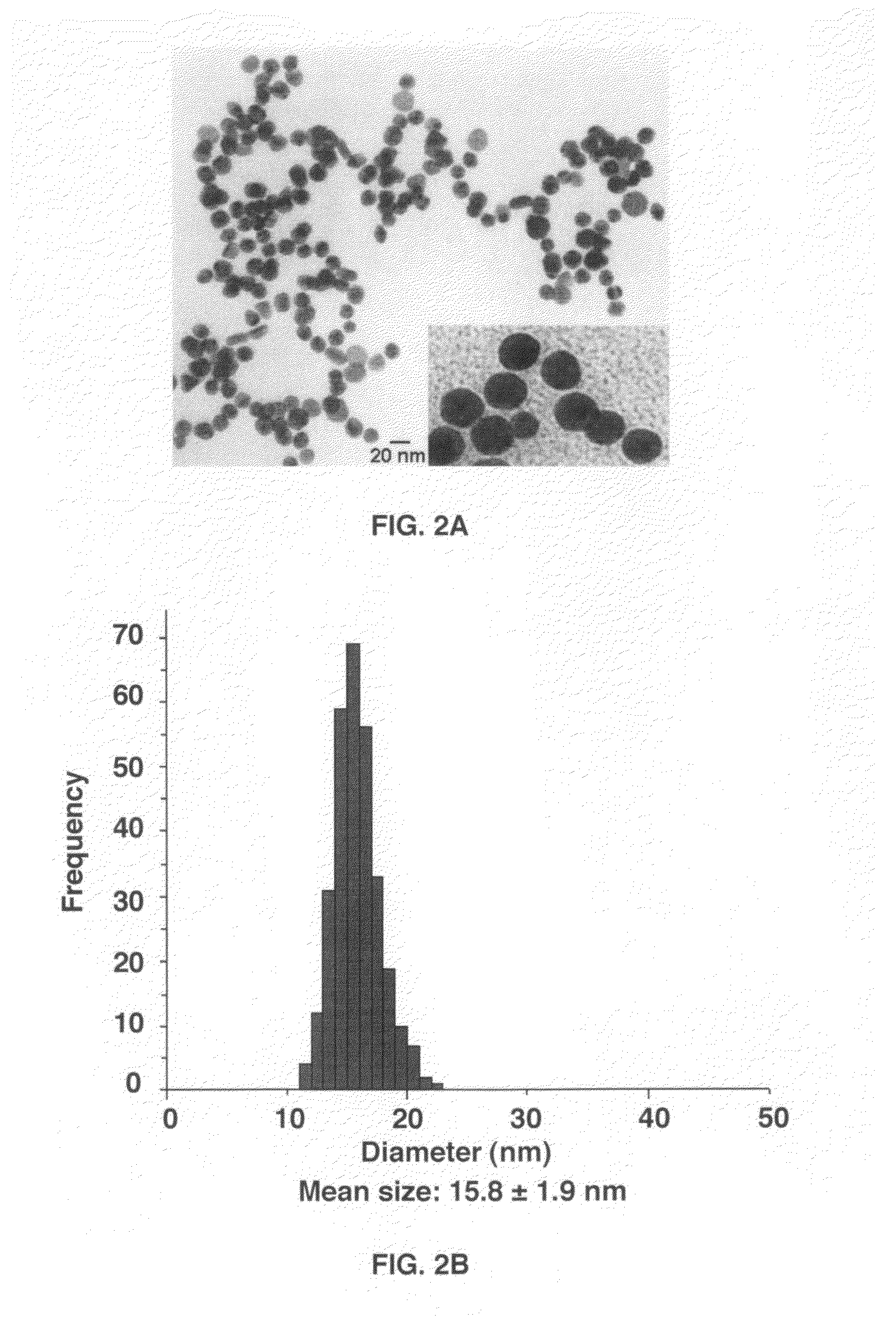
[0040]The synthesis was performed using a modified Frens method[23]. In a 1 L 2-neck-round-bottom flask equipped with a condenser, 500 mL of 0.5 mM HAuCl4 in ultrapure water was brought to boil. Rapid addition of 26 mL of 5% sodium citrate solution (ncitrate / nAu=17.9) to the vigorously stirred gold chloride solution resulted in a series of color change from pale yellow to colorless, then light purple, deeper purple, turning reddish and finally to dark wine red. The color change occurred over 7 minutes. The solution was maintained for 15 minutes at boiling temperature and then removed from the heating bath. Stirring was continued until the solution cooled down to room temperature. The size of the prepared gold nanoparticles was 15.8 nm±1.9 nm according to the TEM measurements.

example 3

Preparation of Concentrated Solution of Lisinopril-Coated Gold Nanoparticles

[0041]Lisinopril dehydrate (330 mg, molar ratio of lisinopril / GNP: 4.02×10⁵) was dissolved in 10 mL of water adjusted to pH 11 using 2 N sodium hydroxide.[24] The resulted lisinopril solution was added to a solution (525 mL) of citrate-stabilized GNP solution previously prepared (15.8 nm, 3.54 nM) while strongly stirring in a 1 L beaker. A certain amount of 2 N NaOH was then added to keep pH around 11. The reaction was kept under strong stirring overnight at room temperature. The lisinopril-capped gold nanoparticles solution was centrifuged several times at 31,000 g for 45 min. at 4° C. and redispersed in basic water (pH 8-9) in order to remove the excess of lisinopril and the exchanged citrate molecules. This purified lisinopril-capped gold nanoparticles solution was used for all the characterization techniques. Concentration of these lisinopril-capped gold nanoparticles was performed by centrifugation at 1...


PUM

PropertyMeasurementUnit
diameteraaaaaaaaaa
circulation timeaaaaaaaaaa
diameteraaaaaaaaaa

Abstract

Overexpression of angiotensin-converting enzyme (ACE) has been associated with a number of pathophysiologies, including those associated with cancer and the cardiovascular system. Thus, targeted imaging of ACE is of crucial importance for monitoring tissue ACE activity as well as treatment efficacy. To this end, lisinopril-coated gold nanoparticles were prepared to provide a new type of probe for targeted molecular imaging of ACE by tuned K-edge computed tomography (CT) imaging.

Description

CROSS-REFERENCE TO RELATED APPLICATION[0001]This nonprovisional application claims benefit of priority under 35 U.S.C. §119(e) of provisional applications U.S. Ser. No. 61 / 260,108, filed Nov. 11, 2009, now abandoned, the entirety of which are hereby incorporated by reference.BACKGROUND OF THE INVENTION[0002]1. Field of the Invention[0003]The present invention relates generally to the fields of nanoparticles and nanoparticle-based molecular imaging. More specifically, the present invention relates to gold nanoparticle imaging agents and uses thereof.[0004]2. Description of the Related Art[0005]Nanoparticle systems are promising new paradigms in pharmacotherapy and are being used in gene therapy, drug delivery, imaging, and drug discovery techniques. A goal of nanodiagnostics is to identify disease at its earliest stage, particularly at the molecular level. Nanoparticle-based molecular imaging has set a unique platform for cellular tracking, targeted diagnostic studies, and image moni...

Claims


Application Information

Patent Timeline
Patent Type & Authority Applications(United States)
IPC IPC(8): A61K49/00C07D207/16A61K49/04A61K49/06B32B5/16B82Y15/00
CPCA61K47/48861A61K49/0428Y10T428/2982B82Y15/00B82Y5/00A61K47/6923
Inventor ARAS, OMERFLEITER, THORSTENJEUDY, JEANDANIEL, MARIE-CHRISTINE
Owner UNIV OF MARYLAND BALTIMORE COUNTY
Why Eureka
Try Eureka