Homing in mesenchymal stem cells
a mesenchymal stem cell and cxcr4 technology, applied in the direction of skeletal/connective tissue cells, drug compositions, biocide, etc., can solve the problem of increasing the gap between the incidence of end-stage heart failure and surgical treatment, and the limited targeting capability of cultured mscs, so as to improve the ability for engraftment
- Summary
- Abstract
- Description
- Claims
- Application Information
AI Technical Summary
Benefits of technology
Problems solved by technology
Method used
Image
Examples
example 1
[0060]Cell culture: hMSCs and HUVECs were purchased from Cambrex BioScience. HMSCs were cultured at 37° C. in a humidified atmosphere of 5% CO2 in Mesenchymal Stem Cell Growth Media (Lonza). HUVECs were cultured in EGM-2 basal media containing growth factors supplied by the manufacturer (Lonza). Passages 2 to 5 of hMSCs or HUVECs were used.
[0061]Spheroids were formed from hMSCs as previously described (Potapova, I. S., 2007, Stem Cells 25:1761-8). Briefly, hMSCs grown to confluence were washed with Dulbecco's phosphate buffered saline (PBS) (Sigma) and dissociated with 0.25% trypsin-EDTA solution (Lonza). The digestion was stopped by addition of trypsin inhibitor solution (Lonza). hMSCs were collected by centrifugation and resuspended in high glucose Dulbecco's Modified Eagle's Medium (DMEM) (Sigma) supplemented with penicillin-streptomycin (Sigma) and 5% fetal bovine serum (Sigma). The cells (250,000 cells in 40 μl) were kept for 3 days in hanging drops. Growth media was changed ev...
example 2
[0071]Expression of Cell Surface Markers by hMSCs
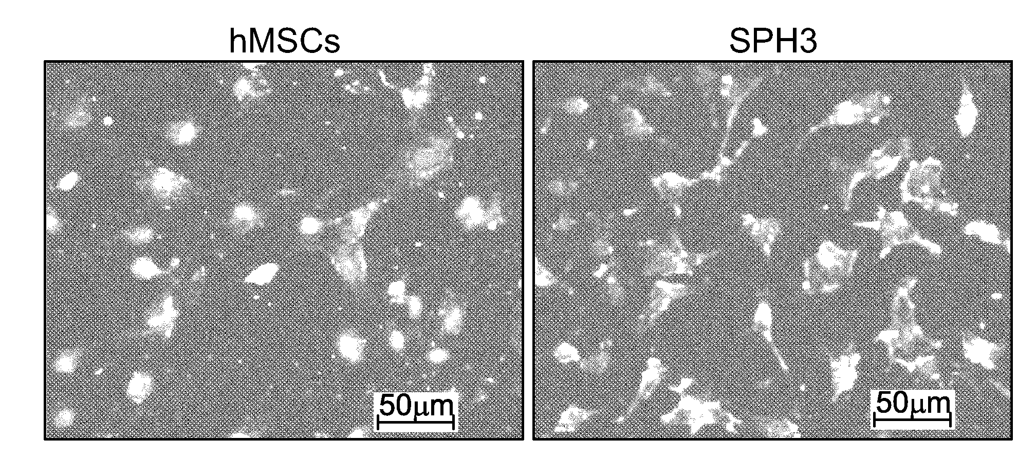
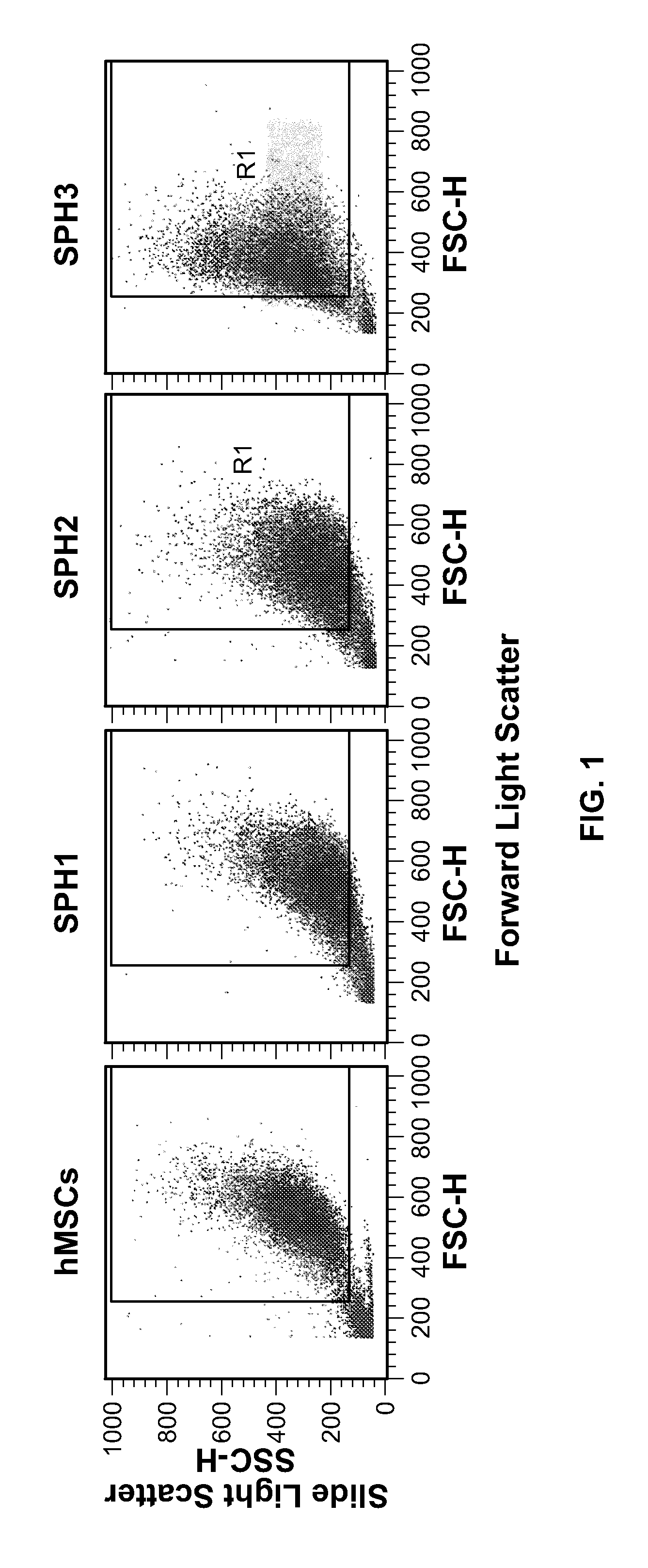
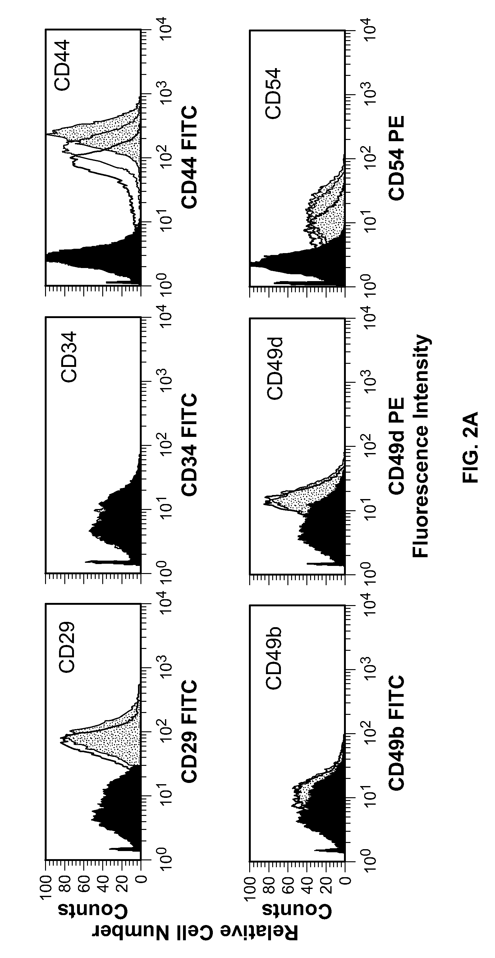
[0072]Cells dissociated from a monolayer or the spheroids gave rise to homogeneous populations. Cells from spheroids had smaller size and higher granularity (FIG. 1). Expression of 19 markers commonly used to characterize hMSCs by flow cytometry (FIG. 2) was examined. HMSCs from a monolayer and the spheroids were positive for CD29, CD44, CD54, CD55, CD73, CD90, CD105, CD166 and HLA-I. Neither cells from a monolayer nor cells from the spheroids were positive for c-met, CD28, CD31, CD34, CD38, CD117 or CD209. The effects of trypsinization on the expression of cell surface markers by hMSCs were also analyzed. Treatment with trypsin for 90 min did not affect the expression of CD49d and CD166 in hMSCs. Detection of CD29, CD44, CD54, CD55, CD90, CD105 and HLA-I was sensitive to trypsin-EDTA treatment. Nevertheless, all antigens that tested positive after 5 min of trypsinization remained positive after 90 min of incubation with trypsin (FIG....
example 3
[0075]Secretion of SDF-1 by hMSCs and hMSC Spheroids
[0076]It has been reported that the expression of CXCR4 by hMSCs inversely correlates with the expression of SDF-1 (Lisignoli, G., 2006, J. Cell. Physiol. 207:364-73). The expression of SDF-1 mRNA was 10-fold down regulated in hMSC spheroids. To determine how formation of the spheroids affects SDF-1 secretion by hMSCs. SDF-1 was measured in media conditioned by a monolayer of hMSCs or 1, 2, or 3 day old hMSC spheroids. The amount of SDF-1 in conditioned media was normalized to the cell number. Relative changes in the secretion of SDF-1 are shown in FIG. 6. There was a statistically significant decline (t-test, p-value<0.05) in SDF-1 secretion by 2 and 3 day old hMSC spheroids in comparison with hMSCs from a monolayer.
PUM
 Abstract
Description
Claims
Application Information
 - R&D
- Intellectual Property
- Life Sciences
- Materials
- Tech Scout
- Unparalleled Data Quality
- Higher Quality Content
- 60% Fewer Hallucinations
Browse by: Latest US Patents, China's latest patents, Technical Efficacy Thesaurus, Application Domain, Technology Topic, Popular Technical Reports.
© 2025 PatSnap. All rights reserved.Legal|Privacy policy|Modern Slavery Act Transparency Statement|Sitemap|About US| Contact US: help@patsnap.com


