Eureka AIR delivers breakthrough ideas for toughest innovation challenges, trusted by R&D personnel around the world.

Methods and arrangements for analysis, diagnosis, and treatment monitoring of vocal folds by optical coherence tomography

a technology analysis method, applied in the field of optical coherence tomography analysis, diagnosis and treatment of vocal fold monitoring, can solve the problems of voice disorders, slp in a vulnerable location, and affecting the health of patients, and achieves the effects of reducing the risk of vocal folds, and reducing the effect of vocal folds

Inactive Publication Date: 2011-09-15
THE GENERAL HOSPITAL CORP
View PDF105 Cites 13 Cited by
  • Summary
  • Abstract
  • Description
  • Claims
  • Application Information

AI Technical Summary

Benefits of technology

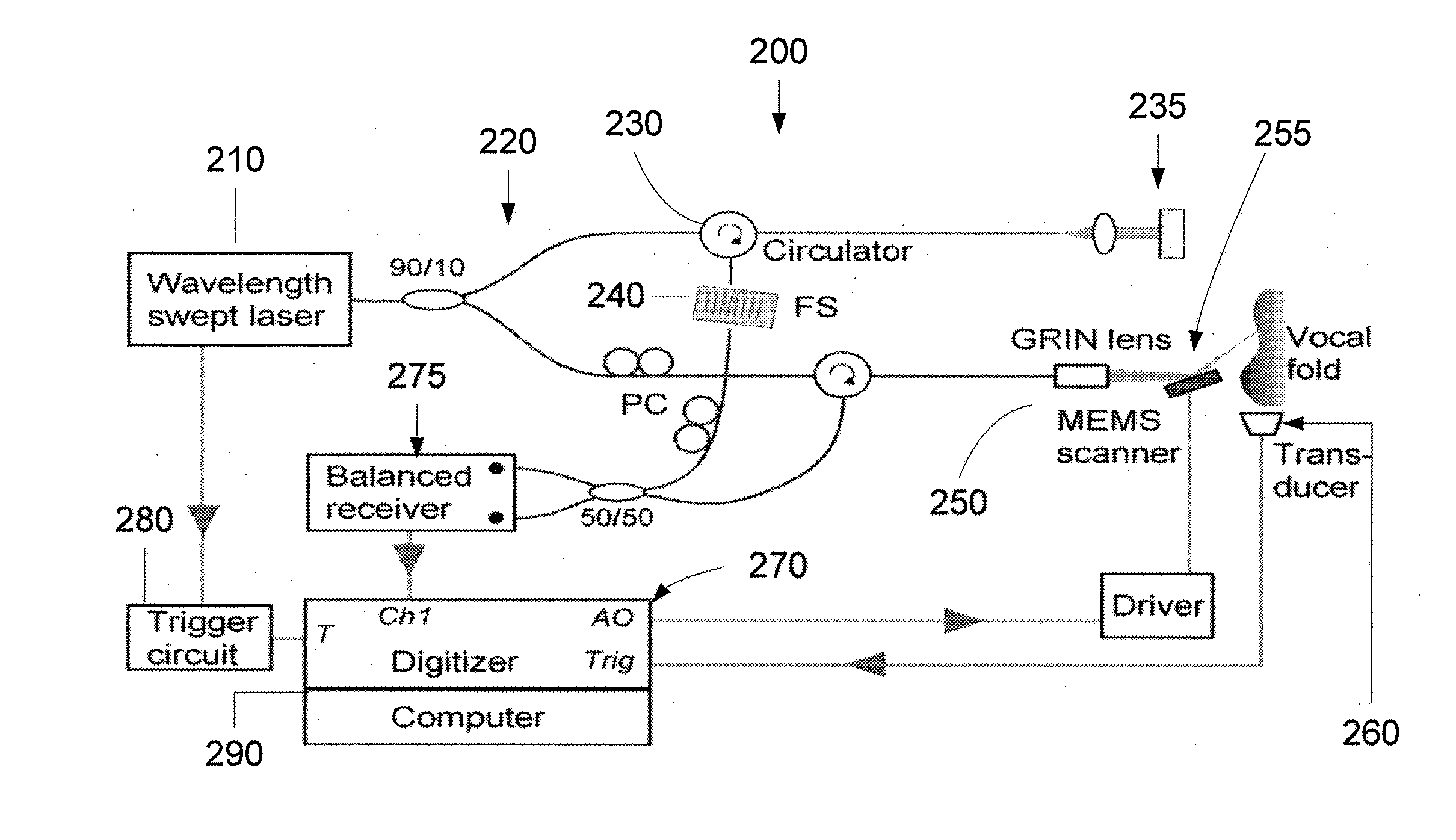
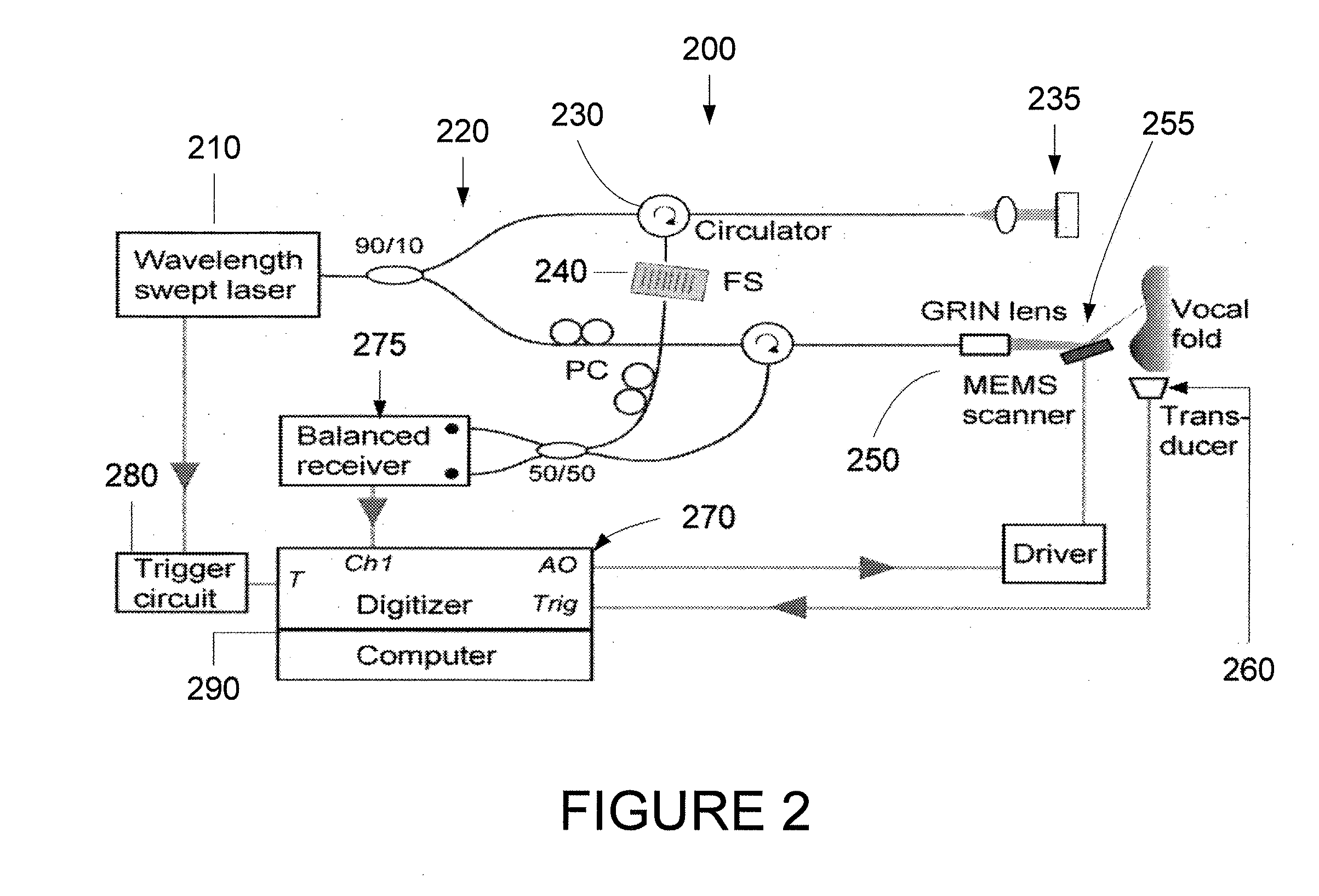
[0013]Exemplary embodiments of the present disclosure can address at least most of the above-described needs and / or issues by facilitating imaging of the vocal fold motion quantitatively with four-dimensional (e.g., 4D: x,y,z and time) resolution. The exemplary embodiments of the present disclosure can utilize Fourier-domain optical coherence tomography (OCT)—herein also referred to as optical frequency domain imaging (OFDI), a procedure that is described in, e.g., S. H., Tearney, G. J., de Boer, J. F., Iftimia, N. & Bouma, B. E., High-speed optical frequency-domain imaging, Optics Express 11, pp. 2953-2963 (2003). An exemplary embodiment of the procedure, system and method according to the present disclosure can facilitate a production of a sequence of high-resolution 3D images of the vocal folds over a full cycle of vibration. In combination with standard laryngeal endoscopes, such exemplary embodiments can be used in a similar way as conventional stroboscopy is used, while facilitating the examination of not only the surface, but also the motion of the entire volume of the essential superficial tissues, quantitatively.
[0014]To rapidly image vibrating vocal folds, according to one exemplary embodiment of the present disclosure, image acquisition methods can be provided which can rely on a use of a voice signal from a microphone, an electroglottograph (EGG) or a subglottic pressure transducer for synchronization. Stable phonation and repeatable triggering, as used in conventional stroboscopy, is necessary. The probe laser beam can be scanned across the vocal fold, acquiring axial profiles at each spatial location and each temporal phase of motion. A subsequent image reconstruction based on the timing synchronization with the voice signal will produce a sequence of high-resolution 3D images of the vocal folds over a full cycle of vibration. A dynamic cross-sectional imaging of vibrating vocal folds can be achieved, which has not been previously obtained demonstrated.
[0016]Improved diagnosis of voice disorders: The exemplary embodiments of the present disclosure can facilitate the clinicians to compare volumetric vocal fold motion of normal and diseased vocal folds quantitatively and observe the location and extent of subsurface pathology in both dynamic and static modes. This can elucidate how pathologies affect vocal fold motion and resulting voice quality, which in turn should lead to improvements in treatment methods.
[0018]Indeed, exemplary embodiments of the present disclosure provide endoscopic technology methods, systems and arrangements can be provided which can facilitate with the diagnosis and treatment of patients with voice disorders. For example, it is possible to use high-speed optical coherence tomography (OCT) methods and systems, combined with physiological triggering, to image vibrating vocal folds with high spatial and temporal resolution. Oscillations of the surface and interior structure of the vocal fold can then be viewed in slow-motion, providing essentially a dynamic histological cross-section. The ability to view previously hidden events and quantitatively capture the motion in three dimensions can indicate that the exemplary embodiments of the present disclosure can be useful and sought after.

Problems solved by technology

Voice disorders can disrupt normal human communication causing far-reaching negative personal and social-economic consequences for those affected.
Thus, diseases or injuries that affect these waves can often result in voice disorders.
A healthy layer of SLP is important to a good voice, but the SLP in a vulnerable location, and is frequently damaged by diseases or trauma.
One problem in the field of Laryngology is how to best treat diseases that affect these thin layers while preserving the mucosal wave and good voice production.
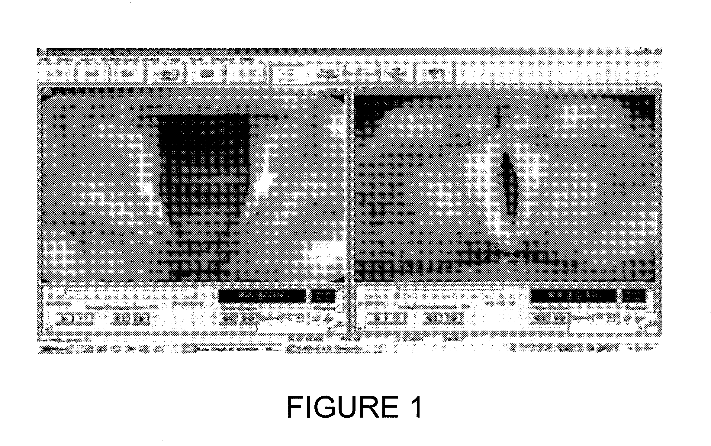
Despite the ubiquity and utility of videostroboscopy, this procedure is highly qualitative, and the data obtained can be quite subjective.
High-speed imaging overcomes some of the limitations of stroboscopy; however, it is still a 2D method limited to viewing the vocal fold surfaces.
Previously, satisfactory method or system for assessing the biomechanics of these materials in situ may be unknown
235-239, 2009) may not be satisfactory due to suboptimal temporal and / or spatial resolution.
However, until recently, OCT procedure has been too slow for providing a comprehensive 3D microscopic imaging, and therefore has been relegated to a point-sampling technique with a field of view comparable to a conventional biopsy.
The image acquisition speed provided by the OFDI techniques, however, may not be fast enough to capture vocal fold motion directly.
Such specification may not currently be attainable due to various technical problems.
Furthermore, it can result in a substantially decreased signal-to-noise ratio (SNR) and clinically unacceptable poor image quality.

Method used


Image

  • Methods and arrangements for analysis, diagnosis, and treatment monitoring of vocal folds by optical coherence tomography
  • Methods and arrangements for analysis, diagnosis, and treatment monitoring of vocal folds by optical coherence tomography
  • Methods and arrangements for analysis, diagnosis, and treatment monitoring of vocal folds by optical coherence tomography

Examples


Embodiment Construction

[0013]Exemplary embodiments of the present disclosure can address at least most of the above-described needs and / or issues by facilitating imaging of the vocal fold motion quantitatively with four-dimensional (e.g., 4D: x,y,z and time) resolution. The exemplary embodiments of the present disclosure can utilize Fourier-domain optical coherence tomography (OCT)—herein also referred to as optical frequency domain imaging (OFDI), a procedure that is described in, e.g., S. H., Tearney, G. J., de Boer, J. F., Iftimia, N. & Bouma, B. E., High-speed optical frequency-domain imaging, Optics Express 11, pp. 2953-2963 (2003). An exemplary embodiment of the procedure, system and method according to the present disclosure can facilitate a production of a sequence of high-resolution 3D images of the vocal folds over a full cycle of vibration. In combination with standard laryngeal endoscopes, such exemplary embodiments can be used in a similar way as conventional stroboscopy is used, while facili...


PUM


Abstract

Exemplary embodiments of an apparatus and a method can be provided. For example, a first information can be obtained for at least one signal that is (i) at least partially periodic and (ii) associated with at least one structure. In addition, a second information associated with the structure can be generated at multiple time points within a single cycle of the at least one signal. The second information can include information for the structure below a surface thereof. Further, it is possible to generate a third information based on the first information and the second information, where the third information is associated with at least one characteristic of the structure.

Description

CROSS-REFERENCE TO RELATED APPLICATION(S)[0001]This application is based upon and claims the benefit of priority from U.S. patent application Ser. No. 61 / 267,780, filed on Dec. 8, 2009, the entire disclosure of which is incorporated herein by reference.FIELD OF THE DISCLOSURE[0002]Exemplary embodiments of the present disclosure relate to the utilization of optical coherence tomography for obtaining information regarding at least one anatomical structure, and more particularly to exemplary methods and arrangements for analysis, diagnosis, and treatment monitoring of vocal folds using optical coherence tomography procedures.BACKGROUND INFORMATION[0003]Voice disorders can disrupt normal human communication causing far-reaching negative personal and social-economic consequences for those affected. It is estimated that about 7.5 million Americans suffer from voice disorders. One of the main causes of voice disorders can be damage to the subepithelial layers of laryngeal vocal fold tissue...

Claims


Application Information

Patent Timeline
Patent Type & Authority Applications(United States)
IPC IPC(8): A61B6/00
CPCA61B1/00172A61B1/2673A61B5/7285A61B5/0084A61B5/6852A61B5/0066
Inventor YUN, SEOK-HYUNKOBLER, JAMES B.
Owner THE GENERAL HOSPITAL CORP
Why Eureka
Eureka Blog
Learn More