Method for simultaneous multi-slice magnetic resonance imaging

a magnetic resonance imaging and simultaneous technology, applied in the field of parallel magnetic resonance imaging, can solve the problems of insufficient speed and efficiency for more advanced imaging applications, reducing the efficiency of conventional epi methods, and inefficient diffusion weighted imaging techniques, etc., to achieve the largest possible separation, reduce the tilt and blurring of pixels, and reduce the effect of aliasing pixels

Active Publication Date: 2011-10-20
THE GENERAL HOSPITAL CORP
View PDF7 Cites 87 Cited by
  • Summary
  • Abstract
  • Description
  • Claims
  • Application Information

AI Technical Summary

Benefits of technology

[0033]The present invention overcomes the aforementioned drawbacks by providing a method for simultaneous multi-slice magnetic resonance imaging (“MRI”) that allows for more reliable separation of aliased pixels than currently available multi-slice MRI methods; the provided method achieves this separation by shifting the aliased pixels by a selected percentage of the imaging field-of-view (“FOV”). In general, the percentage of the FOV to shift the aliased pixels is selected such that the largest possible separation between the aliased pixels is achieved. This selection takes into consideration several factors including the number of slice locations simultaneously acquired, the separation distance of the slice locations, and the in-plane and longitudinal variation of the sensitivity of the RF coil employed to acquire the signal data. Moreover, this FOV shift is achieved while substantially mitigating pixel tilt and blurring, which are problems common to currently available methods. The provided method decreases the amount of time required to acquire the image data from multiple slice locations, and increases the signal-to-noise ratio (“SNR”) achievable compared to currently available multi-slice MRI methods.
[0034]It is an aspect of the invention to provide a method for imaging techniques such as echo planar imaging (“EPI”), in which image data may be simultaneously acquired from multiple slice locations. The EPI pulse sequence employed when practicing the present invention includes a series of magnetic gradient field “blips” applied along the slice-encoding direction contemporaneously with the phase-encoding blips common to EPI sequences. Successive slice-encoding blips are different and designed such that phase accruals along the phase-encoding direction are substantially mitigated, while providing that signal information for every other slice location is shifted by a percentage of the imaging FOV, such as 50 or 33 percent. This percentage FOV shift in the image domain provides for more reliable separation of the aliased signal information. It should be apparent, however, that pulse sequences other than EPI that sample multiple lines of k-space following a single RF excitation can also be employed when practicing the present invention. For example, pulse sequences such as gradient and spin echo (“GRASE”) pulse sequences and fast spin echo (“FSE”) pulse sequences, such as rapid acquisition with relaxation enhancement (“RARE”) sequences.
[0035]It is another aspect of the invention to provide an imaging method, in which imaging speed and sensitivity are improved. While conventional two-dimensional EPI methods are fast (for example, a single slice can be acquired in around 100 milliseconds), it is inefficient and not fast enough for more advanced imaging applications, such as advanced diffusion weighted imaging techniques. In particular, the efficiency of conventional EPI methods is reduced by the two-dimensional slice-by-slice nature of the acquisition. For example, in a standard diffusion imaging tractography application, sixty-four slices are acquired to cover the spatial extent of a patient's head. In this example, the fraction of time the MRI system spends for acquiring each slice is one sixty-fourth of the total imaging time. The provided method improves the efficiency of EPI acquisitions by reducing this fraction by 2-6 fold. Multiple slices are excited simultaneously and reconstructed separately using information from the detector array system. Given successful separation of the simultaneously excited slices, the scan time for a given EPI acquisition is reduced by a factor of up to the number of slices simultaneous excited. This improvement in imaging speed is achieved with only a minor penalty in sensitivity; improving the efficiency of the encoding improves the sensitivity per unit time.
[0036]It is yet another aspect of the invention to provide a method for diffusion weighted imaging in which imaging time is significantly reduced and SNR is significantly increased. These benefits may be achieved using, for example, an EPI pulse sequence modified in the manner described above. As a result, diffusion weighted image data can be acquired for such modern diffusion imaging methods as Q-ball imaging (“QBI”), high angular resolution diffusion imaging (“HARDI”), and diffusion spectrum imaging (“DSI”), in clinically acceptable durations of time. It is contemplated that such decreased in required scan time will provide amenable clinical applications of modern diffusion imaging methods. For example, given the N-fold efficiency gain provided by simultaneously exciting and acquiring image data from N image slice location, scan time can be reduced by employing the desired number of diffusion encoding steps in less time than previously achievable. Similarly, the scan time can be kept the same, but the number of diffusion encoding steps increased. This increase in the number of achievable encoding steps allows more nuanced diffusion information to be obtained.

Problems solved by technology

Moreover, this FOV shift is achieved while substantially mitigating pixel tilt and blurring, which are problems common to currently available methods.
While conventional two-dimensional EPI methods are fast (for example, a single slice can be acquired in around 100 milliseconds), it is inefficient and not fast enough for more advanced imaging applications, such as advanced diffusion weighted imaging techniques.
In particular, the efficiency of conventional EPI methods is reduced by the two-dimensional slice-by-slice nature of the acquisition.

Method used


Image

  • Method for simultaneous multi-slice magnetic resonance imaging
  • Method for simultaneous multi-slice magnetic resonance imaging
  • Method for simultaneous multi-slice magnetic resonance imaging

Examples


Embodiment Construction

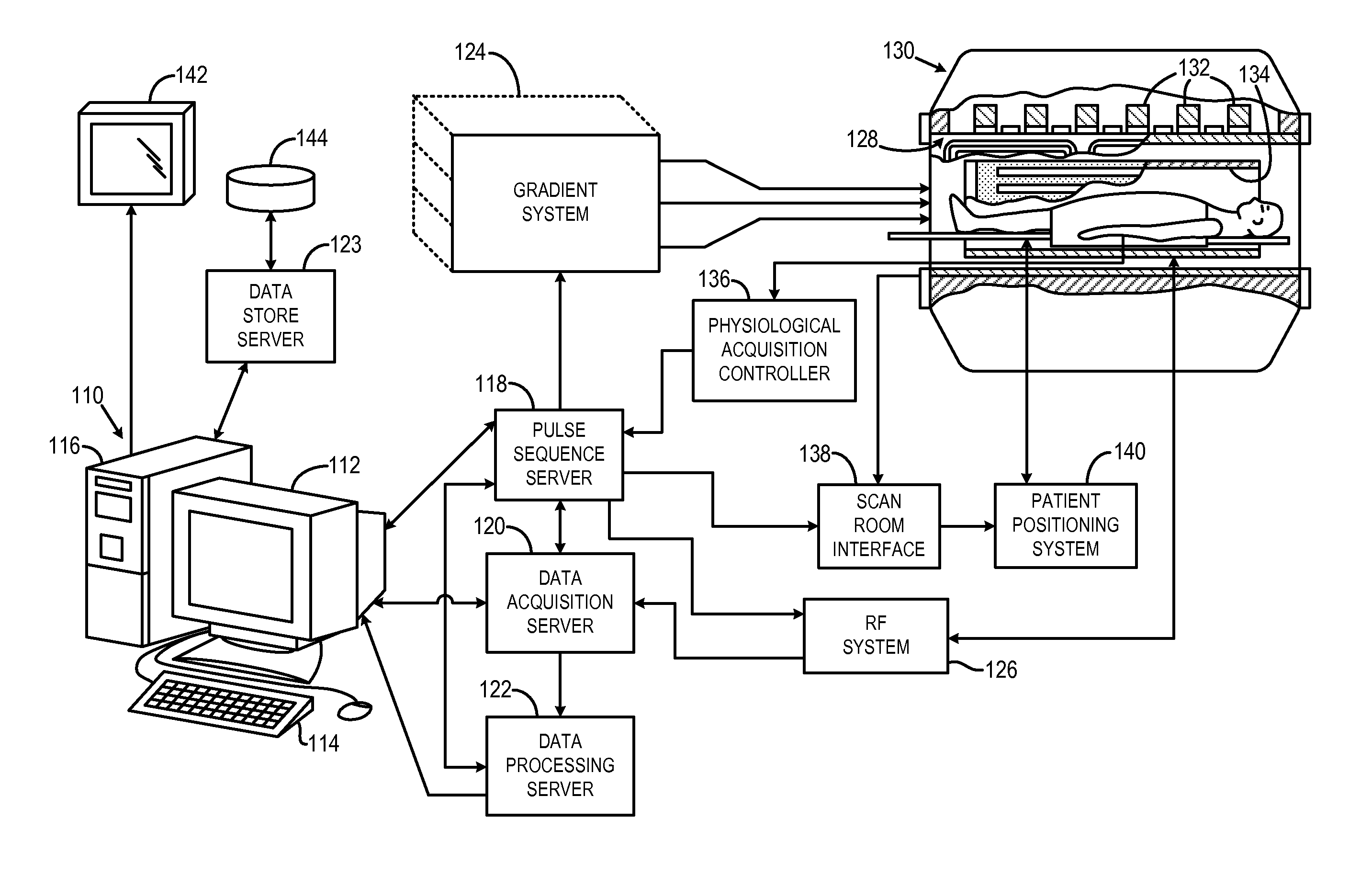
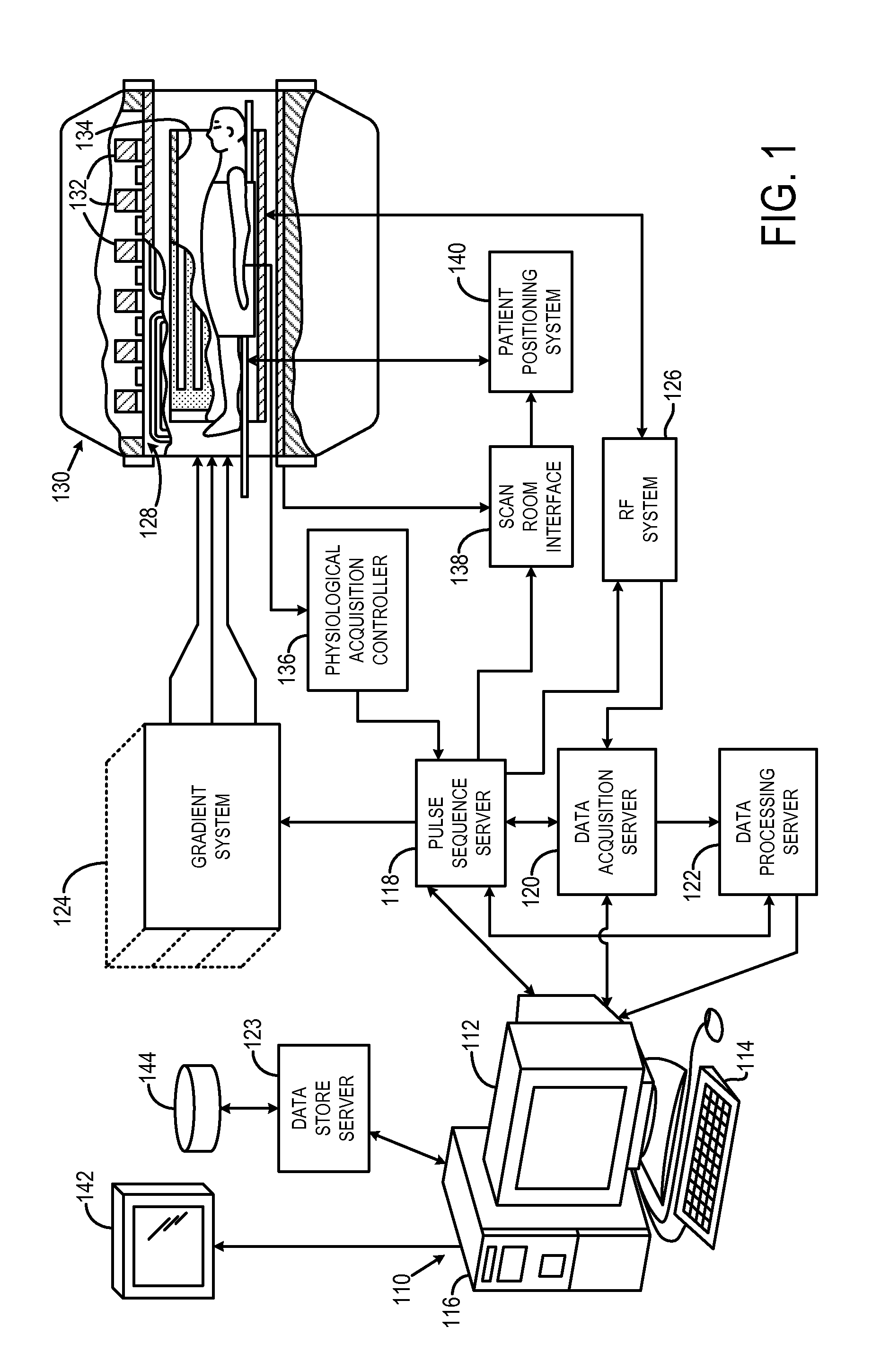
[0048]The succeeding description is provided with reference to the following orthogonal spatial encoding directions common to magnetic resonance imaging (“MRI”): a slice-encoding direction, a phase-encoding direction, and a frequency-encoding direction. By way of example, as referred to herein, the slice-encoding direction corresponds to the z-direction in the image domain, which is aligned along the longitudinal axis of the bore of an exemplary MRI system, and the kz-direction in k-space. In this manner, the obtained images are transverse, or axial, images lying in the x-y plane. Accordingly, as referred to herein, the phase-encoding direction corresponds to the y-direction in the image domain, and the ky -direction in k-space; and the frequency-encoding direction corresponds to the x-direction in the image domain, and the kx-direction in k-space. It will be appreciated by those skilled in the art that the choice of these directions is arbitrary and any suitable permutation of thes...


PUM


Abstract

A method for multi-slice magnetic resonance imaging, in which image data is acquired simultaneously from multiple slice locations using a radio frequency coil array, is provided. By way of example, a modified EPI pulse sequence is provided, and includes a series of magnetic gradient field “blips” that are applied along a slice-encoding direction contemporaneously with phase-encoding blips common to EPI sequences. The slice-encoding blips are designed such that phase accruals along the phase-encoding direction are substantially mitigated, while providing that signal information for each sequentially adjacent slice location is cumulatively shifted by a percentage of the imaging FOV. This percentage FOV shift in the image domain provides for more reliable separation of the aliased signal information using parallel image reconstruction methods such as SENSE. In addition, the mitigation of phase accruals in the phase-encoding direction provides for the substantial suppression of pixel tilt and blurring in the reconstructed images.

Description

STATEMENT REGARDING FEDERALLY SPONSORED RESEARCH[0001]This invention was made with United States government support awarded by the following agency: National Institutes of Health NIH RR1475. The United States government has certain rights in this invention.BACKGROUND OF THE INVENTION[0002]The field of the invention is methods and systems for magnetic resonance imaging. More particularly, the invention relates to methods and systems for parallel magnetic resonance imaging, in which k-space data is acquired from multiple slice locations substantially contemporaneously.[0003]Magnetic resonance imaging (“MRI”) uses the nuclear magnetic resonance (“NMR”) phenomenon to produce images. When a substance such as human tissue is subjected to a uniform magnetic field (polarizing field B0), the individual magnetic moments of the nuclei in the tissue attempt to align with this polarizing field, but precess about it in random order at their characteristic Larmor frequency. If the substance, or ti...

Claims


Application Information

Patent Timeline
Patent Type & Authority Applications(United States)
IPC IPC(8): G01R33/48
CPCG01R33/4835G01R33/543G01R33/5616G01R33/5611
Inventor SETSOMPOP, KAWINWALD, LAWRENCE L.
Owner THE GENERAL HOSPITAL CORP
Why Eureka
Try Eureka