Hard tissue anchors and delivery devices

a technology of hard tissue and anchors, which is applied in the direction of ligaments, surgical staples, therapy, etc., can solve the problems of limiting the area and types of tissue available for suturing thereto, migration and pull-out, and achieve the effect of reducing the migration of the device and strengthening the attachmen

Active Publication Date: 2011-10-20
TC1 LLC
View PDF4 Cites 13 Cited by
  • Summary
  • Abstract
  • Description
  • Claims
  • Application Information

AI Technical Summary

Benefits of technology

[0007]The present invention provides devices, systems and methods for anchoring medical devices to hard tissues, such as bones or bony structures, particularly vertebrae. A variety of medical devices are used to treat portions of the anatomy which reside near bones or bony structures within the body of a patient. The devices and systems of the present invention are suitable for use with many of such medical devices and specialized devices used for particular treatments. By anchoring these directly to the surrounding hard tissue, the devices are anchored closer to the source of treatment. This provides additional stability and reduces migration of the device at the treatment site. Also, by attaching to hard tissue rather than soft tissue, a stronger attachment is often able to be made.
[0009]In some embodiments, the head includes a channel connected to the aperture, wherein the channel is configured allow passage of the element from outside of the head to the aperture. In some instances, the head is adjustable to close the channel, such as by deformation of the head. Optionally, deformation of the head may secure the element within the aperture. In some embodiments, the head further comprises a grommet disposed within the aperture. The grommet may assist in holding the element within the aperture.
[0013]In some instances, the method further comprises implanting the element in the body. Such implanting may occur before the positioning step of positioning the element within the aperture. This allows the hard tissue anchors to be utilized with existing implanted systems.

Problems solved by technology

Thus, the device may only be sutured at these locations, limiting the areas and types of tissue available for suturing thereto.
Such distance and instability of anchoring tissue can contribute to lead migration and pull-out.
And, lead migration and pull-out are common problems associated with SCS.

Method used


Image

  • Hard tissue anchors and delivery devices
  • Hard tissue anchors and delivery devices
  • Hard tissue anchors and delivery devices

Examples


Embodiment Construction

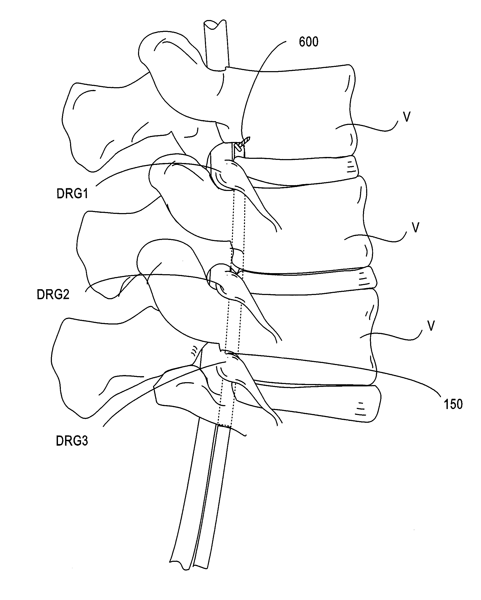
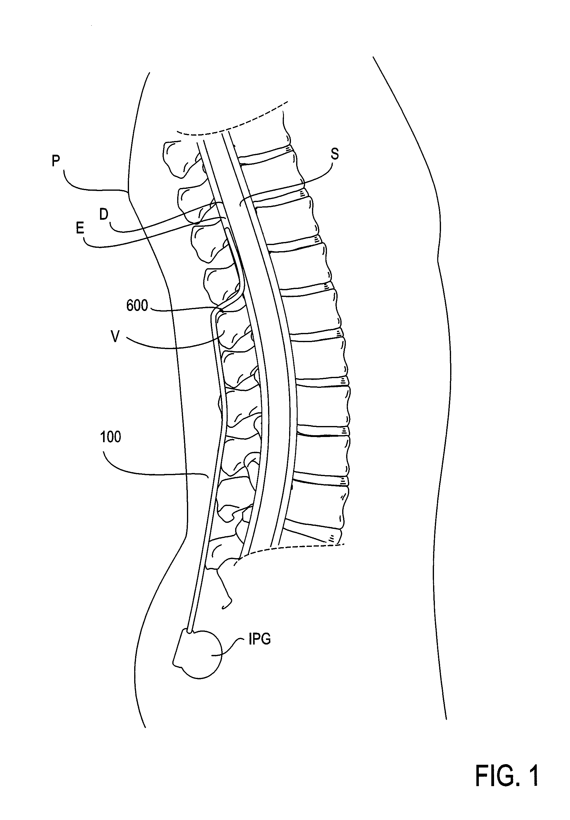
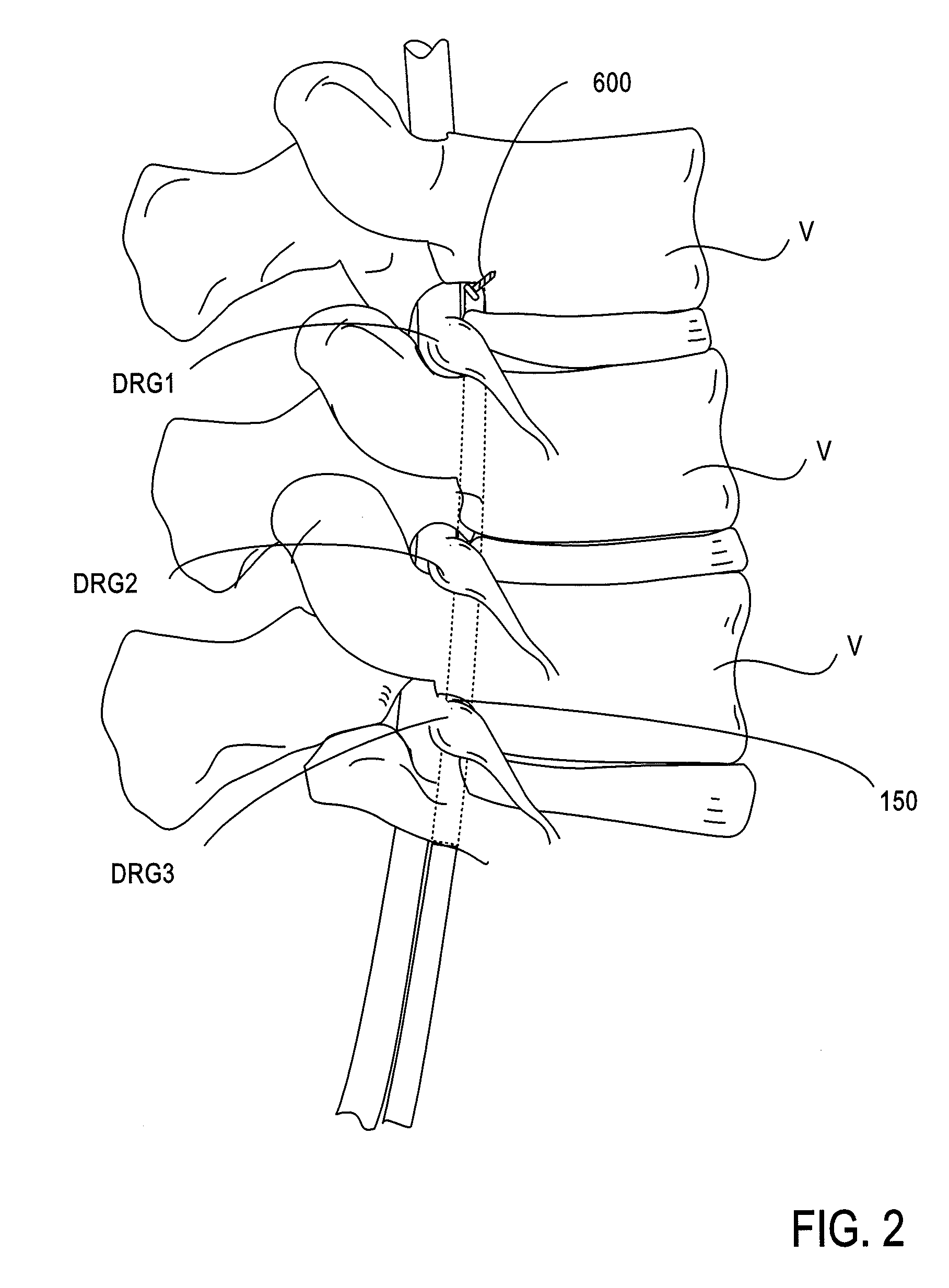
[0039]The present invention provides devices, systems and methods for anchoring medical devices to hard tissues, such as bones or bony structures, particularly vertebrae. A variety of medical devices are used to treat portions of the anatomy which reside near bones or bony structures within the body of a patient. For example, spinal cord stimulators (SCS) are positioned along the spinal column to treat pain. FIG. 1 illustrates a conventional SCS system comprising an implantable lead 100 and an implantable power source or implantable pulse pulse generator IPG. Using fluoroscopy, the lead 100 is implanted into the epidural space E of the spinal column S and positioned against the dura layer of the spinal cord. The lead 100 is implanted either through the skin via an epidural needle (for percutaneous leads) or directly and surgically through a mini laminotomy operation (for paddle leads). In either case, the leads 100 extend from the spinal column S to the IPG which is remotely implant...


PUM


Abstract

The present invention provides devices, systems and methods for anchoring medical devices to hard tissues, such as bones or bony structures, particularly vertebrae. By anchoring these medical devices directly to the surrounding hard tissue, the devices are anchored closer to the source of treatment. This provides additional stability and reduces migration of the device at the treatment site. Also, by attaching to hard tissue rather than soft tissue, a stronger attachment is often able to be made.

Description

CROSS-REFERENCES TO RELATED APPLICATIONS[0001]This application is a continuation of U.S. patent application Ser. No. 11 / 952,081, filed on Dec. 6, 2007, which claims priority of U.S. Provisional Patent Application No. 60 / 873,549, filed on Dec. 6, 2006, which is incorporated herein by reference for all purposes.STATEMENT AS TO RIGHTS TO INVENTIONS MADE UNDER FEDERALLY SPONSORED RESEARCH AND DEVELOPMENT[0002]NOT APPLICABLEREFERENCE TO A “SEQUENCE LISTING,” A TABLE, OR A COMPUTER PROGRAM LISTING APPENDIX SUBMITTED ON A COMPACT DISK[0003]NOT APPLICABLEBACKGROUND OF THE INVENTION[0004]A variety of implantable medical devices are used to treat portions of the anatomy which reside near bones or bony structures within the body of a patient. Such devices are typically anchored in place by suturing portions of the device to surrounding soft tissue. Often the device includes suture holes designed specifically for this purpose at predetermined locations along the device. Thus, the device may onl...

Claims


Application Information

Patent Timeline
Patent Type & Authority Applications(United States)
IPC IPC(8): A61B17/56
CPCA61B17/0642A61B17/70A61B17/86A61N1/0558A61B17/8891A61B17/92A61B2017/0647A61B17/8883A61B17/861A61B17/864A61B2017/0648A61B17/8886A61B17/8875
Inventor BURDULIS, ALBERT G.
Owner TC1 LLC
Why Eureka
Try Eureka