Monitoring, Managing and/or Protecting System and Method for Non-Targeted Tissue

a non-targeted tissue and monitoring system technology, applied in the field of medical systems for monitoring and protecting non-targeted esophageal tissue, can solve the problems of non-targeted tissue tissue necrosis, known endocardial catheter pulmonary vein ostia ablation systems relating to monitoring, maintenance, and/or control, and the effect of treat the disease, and not being able to detect the diseas

Active Publication Date: 2011-11-10
ST JUDE MEDICAL ATRIAL FIBRILLATION DIV
View PDF13 Cites 47 Cited by
  • Summary
  • Abstract
  • Description
  • Claims
  • Application Information

AI Technical Summary

Benefits of technology

[0021]In accordance with another aspect of the present disclosure, a method of monitoring, managing and protecting a region of non-targeted tissue during an ablation procedure performed on targeted tissue proximate the region of non-targeted tissue is provided. The method comprises a first step of inserting an esophageal catheter in the esophagus such that an electrode on the esophageal catheter is aligned with and facing the posterior wall of the atrium. Next, an anchoring means is deployed such that the anterior surface of the anchoring means is facing the posterior wall of the atrium. After inserting an ablation electrode into the endocardial chamber, an electrical response assessment system or component is operatively connecting to the electrode on the esophageal catheter and may be operatively connected to the electrode in the endocardial chamber. The electrical response assessment system may be separate from, replace, or be incorporated into an electronic control system (ECS), which may include an electronic control unit (ECU) for assistance in controlling an RCGS and the ablation catheter. Once connected, the electrical characteristics of the tissue of the esophagus or the tissue between the esophageal and ablation electrodes can be measured and managed, including, during ablation of the posterior atrial wall, the electrical characteristics measured by the electrical response assessment system or component may be used to control or prevent ablation energy being delivered to the endocardial electrode. The monitoring, managing and / or protecting system or component can be used in conjunction with the RCGS, or separate from the RCGS and independent of the ablation catheter.

Problems solved by technology

However, these known devices are not without their drawbacks.
For instance, a drawback in known endocardial catheter pulmonary vein ostia ablation systems relates to the monitoring, maintenance, and / or control of the temperature in non-targeted tissue proximate the targeted ablation site during the ablation procedure.
More particularly, when certain heart tissue is being ablated, the energy emitted from the ablating device may be strong enough or generate a high enough temperature to cause tissue necrosis in non-targeted tissue.
As an example, since the esophagus which is generally located posterior to the atrium, RF ablation on the posterior atrial wall has been known to cause serious complications such as esophageal fistula.
Such complications have even led to death of patients following RF ablation for treatment of Atrial Fibrillation (AF).
Such complications are created by thermally-mediated damage to the esophagus due to overheating of the esophagus from uncontrollable application of RF energy during ablation on the posterior atrial wall.
However, thermal monitoring of the luminal wall of the esophagus is generally inadequate in preventing such esophageal complications caused by ablation of the atrial wall.
At the outset, the temperature measurement of the esophageal wall is generally unreliable due the difficulty in accurately positioning the thermal probe in the esophagus relative to the ablation site on the atrial wall.
Even if the thermal probes are properly positioned in the esophagus, the threshold cut-off temperature to prevent esophageal complications cannot be set a priori.
Further, the electric field and thermal properties of these tissue layers are not readily known.
Based on these unknown variables, the thermal gradient from the endocardial site to the esophageal wall often cannot be readily determined.
Consequently, the maximum temperature that can be allowed on the endocardial wall without creating dangerously high temperatures on the esophagus cannot be readily determined.
The difficulty of reliably determining such temperatures on the endocardial side is further compounded by the unreliability of determining the endocardial tissue temperature during ablation.
If the thermal probes could reliably measure temperature of the esophageal luminal wall, that thermal information arrives after the fact that the esophageal wall has already attained potentially harmful temperature.
Thus, by the time the thermal probe senses a temperature rise in the luminal wall of the esophagus, it is usually too late for any preventative or corrective action.
Because of the aforementioned limitations, thermal monitoring of the esophageal luminal wall fails to provide a priori information that can be reliably used to adequately protect and prevent thermally-mediated esophageal injury during endocardial RF ablation of the posterior atrial wall during atrial fibrillation treatment.

Method used


Image

  • Monitoring, Managing and/or Protecting System and Method for Non-Targeted Tissue
  • Monitoring, Managing and/or Protecting System and Method for Non-Targeted Tissue
  • Monitoring, Managing and/or Protecting System and Method for Non-Targeted Tissue

Examples


Embodiment Construction

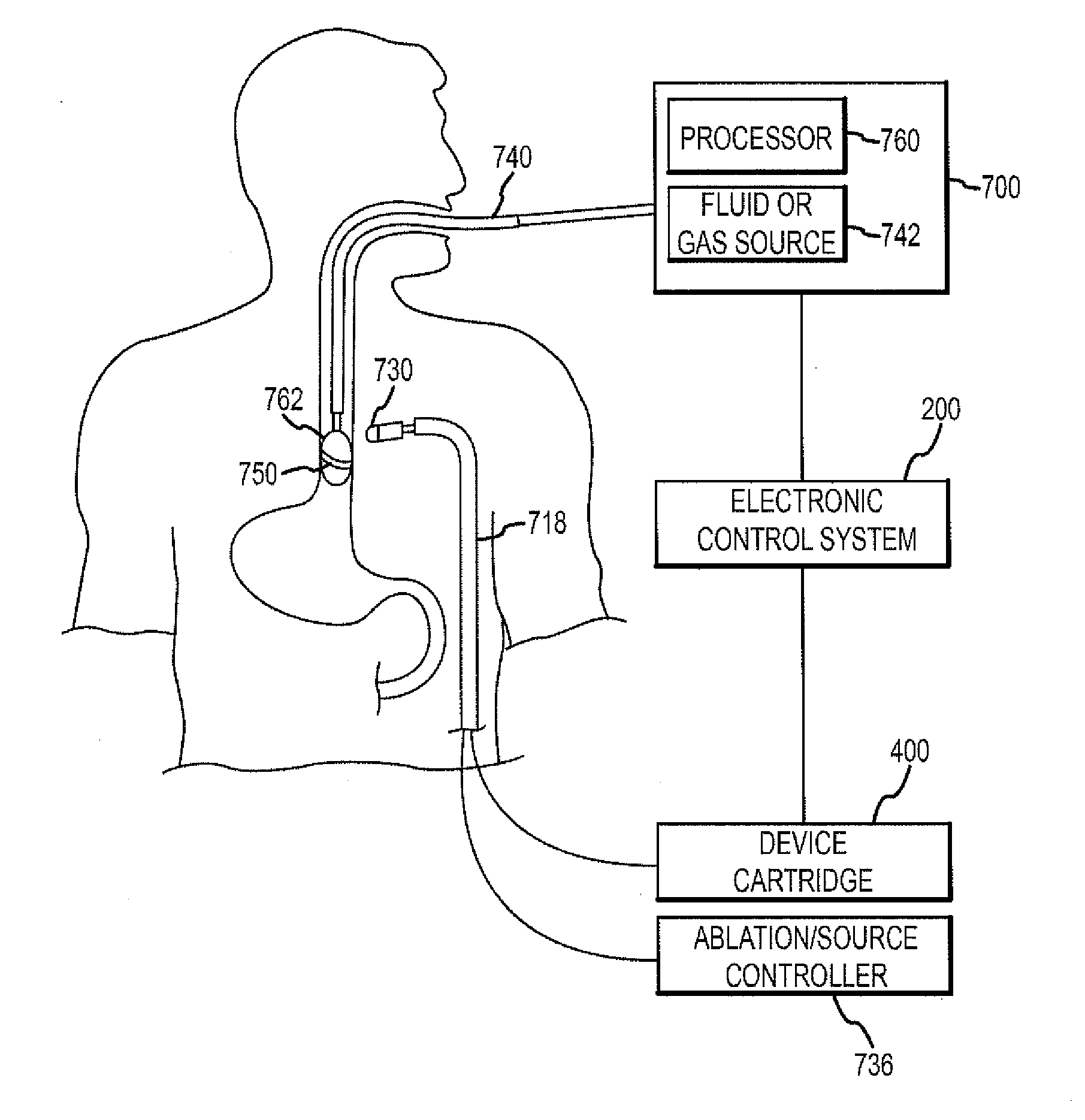
[0086]The present disclosure is directed to a monitoring, managing and / or protecting system or component for use with various medical procedures, such as ablation procedures, to monitor and protect non-targeted tissue proximate to the location of the ablation procedure. The present invention is of aid in a number of procedure types, whether doctor controlled or automatically controlled, e.g., by a robotic system or magnetic system. The inventions monitor, manage and protect the non-targeted tissue including notifying the practitioner or the system performing the ablation procedure when the non-targeted tissue may be damaged and / or controlling or stopping the ablation procedure when necessary to protect the non-targeted tissue.

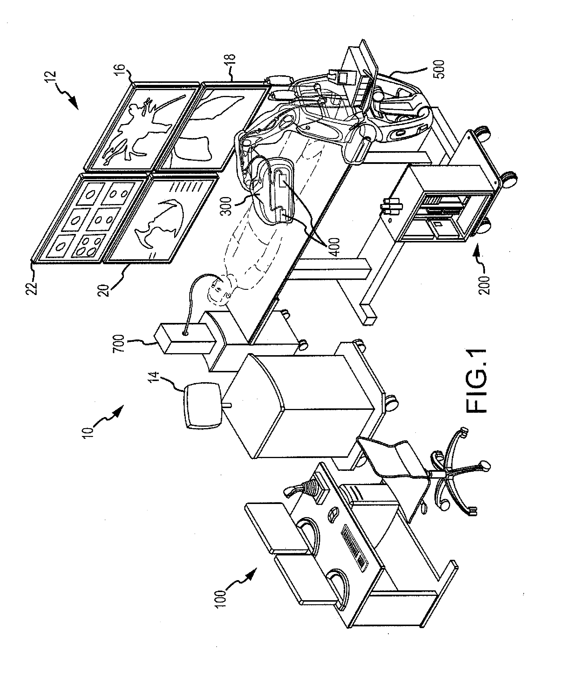
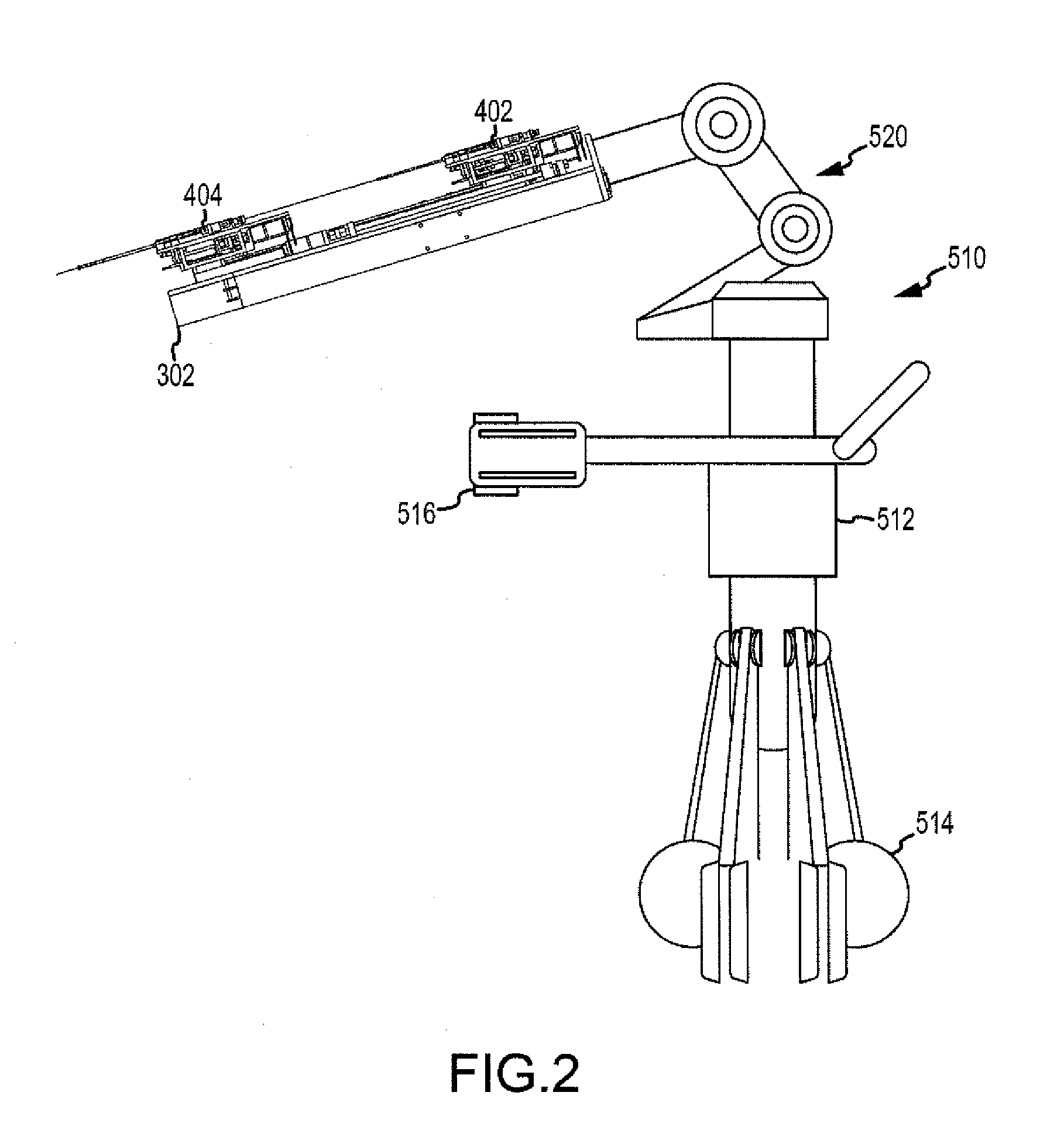
[0087]Before proceeding to a detailed description of the non-targeted tissue monitoring, managing and protecting system, a brief overview (for context) of one possible robotic control and guidance system (RCGS) for manipulating a medical device, such an ablatio...


PUM


Abstract

A monitoring, managing and protecting system is provided that includes a monitoring probe working in conjunction with an ablating device. The probe is configured to be positioned in close proximity to a region of non-targeted tissue proximate an ablation site of targeted tissue and to be operatively connected to an electrical response assessment system or component. The probe includes an elongate shaft having proximal and distal ends, with a handle disposed at the proximal end thereof and a tissue monitoring and protecting apparatus disposed at the distal end thereof. The ablating device includes an elongate shaft having proximal and distal ends, with a handle mounted at the proximal end thereof and an ablation element mounted at the distal end thereof. The monitoring probe measures electrical characteristics of the non-targeted tissue and/or of the tissue between the monitoring electrode and the ablation electrode. The electrical response assessment system determines whether the tissue is being damaged based on the electrical measurements. The monitoring, managing and protecting system can notify a practitioner based on the determination, or modify or stop the ablation procedure.

Description

CROSS-REFERENCE TO RELATED APPLICATIONS[0001]This application claims the priority of U.S. provisional application No. 61 / 331,700, filed May 5, 2010, which is incorporated herein by reference as though fully set forth herein.BACKGROUND OF THE INVENTION[0002]a. Field of the Invention[0003]The present disclosure relates generally to medical systems for monitoring and protecting non-targeted tissue during the performance of medical procedures or therapeutic functions, such as, for example, ablation procedures. More particularly, the present disclosure relates to an esophageal monitoring, managing and protecting system for protecting non-targeted esophageal tissue during proximate ablation procedures, such as ablation procedures in the atrium.[0004]b. Background Art[0005]It is known to use minimally invasive surgical devices or ablating tools to perform ablation procedures in, for example, the heart. For instance, in treating a condition known as atrial fibrillation, it is known to advan...

Claims


Application Information

Patent Timeline
Patent Type & Authority Applications(United States)
IPC IPC(8): A61B18/12
CPCA61B18/1492A61B2019/2211A61B19/2203A61B19/40A61B2018/0022A61B2018/00285A61B2018/00642A61B2018/00696A61B2018/00714A61B2018/00779A61B2018/00869A61B2018/00875A61B2018/00898A61B2018/00904A61B18/18A61B34/30A61B90/04A61B2034/301
Inventor HEIMBECHER, REED R.PAUL, SAURAVBERNS, JOHN M.SCHWEITZER, JEFFREY A.
Owner ST JUDE MEDICAL ATRIAL FIBRILLATION DIV
Why Eureka
Try Eureka