Medical Image Processing and Registration System

a medical image and registration system technology, applied in the field of image data subtraction system, can solve the problems of labor and time-consuming operation, inability to accurately subtract anatomical structures from a displayed composite image, and inability to accurately register between frames, etc., to achieve enhanced visualization of vessels and enhance visualization of vessels

Inactive Publication Date: 2011-12-01
SIEMENS HEALTHCARE GMBH
View PDF6 Cites 37 Cited by
  • Summary
  • Abstract
  • Description
  • Claims
  • Application Information

AI Technical Summary

Benefits of technology

[0005]A system automatically calculates optimal horizontal & vertical shift values for each frame of a subtracted medical X-ray image in order to correct linear mis-registration (mis-alignment) between mask and fill frames. An image data subtraction system enhances visualization of vessels subject to movement using an imaging system. The imaging system acquires data representing a first image set comprising multiple temporally sequential individual mask images of vessels of a portion of patient anatomy during multiple heart cycles in the absence of a contrast agent. The imaging system also acquires data representing a second image set comprising multiple temporally sequential individual contrast enhanced images of vessels of the portion of patient anatomy during multiple heart cycles in the presence of a contrast agent. An image data processor automatically identifies temporally corresponding pairs of images comprising a mask image and a contrast enhanced image and for the corresponding pairs, automatically determines a shift of a contrast enhanced image relative to a mask image to compensate for motion induced image mis-alignment. The image data processor automatically shifts a contrast enhanced image relative to a mask image in response to the determined shift. The image data processor subtracts data representing a mask image of a corresponding pair from a shifted contrast enhanced image of the corresponding pair, to provide multiple subtracted images showing enhanced visualization of vessels.

Problems solved by technology

If a patient being X-rayed moves during the time between the acquisition of a mask frame and acquisition of a fill frame, the registration between the frames may be rendered incorrect and the anatomical structures may not be accurately subtracted out of a displayed composite image.
This is a labor and time-intensive operation.

Method used


Image

  • Medical Image Processing and Registration System
  • Medical Image Processing and Registration System
  • Medical Image Processing and Registration System

Examples


Embodiment Construction

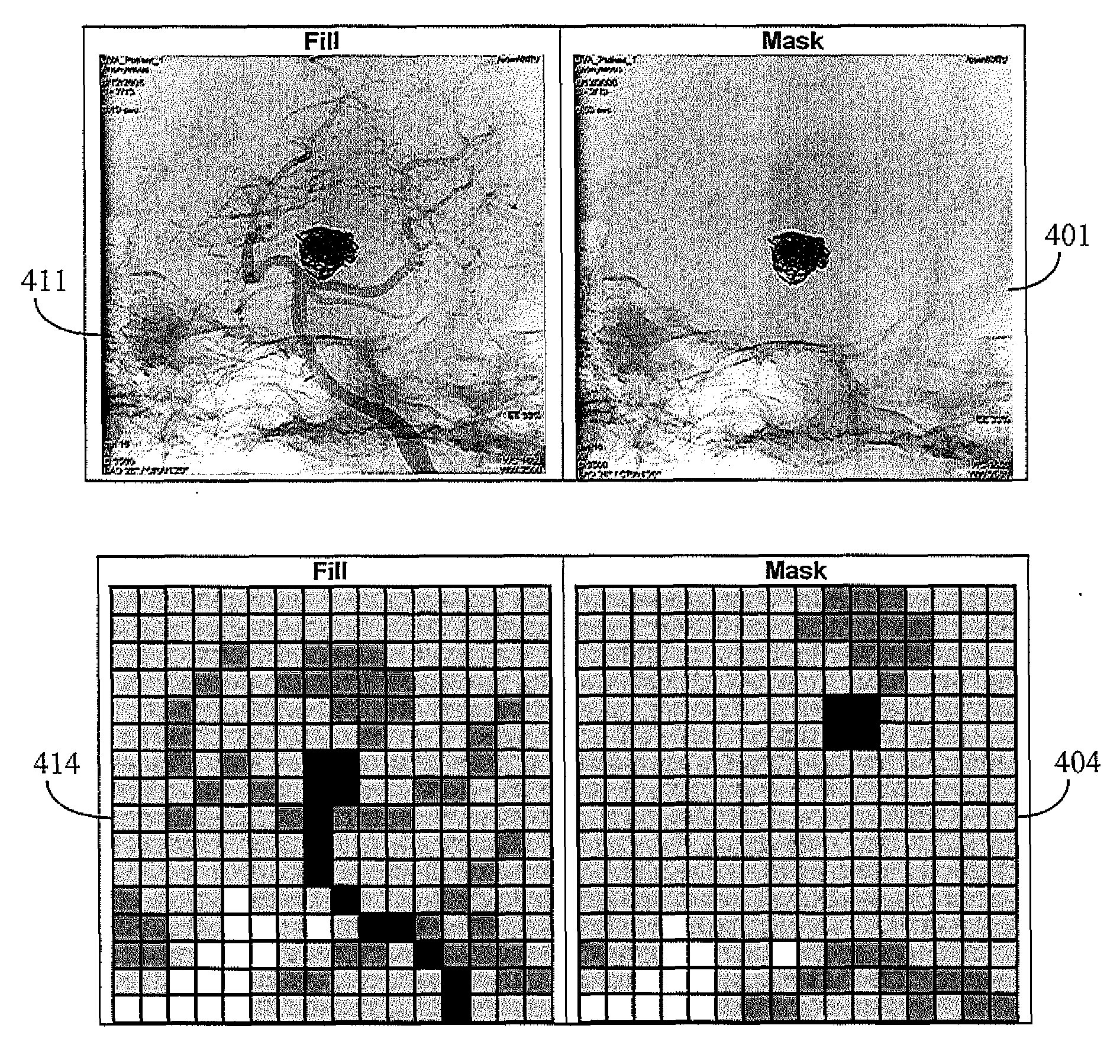
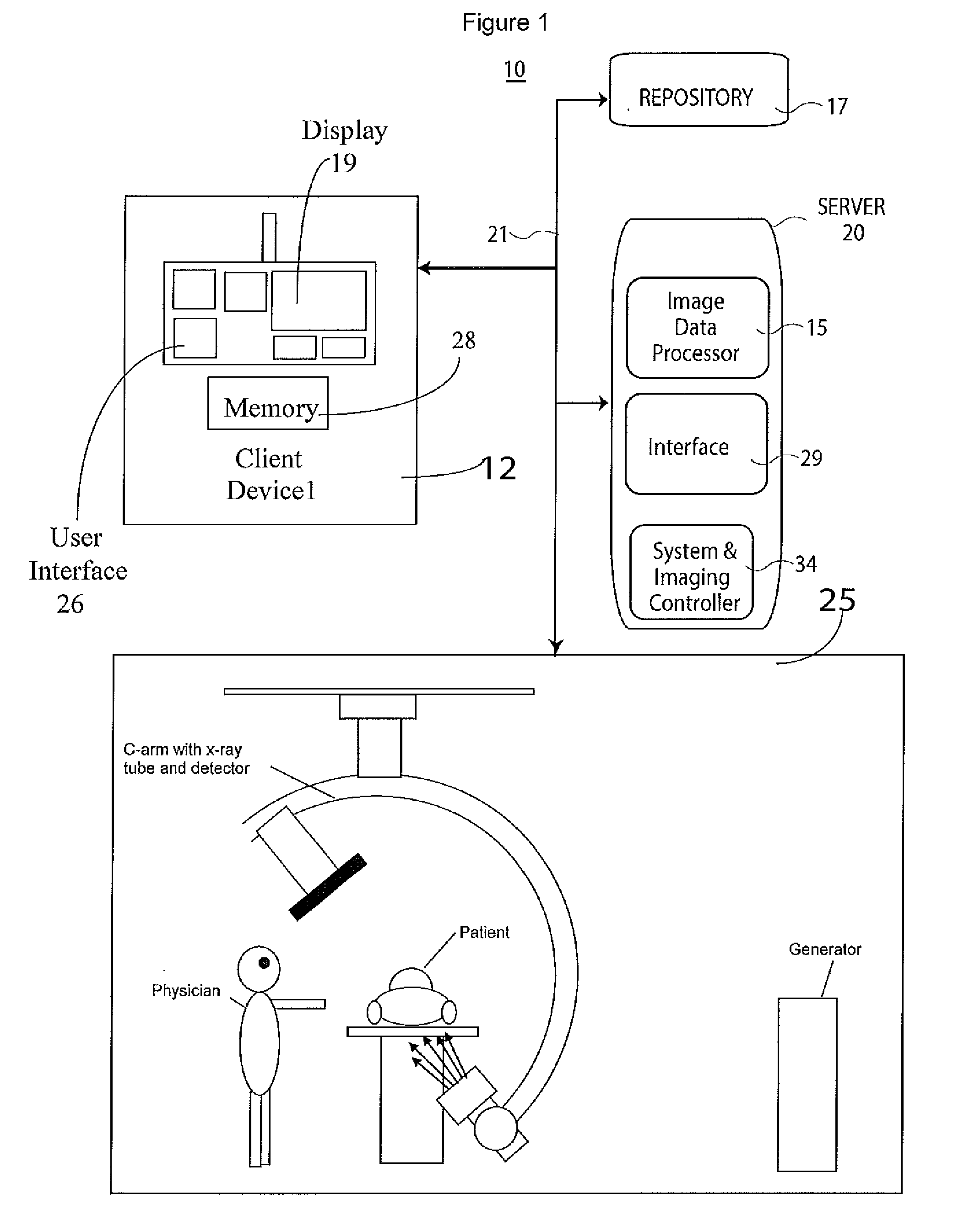
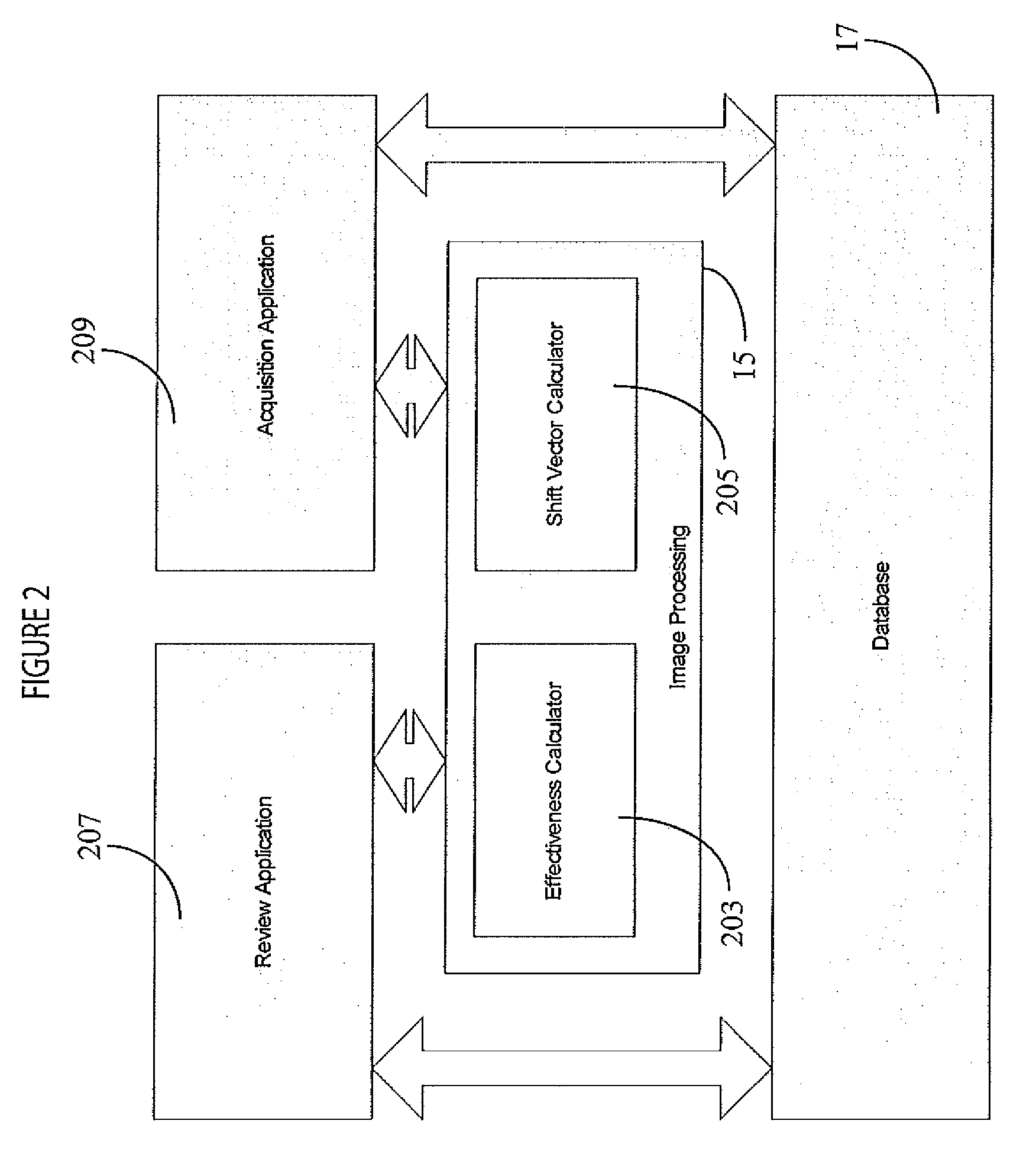
[0019]A system automatically calculates optimal horizontal and vertical shift values for pixels of each frame of a subtracted medical X-ray image in order to correct for a linear registration (alignment) error between a mask and fill frame due to patient movement occurring between acquisition of these frames. The system automatically corrects for the registration error before or during review of an image following image acquisition. The system linearly shifts a mask flame before subtraction from a fill frame to correct registration between the mask and fill frames to improve the result of subtraction and the clarity of a displayed composite image. The distance to shift the mask frame is either manually set by a user or automatically calculated by a reviewing application. This shift distance may be different for individual mask-fill image pairs in a multi-frame sequence of X-ray images as the patient can move at any point during image acquisition. The system fully automates the selec...


PUM


Abstract

An image data subtraction system enhances visualization of vessels subject to movement using an imaging system. The imaging system acquires data representing first and second anatomical image sets comprising multiple temporally sequential individual mask (without contrast agent) and fill images (with contrast agent) of vessels respectively, during multiple heart cycles. An image data processor automatically identifies temporally corresponding pairs of images comprising a mask image and a contrast enhanced image and for the corresponding pairs, automatically determines a shift of a contrast enhanced image relative to a mask image to compensate for motion induced image mis-alignment. The image data processor automatically shifts a contrast enhanced image relative to a mask image in response to the determined shift. The image data processor subtracts data representing a mask image of a corresponding pair from a shifted contrast enhanced image of the corresponding pair, to provide multiple subtracted images showing enhanced visualization of vessels.

Description

[0001]This is a non-provisional application of provisional application Ser. No. 61 / 347,855 filed 25 May, 2010, by M. Pajeau.FIELD OF THE INVENTION[0002]This invention concerns an image data subtraction system to enhance visualization of vessels subject to movement by compensating for a motion induced shift of a contrast enhanced image relative to a mask image.BACKGROUND OF THE INVENTION[0003]Subtracted X-ray images are derived by subtracting a mask frame (i.e. an image without contrast agent) from one or more fill frames (i.e. an image with contrast agent) to hide (by “subtracting” out) uninteresting anatomical structures (i.e. bones during a vascular study). In order to perform this subtraction the anatomical structures in the mask frame need to be registered (aligned) with the corresponding structures in the fill frames. If a patient being X-rayed moves during the time between the acquisition of a mask frame and acquisition of a fill frame, the registration between the frames may ...

Claims


Application Information

Patent Timeline
Patent Type & Authority Applications(United States)
IPC IPC(8): G06K9/00G06V30/144
CPCG06T5/50G06T2207/30101G06T2207/20224G06T2207/10116
Inventor PAJEAU, MICHAEL
Owner SIEMENS HEALTHCARE GMBH
Why Eureka
Try Eureka