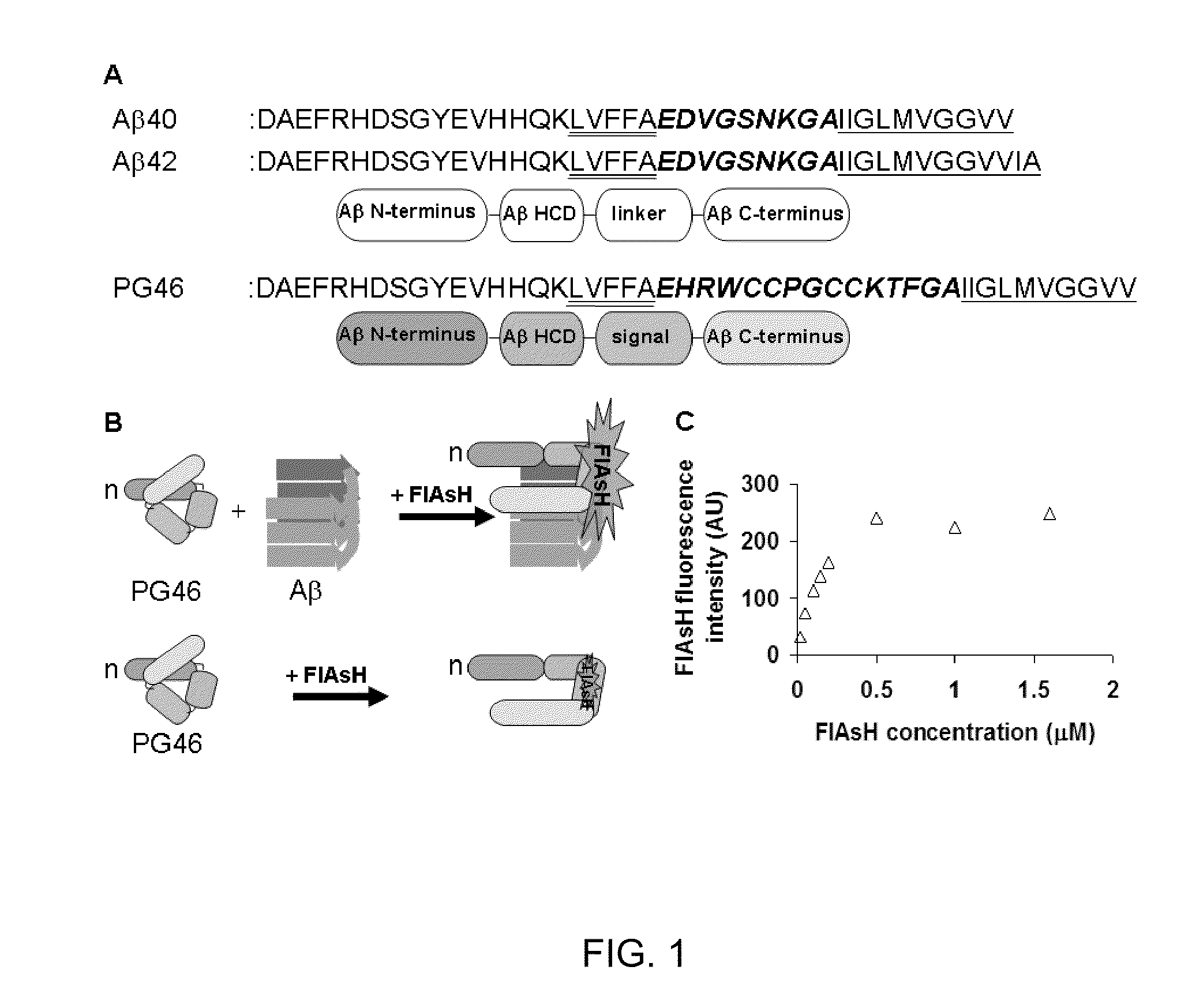
[0050]Design of a peptide probe prototype. The desired property of peptide probes is the ability to modulate fluorescence signals through association with AR species, in particular oligomers (FIG. 1 B). Peptide probes contain the N-terminus (D1-K16), HCD (L17-A21) and the C-terminus (I31-V40) of Aβ3, and the ‘signal domain’ (FIG. 1A). The Aβ N-terminus is included as its presence may reduce the self-assembly of peptide probes (Pike, C. J., et al., Amino-terminal deletions enhance aggregation of beta-amyloid peptides in vitro, J Biol Chem 270, 23895-23898, 1995). The HCD and C-terminal domains of Aβ undergo conformational changes during Aβ self-assembly (Teplow, D. B., et al., Elucidating amyloid beta-protein folding and assembly: A multidisciplinary approach, Acc Chem Res 39, 635-645, 2006; Lazo, N. D., et al., On the nucleation of amyloid beta-protein monomer folding, Protein Sci 14, 1581-1596, 2005; Tjernberg, L. O., et al., Arrest of beta-amyloid fibril formation by a pentapeptide ligand, J Biol Chem 271, 8545-8548, 1996; Soto, C., et al., Beta-sheet breaker peptides inhibit fibrillogenesis in a rat brain model of amyloidosis: implications for Alzheimer's therapy, Nat Med 4, 822-826, 1998; Liu, R., et al., Residues 17-20 and 30-35 of beta-amyloid play critical roles in aggregation, J Neurosci Res 75, 162-171, 2004; Christopeit, T., et al., Mutagenic analysis of the nucleation propensity of oxidized Alzheimer's beta-amyloid peptide, Protein Sci 14, 2125-2131, 2005; Chimon, S., et al., Evidence of fibril-like beta-sheet structures in a neurotoxic amyloid intermediate of Alzheimer's beta-amyloid, Nat Struct Mol Biol, 2007; Losic, D., et al., High resolution scanning tunnelling microscopy of the beta-amyloid protein (Abeta1-40) of Alzheimer's disease suggests a novel mechanism of oligomer assembly, J Struct Biol 155, 104-110, 2006; Mastrangelo, I. A., et al., High-resolution atomic force microscopy of soluble Abeta42 oligomers, J Mol Biol 358, 106-119, 2006; Petkova, A. T., et al., A structural model for Alzheimer's beta-amyloid fibrils based on experimental constraints from solid state NMR, Proc Natl Acad Sci USA 99, 16742-16747, 2002; Luhrs, T., et al., 3D structure of Alzheimer's amyloid-beta(1-42) fibrils, Proc Natl Acad Sci USA 102, 17342-17347, 2005). These conformational changes are utilized in a peptide probe for its functional coupling between binding to Aβ and fluorescence signaling. The HCD and C-terminal domains will also provide a peptide probe with binding affinity toward Aβ, as they are critical in Aβ self-assembly (Tjernberg, L. O., et al., Arrest of beta-amyloid fibril formation by a pentapeptide ligand, J Biol Chem 271, 8545-8548, 1996; Soto, C., et al., Beta-sheet breaker peptides inhibit fibrillogenesis in a rat brain model of amyloidosis: implications for Alzheimer's therapy, Nat Med 4, 822-826, 1998; Liu, R., et al., Residues 17-20 and 30-35 of beta-amyloid play critical roles in aggregation, J Neurosci Res 75, 162-171, 2004; Christopeit, T., et al., Mutagenic analysis of the nucleation propensity of oxidized Alzheimer's beta-amyloid peptide, Protein Sci 14, 2125-2131, 2005). The signal domain is responsible for conformation-dependent fluorescence generation. A tetracystein motif such as CCXXCC (X: a noncystein amino acid) is included in the signal domain (FIG. 1A-B). The signal domain forms the conditional binding site of a nontoxic, membrane-permeable biarsenical fluorescent dye, FlAsH (Adams, S. R., et al., New biarsenical ligands and tetracysteine motifs for protein labeling in vitro and in vivo: synthesis and biological applications, J Am Chem Soc 124, 6063-6076, 2002). FlAsH becomes fluorescent (>50,000x) very rapidly, within a few seconds to minutes, upon binding to the tetracystein motif (Id.). The structures of adjacent flanking sequences would affect the conformation of the tetracystein motif and, therefore, differentiate FlAsH binding and fluorescence (Id.; Ignatova, Z., et al., Monitoring protein stability and aggregation in vivo by real-time fluorescent labeling, Proc Natl Acad Sci USA 101, 523-528, 2004; Madani, F., et al., Hairpin structure of a biarsenical-tetracysteine motif determined by NMR spectroscopy, J Am Chem Soc 131, 4613-4615, 2009; Martin, B. R., et al., Mammalian cell-based optimization of the biarsenical-binding tetracysteine motif for improved fluorescence and affinity, Nat Biotechnol 23, 1308-1314, 2005). Based on these findings, we reasoned that functional coupling between Aβ recognition and fluorescence signaling could be achieved by the conformational change of a peptide probe, particularly in the signal domain and the neighboring HCD and C-terminal domains, upon binding to A. Structural arrangements of HCD, the C-terminus and the linker region of Aβ are different in monomers, oligomers and fibrils (Teplow, D. B., et al., Elucidating amyloid beta-protein folding and assembly: A multidisciplinary approach, Acc Chem Res 39, 635-645, 2006; Fernandez-Busquets, X., et al., Recent structural and computational insights into conformational diseases, Curr Med Chem 15, 1336-1349, 2008; Lazo, N. D., et al., On the nucleation of amyloid beta-protein monomer folding, Protein Sci 14, 1581-1596, 2005; Wetzel, R., et al., Plasticity of amyloid fibrils, Biochemistry 46, 1-10, 2007; Zhang, S., et al., The Alzheimer's peptide a beta adopts a collapsed coil structure in water, J Struct Biol 130, 130-141, 2000; Lee, J. P., et al., 1H NMR of A beta amyloid peptide congeners in water solution. Conformational changes correlate with plaque competence, Biochemistry 34, 5191-5200, 1995; Riek, R., et al., NMR studies in aqueous solution fail to identify significant conformational differences between the monomeric forms of two Alzheimer peptides with widely different plaque-competence, A beta(1-40)(ox) and A beta(1-42)(ox), Eur J Biochem 268, 5930-5936, 2001; Hou, L., et al., Solution NMR studies of the A beta(1-40) and A beta(1-42) peptides establish that the Met35 oxidation state affects the mechanism of amyloid formation, JAm Chem Soc 126, 1992-2005, 2004; Lansbury, P. T., Jr., Evolution of amyloid: what normal protein folding may tell us about fibrillogenesis and disease, Proc Natl Acad Sci USA 96, 3342-3344, 1999; Walsh, D. M., et al., Amyloid beta-protein fibrillogenesis. Structure and biological activity of protofibrillar intermediates, J Biol Chem 274, 25945-25952, 1999; Huang, T. H., et al., Structural studies of soluble oligomers of the Alzheimer beta-amyloid peptide, J Mol Biol 297, 73-87, 2000; Nichols, M. R., et al., Growth of beta-amyloid(1-40) protofibrils by monomer elongation and lateral association, Characterization of distinct products by light scattering and atomic force microscopy, Biochemistry 41, 6115-6127, 2002; Barghorn, S., et al., Globular amyloid beta-peptide oligomer—a homogenous and stable neuropathological protein in Alzheimer's disease, J Neurochem 95, 834-847, 2005; Chimon, S., et al., Capturing intermediate structures of Alzheimer's beta-amyloid, Abeta(1-40), by solid-state NMR spectroscopy, J Am Chem Soc 127, 13472-13473, 2005; Chimon, S., et al., Evidence of fibril-like beta-sheet structures in a neurotoxic amyloid intermediate of Alzheimer's beta-amyloid, Nat Struct Mol Biol, 2007; Losic, D., et al., High resolution scanning tunnelling microscopy of the beta-amyloid protein (Abeta1-40) of Alzheimer's disease suggests a novel mechanism of oligomer assembly, J Struct Biol 155, 104-110, 2006; Mastrangelo, I. A., et al., High-resolution atomic force microscopy of soluble Abeta42 oligomers, J Mol Biol 358, 106-119, 2006; Hoyer, W., et al., Stabilization of a beta-hairpin in monomeric Alzheimer's amyloid-beta peptide inhibits amyloid formation, Proc Natl Acad Sci USA 105, 5099-5104, 2008; Habicht, G., et al., Directed selection of a conformational antibody domain that prevents mature amyloid fibril formation by stabilizing Abeta protofibrils, Proc Natl Acad Sci USA 104, 19232-19237, 2007; Petkova, A. T., et al., A structural model for Alzheimer's beta—amyloid fibrils based on experimental constraints from solid state NMR, Proc Natl Acad Sci USA 99, 16742-16747, 2002; Luhrs, T., et al., 3D structure of Alzheimer's amyloid-beta(1-42) fibrils, Proc Natl Acad Sci USA 102, 17342-17347, 2005; Whittemore, N. A., et al., Hydrogen-deuterium (H / D) exchange mapping of Abeta 1-40 amyloid fibril secondary structure using nuclear magnetic resonance spectroscopy, Biochemistry 44, 4434-4441, 2005; Williams, A. D., et al., Alanine scanning mutagenesis of Abeta(1-40) amyloid fibril stability, J Mol Biol 357, 1283-1294, 2006; Williams, A. D., et al., Mapping abeta amyloid fibril secondary structure using scanning proline mutagenesis, J Mol Biol 335, 833-842, 2002; Torok, M., et al., Structural and dynamic features of Alzheimer's Abeta peptide in amyloid fibrils studied by site-directed spin labeling, J Biol Chem 277, 40810-40815, 2002; Grant, M. A., et al., Familial Alzheimer's disease mutations alter the stability of the amyloid beta-protein monomer folding nucleus, Proc Natl Acad Sci USA 104, 16522-16527, 2007; Baumketner, A., et al., Amyloid beta-protein monomer structure: a computational and experimental study, Protein Sci 15, 420-428, 2006; Baumketner, A., et al., The structure of the Alzheimer amyloid beta 10-35 peptide probed through replica-exchange molecular dynamics simulations in explicit solvent, J Mol Biol 366, 275-285, 2007; Triguero, L., et al., Molecular dynamics study to investigate the effect of chemical substitutions of methionine 35 on the secondary structure of the amyloid beta (Abeta(1-42)) monomer in aqueous solution, J Phys Chem B 112, 2159-2167, 2008; Borreguero, J. M., et al., Folding events in the 21-30 region of amyloid beta-protein (Abeta) studied in silico, Proc Natl Acad Sci USA 102, 6015-6020, 2005). For these reasons, it was hypothesized that the binding of a peptide probe to distinct Aβ species would produce different levels of FlAsH fluorescence. The initial peptide probe, PG46 (FIG. 1A), contains CCPGCC, the most effective tetracystein sequence for FlAsH fluorescence (Adams, S. R., et al., New biarsenical ligands and tetracysteine motifs for protein labeling in vitro and in vivo: synthesis and biological applications, J Am Chem Soc 124, 6063-6076, 2002; Martin, B. R., et al., Mammalian cell-based optimization of the biarsenical-binding tetracysteine motif for improved fluorescence and affinity, Nat Biotechnol 23, 1308-1314, 2005). Additional residues (HRW and KTF) were introduced on both ends of the tetracystein motif in PG46 to improve FlAsH binding and fluorescence (Martin, B. R., et al., Mammalian cell-based optimization of the biarsenical-binding tetracysteine motif for improved fluorescence and affinity, Nat Biotechnol 23, 1308-1314, 2005).

