Method For Detecting And Distinguishing Intrahepatic Cholangiocarcinoma

a cholangiocarcinoma and intrahepatic technology, applied in the field of detecting and distinguishing intrahepatic cholangiocarcinoma, can solve the problems of difficult differentiation of hepatocellular carcinoma from hepatic cholangiocarcinoma, low survival rate, etc., and achieves accurate detection, high precision, and simple procedure

Active Publication Date: 2012-03-15
NAT INST OF ADVANCED IND SCI & TECH +1
View PDF1 Cites 24 Cited by
  • Summary
  • Abstract
  • Description
  • Claims
  • Application Information

AI Technical Summary

Benefits of technology

[0060]The present invention provides a method for early detecting and distinguishing intrahepatic cholangiocarcinoma, difficult to differentiate from hepatocellular carcinoma and difficult to early and accurately detect, with a performance clinically acceptable in terms of applicability, sensitivity and precision and, in addition, a method for detecting intrahepatic cholangiocarcinoma in a way applicable on a clinical site by a simple procedure. Particularly, the method for detecting intrahepatic cholangiocarcinoma according to the present invention uses a clinical sample or clinical section containing bile as a test specimen for detecting intrahepatic cholangiocarcinoma and enables the detection of intrahepatic cholangiocarcinoma with high precision; thus, it permits the early detection of the occurrence of intrahepatic cholangiocarcinoma with simplicity and high precision on a clinical site, which has been difficult when a conventional gene marker is used. In addition, the kit for detecting and / or distinguishing intrahepatic cholangiocarcinoma provided for performing the method for detecting intrahepatic cholangiocarcinoma according to the present invention is a kit excellent in clinical application, which is simple and precise in the detection of intrahepatic cholangiocarcinoma and can be easily applied on a clinical site.
[0061]Among candidate marker molecules for specifically detecting intrahepatic cholangiocarcinoma, MY.1E12-binding MUC1, CA125, N-CAM-L1, and Maspin for which intrahepatic cholangiocarcinoma-specific staining is observed provide useful tissue markers for the differential diagnosis of intrahepatic cholangiocarcinoma. In conventional biopsy, the results are influenced by a method for collecting bile and a preparation production procedure as well as widely varying among diagnosing persons differentiating between shapes of cells, reducing the sensitivity thererof. Against such problems, in addition to tissue staining, cell staining is performed using a molecule including WFA to enable the utilization thereof as a marker for biopsy capable of differentiation with high precision compared to conventional biopsy.

Problems solved by technology

Particularly, intrahepatic cholangiocarcinoma requires early detection and accurate diagnosis because it has a low survival rate after surgical resection compared to hepatocellular carcinoma.
However, intrahepatic cholangiocarcinoma is difficult to early detect.
In addition, it is often experienced that even when this carcinoma can be detected by diagnostic imaging such as CT scan, the differentiation thereof from hepatocellular carcinoma is difficult.

Method used


Image

  • Method For Detecting And Distinguishing Intrahepatic Cholangiocarcinoma
  • Method For Detecting And Distinguishing Intrahepatic Cholangiocarcinoma
  • Method For Detecting And Distinguishing Intrahepatic Cholangiocarcinoma

Examples


example 1

[0079][Selection of Intrahepatic Cholangiocarcinoma-Specific Glycan-Recognizing Lectin Using Lectin Array]

[0080](Test Method)

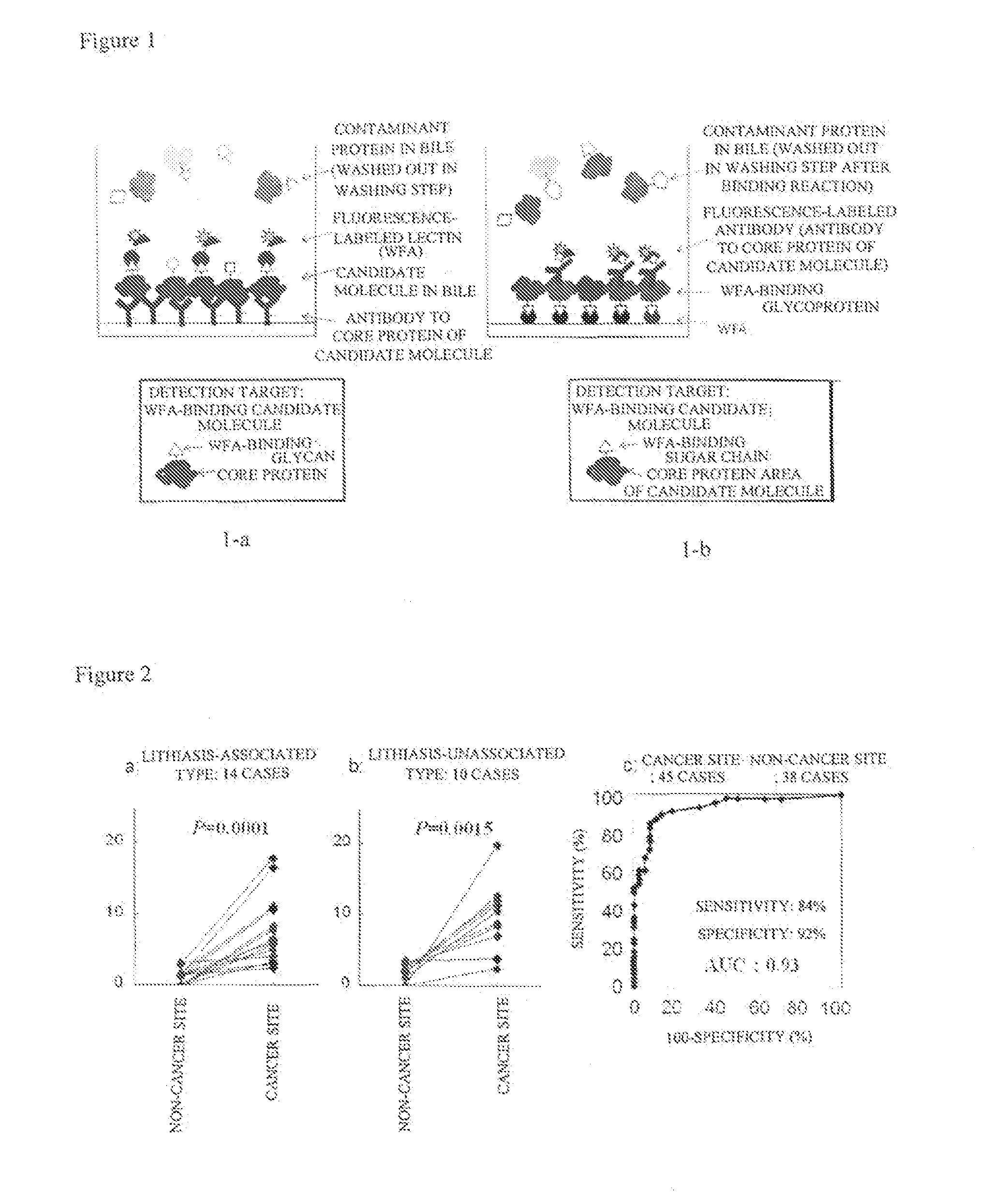
[0081]A lectin microarray has more than 40 plant lectins exhibiting different binding specificity, which are immobilized on the same glass plate, and can be used to analyze simultaneously the interactions thereof with a sugar chain on the glycoprotein to be analyzed (Kuno et al., Nature Methods 2: 851-856, 2005). This system was used to choose suitable lectins to perform intrahepatic cholangiocarcinoma-specific staining. In this experiment, patients associated with lithiasis (cancer and non-cancer areas in the same sections), 10 patients unassociated with lithiasis (cancer and non-cancer areas in the same sections), 21 cancer areas from patients unassociated with lithiasis, and 14 non-cancer areas from patients unassociated with lithiasis were used. In brief, the total number of analyses was 45 for cancer areas and 38 for non-cancer areas. The protocol from th...

example 2

[0093][Detection of Intrahepatic Cholangiocarcinoma Using Intrahepatic Bile Duct]

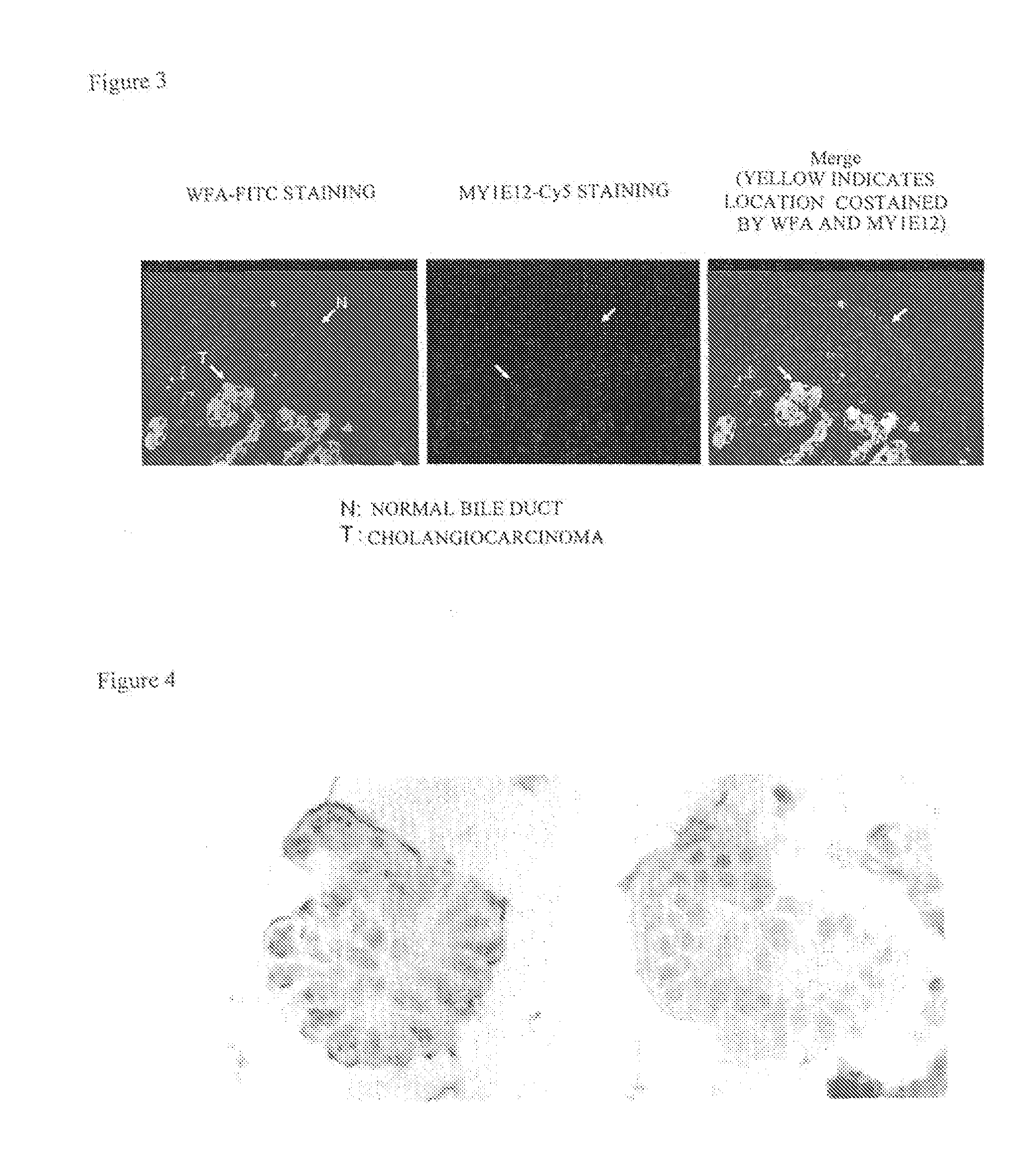
[0094]As shown in Example 1, it is possible to find out intrahepatic cholangiocarcinoma in a tissue section by WFA staining. In next, surgical specimen from intrahepatic cholangiocarcinoma patients but for lithiasis was used to perform as follows.

[0095](Experimental Method)

[0096]Tissue staining with WFA was carried out as follows using a staining kit, Histo-fine (Nichirei Corporation). Formalin-fixed and paraffin embedded materials from intrahepatic cholangiocarcinoma patient were sliced (5 μm thick). Deparaffinized and rehydrated sections were washed with PBS, air-dried, immersed in a 10 mM citrate buffer (pH 6.0), and autoclaved at 121° C. for 15 minutes to dissociate intermolecular (intramolecular) crosslinking due to formalin. The treated section was allowed to stand for a while at room temperature and then repeatedly immersed for 5 minutes three times, and the tissue surface was washed. Then, the b...

example 3

[0101][Detection of Intrahepatic Cholangiocarcinoma Using Cells in Bile]

[0102]Bile duct drainage was conducted to perform releasing obstructive jaundice, and the bile collected at the time was used as a sample for analysis. When WFA staining was performed by the same procedure as in Example 2, WFA-positive cells were identified in a sample in which the presence of cancer was confirmed by histologic diagnosis and biopsy, resulting in cytologically determined cancer. However, no cells stained by WFA were identified in a sample in which histologic diagnosis and biopsy for cancer was negative (FIG. 4: the results of histochemical analysis by WFA staining in a biopsy preparation).


PUM

PropertyMeasurementUnit
binding constantaaaaaaaaaa
thickaaaaaaaaaa

Abstract

Disclosed are a method for early, sensitively and reliably detecting and distinguishing intrahepatic cholangiocarcinoma in a malignant tumor occurring primarily in the liver in a simple way, and a kit therefor. In the method, a glycan biomarker consisting of a lectin WFA (Wisteria floribunda Agglutinin)-binding glycoprotein derived from intrahepatic cholangiocarcinoma is used as a cancer marker to detect intrahepatic cholangiocarcinoma by detecting the cancer marker in a test specimen. The method for detecting intrahepatic cholangiocarcinoma can clearly differentiate intrahepatic cholangiocarcinoma from hepatocellular carcinoma and enables early detection and determination with a performance clinically acceptable in terms of applicability, sensitivity and precision.

Description

TECHNICAL FIELD[0001]The present invention relates to the provision of a method for early, sensitively and reliably detecting and distinguishing intrahepatic cholangiocarcinoma as a malignant tumor occurring primarily in the liver in a simple way and a kit therefor, and more specifically to the provision of a method for detecting and distinguishing intrahepatic cholangiocarcinoma employing a clinically applicable and simple device having performance clinically acceptable in terms of applicability, sensitivity and precision by using a glycan biomarker consisting of a lectin WFA (Wisteria floribunda Agglutinin)-binding glycoprotein as a cancer marker to detect a intrahepatic cholangiocarcinoma-specific glycan structure in the glycan biomarker as a device for early and specifically detecting intrahepatic cholangiocarcinoma, and a kit therefor.BACKGROUND ART[0002]Cancer is in the first place among death cause diseases in Japan, and fatalities therefrom are way ahead of those from other ...

Claims


Application Information

Patent Timeline
Patent Type & Authority Applications(United States)
IPC IPC(8): G01N33/574C40B30/04
CPCG01N21/648G01N33/57446G01N33/57438G01N33/57407
Inventor KUNO, ATSUSHIHIRABAYASHI, JUNMATSUDA, ATSUSHINARIMATSU, HISASHIIKEHARA, YUZURUSHODA, JUNICHIKAWAMOTO, TORU
Owner NAT INST OF ADVANCED IND SCI & TECH
Why Eureka
Try Eureka