Methods and devices for repairing bone defects

a technology for bone defects and bone adhesives, applied in the field of bone repair, can solve the problems of flexural, shear properties, and removal of bone parts that also need replacement, and achieve the effect of improving the handling and delivery of bone adhesives

Inactive Publication Date: 2012-03-15
DOCTORS RES GROUP
View PDF2 Cites 0 Cited by
  • Summary
  • Abstract
  • Description
  • Claims
  • Application Information

AI Technical Summary

Benefits of technology

[0010]According to the present invention, a method for repairing a bone defect includes preparing a biocompatible adhesive, suspending polymerization of the biocompatible adhesive prior to full cure, delivering the biocompatible adhesive to the bone defect while polymerization is substantially suspended and accelerating polymerization of the biocompatible adhesive to achieve full cure. Polymerization of the biocompatible adhesive may be effectively suspended over at least a three (3) month time period and, more preferably, over a two (2) year time period. The method provides an implanted bone defect filler with a malleable osteoconductive structure without compromising the bone adhesive's physical characteristics. Additionally, the method improves handling and delivery of the bone adhesive. Polymerization of the biocompatible adhesive may be substantially suspended by freezing the biocompatible adhesive.

Problems solved by technology

For instance, a bone fracture may result in a portion of missing bone that must be replaced.
Similarly, an infection may result in the removal of a portion of bone also requiring replacement.
However, conventional synthetic bone defect fillers are problematic because they have poor tensile, flexural, and shear properties and may adhere poorly to the surrounding bone, which can result in washout of the bone defect filler from the bone defect prior to ingrowth of new bone into the bone defect filler.
However, these permanent bone defect fillers are problematic because, inter alia, they are not resorbable and / or cannot be molded and shaped for in situ curing.
However, collagen fiber bone defect fillers, while shapeable, are problematic because they may have poor tensile and shear properties and may adhere poorly to the surrounding bone.
Additionally, the degree of porosity provided by the collagen fiber structure may be adversely affected through the handling and application thereof such as during mixing of the material, when dispensed through a syringe or similar device, when packed into a surgical site or due to compression of by adjacent tissue or the like.
Additionally, like collagen fiber structures, the degree of porosity provided by cement bone defect fillers may be adversely affected through the handling and application thereof such as during mixing of the material, when dispensed through a syringe or similar device, when packed into a surgical site or due to compression by adjacent tissue or the like.
However, like shapeable bone defect fillers, the degree of porosity provided by polyurethane biocompatible adhesives may be adversely affected through the handling and application thereof such as during mixing of the material, when dispensed through a syringe or similar device, when packed into a surgical site or due to compression of by adjacent tissue or the like.
Additionally, due to their improved adhesive characteristics, polyurethane adhesives may be difficult for a doctor to handle during polymerization thereof, since the polyurethane adhesives pass through a phase in which they are highly adhesive and may stick to unwanted surfaces.
However, this technique avoids optimal adhesion between the polyurethane adhesives and the surrounding bone since the polyurethane adhesive is not applied when most adhesive.
However, these techniques are also problematic since the former requires the polyurethane adhesive to reach a state of polymerization before instrument use or removal and the later operates under the expectation that the instrument will become coated in the polyurethane adhesive and ruined.
However, wearing and removing multiple pairs of gloves may be time consuming and, therefore, expensive for a surgeon to have to undertake during a surgical procedure.
Additionally, this technique may be frustrating for the user and is wasteful, since it operates under the expectation that the gloves will become coated and in the polyurethane adhesive and ruined.
However, this technique is problematic in that it reduces adhesion of the polyurethane adhesive to its intended surfaces and may contaminate the polyurethane adhesive, which may have potentially adverse affects on the polymerization chemistry, such as increased expansion, decreased adhesive characteristics and / or decreased mechanical strength.

Method used


Image

  • Methods and devices for repairing bone defects
  • Methods and devices for repairing bone defects
  • Methods and devices for repairing bone defects

Examples


Embodiment Construction

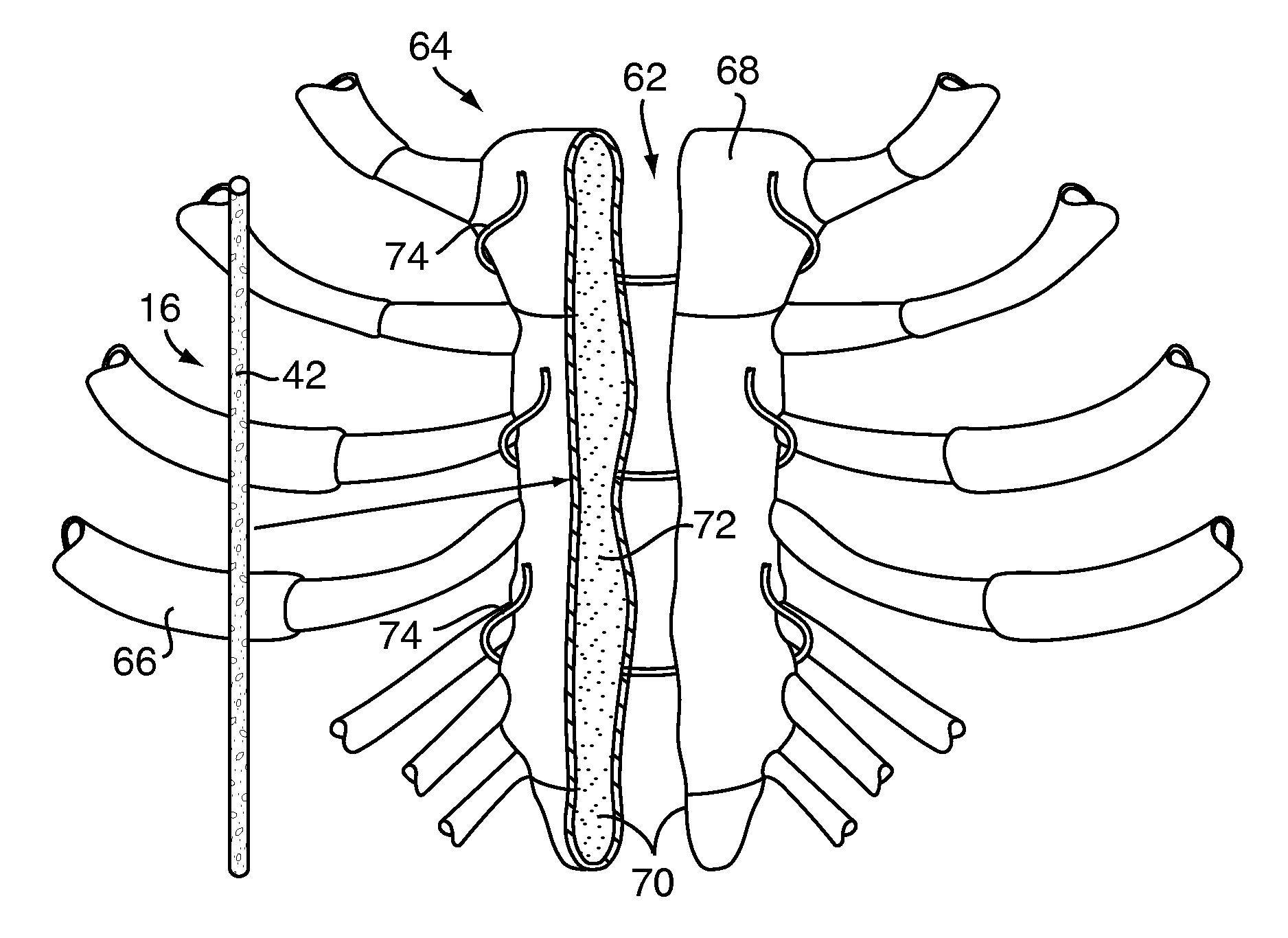
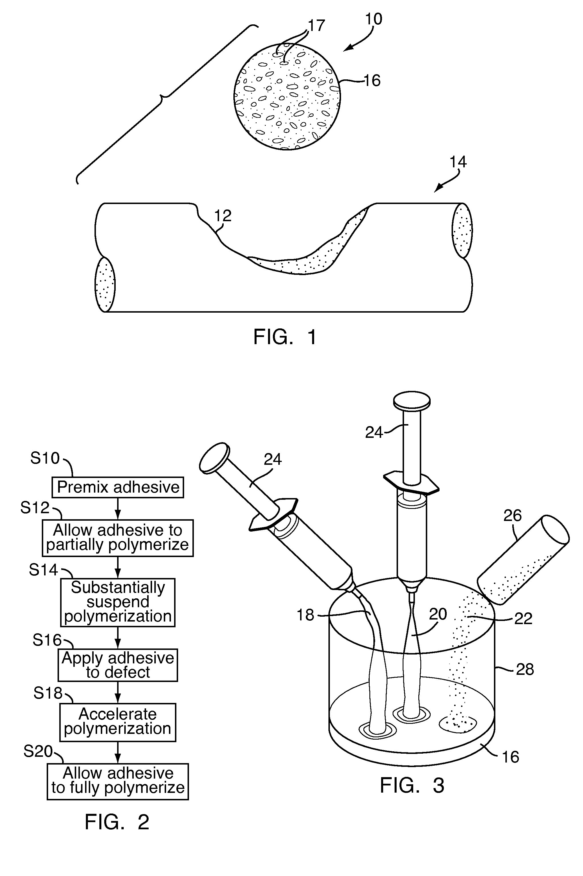
[0043]Referring to FIG. 1, a bone defect filler 10, for repairing a defect 12 in a bone 14 of a patient, includes a biocompatible adhesive 16 that is partially polymerized. The biocompatible adhesive 16 may have pores 17 partially formed therein during the partial polymerization, as will be discussed in greater detail below. Polymerization of the biocompatible adhesive 16 has been substantially suspended, as will also be discussed in greater detail below, to provide for improved handling of the bone defect filler 10, to reduce the likelihood of contamination of the bone defect filler 10 and to ease delivery of the bone defect filler 10 to the defect 12.

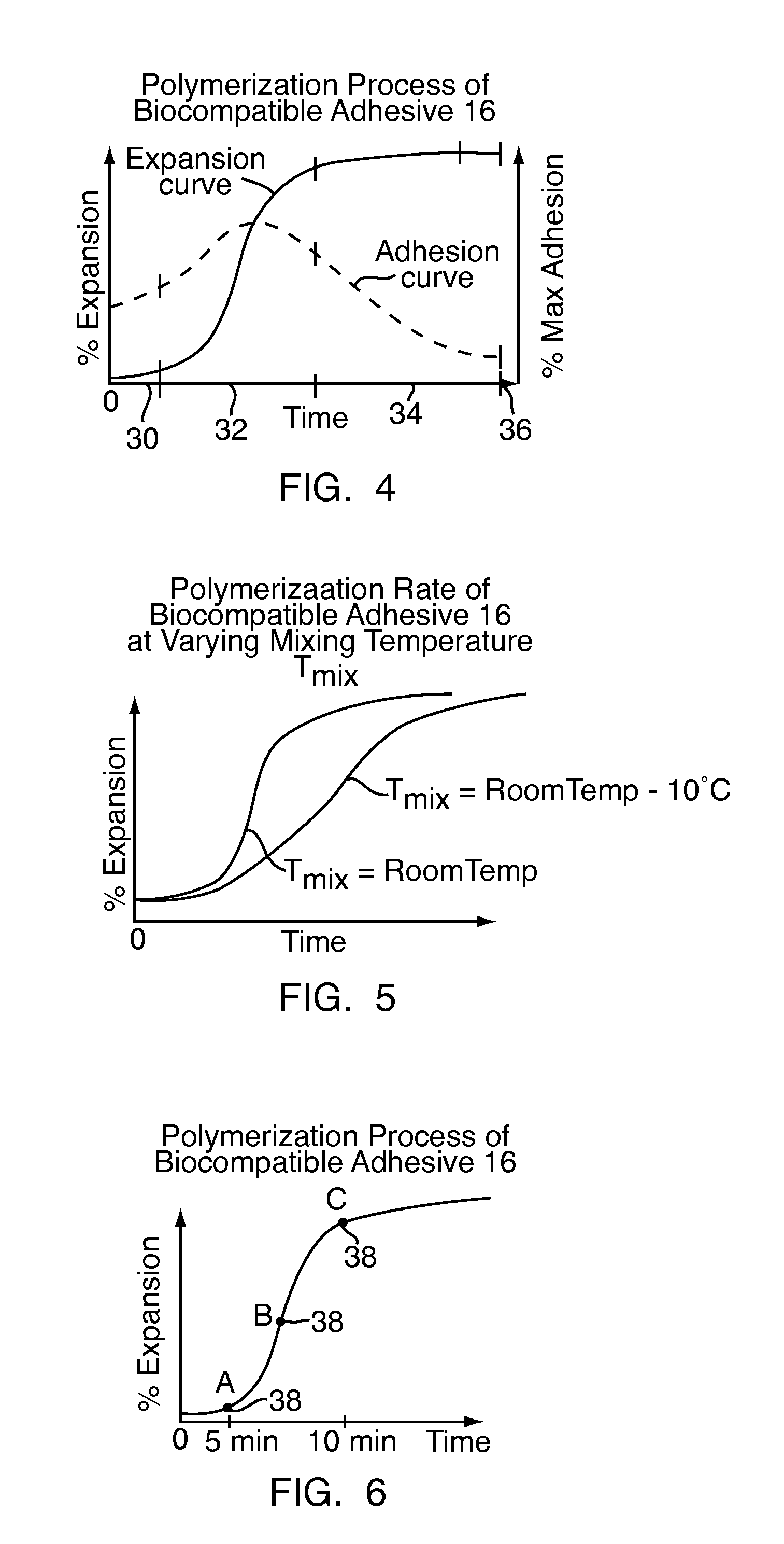
[0044]Referring to FIG. 2, to form the bone defect filler 10, shown in FIG. 1, the biocompatible adhesive 16, shown in FIG. 1, is initially mixed at step S10. Once mixed, the biocompatible adhesive 16, shown in FIG. 1, is allowed to partially polymerize at step S12. Polymerization of the biocompatible adhesive 16, shown in FIG. 1, is ...


PUM

PropertyMeasurementUnit
temperatureaaaaaaaaaa
diameteraaaaaaaaaa
pore sizeaaaaaaaaaa

Abstract

A bone repair apparatus includes a partially polymerized biocompatible adhesive provided in a state wherein polymerization of the biocompatible adhesive is substantially suspended. A method for repairing a bone defect includes preparing the biocompatible adhesive, suspending polymerization of the biocompatible adhesive prior to full cure, delivering the biocompatible adhesive to the bone defect while polymerization is substantially suspended and accelerating polymerization of the biocompatible adhesive to achieve full cure. The method provides an implanted biocompatible adhesive with a malleable osteoconductive structure without compromising the physical characteristics of the biocompatible adhesive, thereby improving handling and delivery of the bone adhesive.

Description

FIELD OF THE INVENTION[0001]The present invention relates to methods and devices for use in bone repair and, more particularly, to bone repair compositions and methods of implantation thereof.BACKGROUND OF THE INVENTION[0002]There are many situations in which defects in bones or portions of bones must be repaired or replaced, including fractures, joint degeneration, abnormal bone growth, infection and the like. For instance, a bone fracture may result in a portion of missing bone that must be replaced. Similarly, an infection may result in the removal of a portion of bone also requiring replacement.[0003]Conventional bone replacement technologies have developed a variety of bone defect fillers for use in orthopedic surgery to repair bones by filling bone voids, gaps, cracks and the like. These bone defect fillers may include a porous structure, which preferably has a high degree of pore interconnectivity, to provide an osteoconductive structure for bone ingrowth after the surgery. F...

Claims


Application Information

Patent Timeline
Patent Type & Authority Applications(United States)
IPC IPC(8): A61B17/58A61K31/765A61P19/08A61K31/74
CPCA61B17/68A61K31/765A61K31/74A61B17/823A61P19/08
Inventor DESLAURIERS, RICHARD J.KOLB, ERICTOMICH, JOHN A.BOXBERGER, JOHNAKKARAPAKA, NARESH
Owner DOCTORS RES GROUP
Why Eureka
Try Eureka